Function of autophagy genes in cell death
a cell death and autophagy gene technology, applied in the field of cell death, can solve the problems of cell metabolism and integrity compromise, cell death, proteolytic activation, etc., and achieve the effects of increasing autophagic cell death, inhibiting cell death, and reducing expression of receptor interacting protein (rip)
- Summary
- Abstract
- Description
- Claims
- Application Information
AI Technical Summary
Benefits of technology
Problems solved by technology
Method used
Image
Examples
example 1
Autophagy and ATG Genes are Required for zVAD-Induced Cell Death.
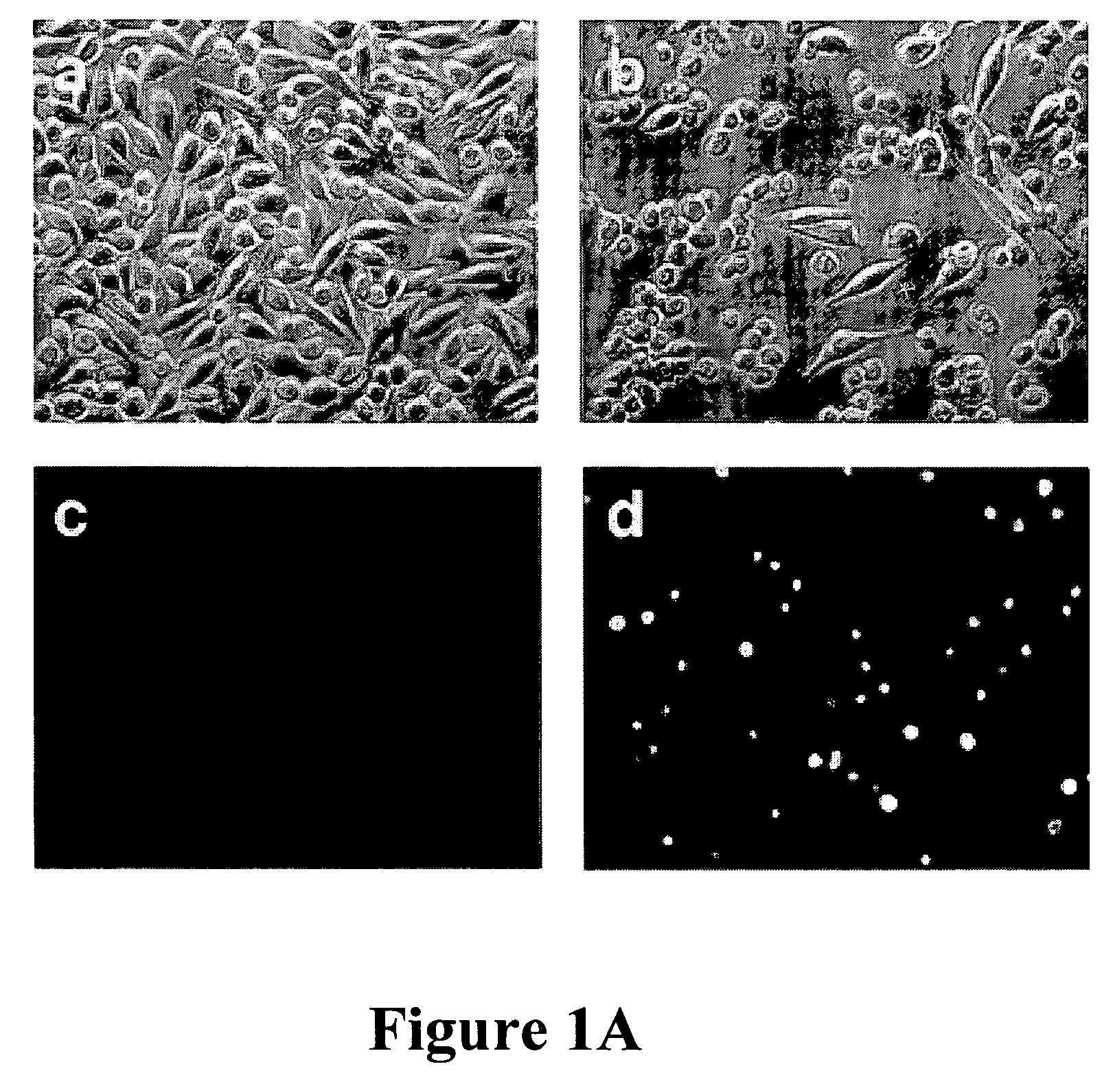
[0101]In mouse L929 fibroblastic cells, tumor necrosis factor (TNF), oxidants, ceramide, and radiation can induce caspase-independent death (11). However, benzyloxycarbonyl-Valyl-Alanyl-Aspartic-acid (O-methyl)-fluoromethylketone (zVAD), a caspase inhibitor with broad specificity, also directly induced the death of L929 cells. L929 cells were treated with 1 ul of dimethyl sulfoxide (DMSO) FIG. 1A (a and c) or 20 uM zVAD (b and d) for 24 hours and examined by phase contrast microscopy (a and b); or 4′,6′-diamidino-2-phenylindole-staining and fluorescent microscopy (c and d). Magnification: 200×. Death began at 12 hours after zVAD treatment and was complete after 40 hrs as shown in FIG. 1B. Transmission electron microscopy (TEM) revealed intact mitochondria and endoplasmic reticulum, condensed osmophilic cytoplasm, and numerous large cytoplasmic inclusions that were membrane-bound vacuoles characteristic of autophagy as ...
example 2
Requirement of RIP and JHK Signaling Pathways for Autophagic Death
[0111]Death receptors can elicit nonapoptotic death through the “receptor-interacting protein” (RIP), a death-domain-containing, serine-threonine kinase (6, 7). RIP expression was reduced by RNAi and decreased autophagy and cell death was observed. For example, cells were treated with zVAD after transfection with RIP RNAi or nonspecific (NS) oligonucleotides. Reduction in cell number (solid bars) and the fractions of cells with autophagic features based on TEM (open bars) are shown in FIG. 2A. FIG. 2B is a western blot for phospho-JNK (left lane) or total JNK protein (right lane) after zVAD treatment. zVAD activated c-Jun N-terminal kinase (JNK) that is also activated by RIP in response to cytokines.
[0112]L929 cells were pretreated with 1 ug / ml JNK inhibitor II, 1 ug / ml p38 inhibitor SB 203580, or 1 ug / ml Erk inhibitor PD 98059 for 1 hour, and were then treated with 20 uM zVAD for 40 hours, with zVAD dissolved in DMSO...
example 3
Inhibition of the Autophagic Death Pathway by Caspase-8
[0114]Finally, the mechanism as to how zVAD induced autophagic cell death was investigated. RNAi was used to progressively reduce caspase-8 expression over time. FIG. 3A shows the time course of viability of L929 cells transfected with either nonspecific (NS) (open bars) or caspase 8-specific (solid bars) RNAi at 24, 96, and 110 hours after transfection. It was found that cell death was correspondingly increased by inhibiting the expression of caspase 8 and thereby inducing the autophagic pathway. Inset panels show the abundance of caspase-8 protein by Western blot.
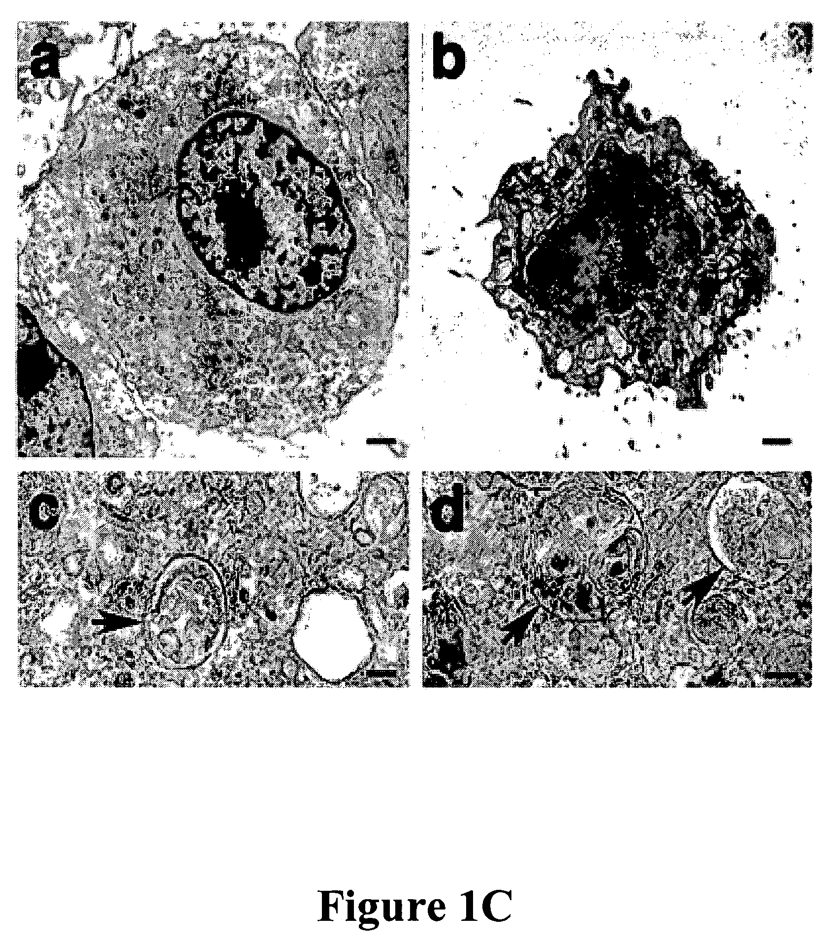
[0115]Representative TEM pictures and quantification of the cells treated with either nonspecific or caspase-8 specific RNAi showed features of autophagy. Cells were harvested at 96 hours after RNAi transfection (a) NS control cell, (b to d) Caspase-8 RNAi at different magnifications. The arrows in c and d show double-membrane autophagic vacuoles. FIG. 3B(e) shows the...
PUM
| Property | Measurement | Unit |
|---|---|---|
| temperatures | aaaaa | aaaaa |
| maintenance time | aaaaa | aaaaa |
| temperature | aaaaa | aaaaa |
Abstract
Description
Claims
Application Information
 - R&D
- Intellectual Property
- Life Sciences
- Materials
- Tech Scout
- Unparalleled Data Quality
- Higher Quality Content
- 60% Fewer Hallucinations
Browse by: Latest US Patents, China's latest patents, Technical Efficacy Thesaurus, Application Domain, Technology Topic, Popular Technical Reports.
© 2025 PatSnap. All rights reserved.Legal|Privacy policy|Modern Slavery Act Transparency Statement|Sitemap|About US| Contact US: help@patsnap.com


