Proteins having effects of controlling cell migration and cell death
- Summary
- Abstract
- Description
- Claims
- Application Information
AI Technical Summary
Benefits of technology
Problems solved by technology
Method used
Image
Examples
example 1
Isolation of FILIP cDNA and Localization of FILIP
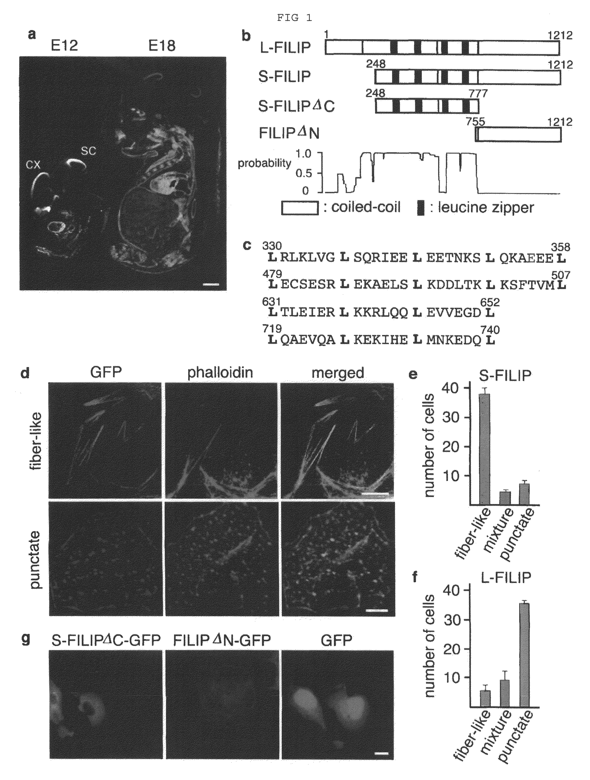
[0124]Although it has been known that Filamin 1 (ABP-280), an actin binding protein, is an essential component of the radial migratory machinery for obtaining postmitotic neocortical neurons (Neuron 21, 1315-1325, 1998), its expression in migratory and postmigratory neurons involved in the development of neocortex, in the region from the intermediate zone to the cortical plate (Neuron 21, 1315-1325, 1998), suggests that the start of migration out of the ventricular zone is possibly controlled by other system. In order to elucidate the molecules concerning controlling the start of neuronal migration, mRNA differential display, in situ hybridization histochemistry, and screening of rat cDNA library were conducted according to the method written in Mol. Brain. Res. 62, 187-195, 1998. First, genes that expressed more abundantly in the neocortics of Wistar rats on embryonic day 11 to 12 (E11 to 12) compared with Wistar rats on embryonic da...
example 2
Interaction of FILIPs with Actin Binding Protein Filamin 1
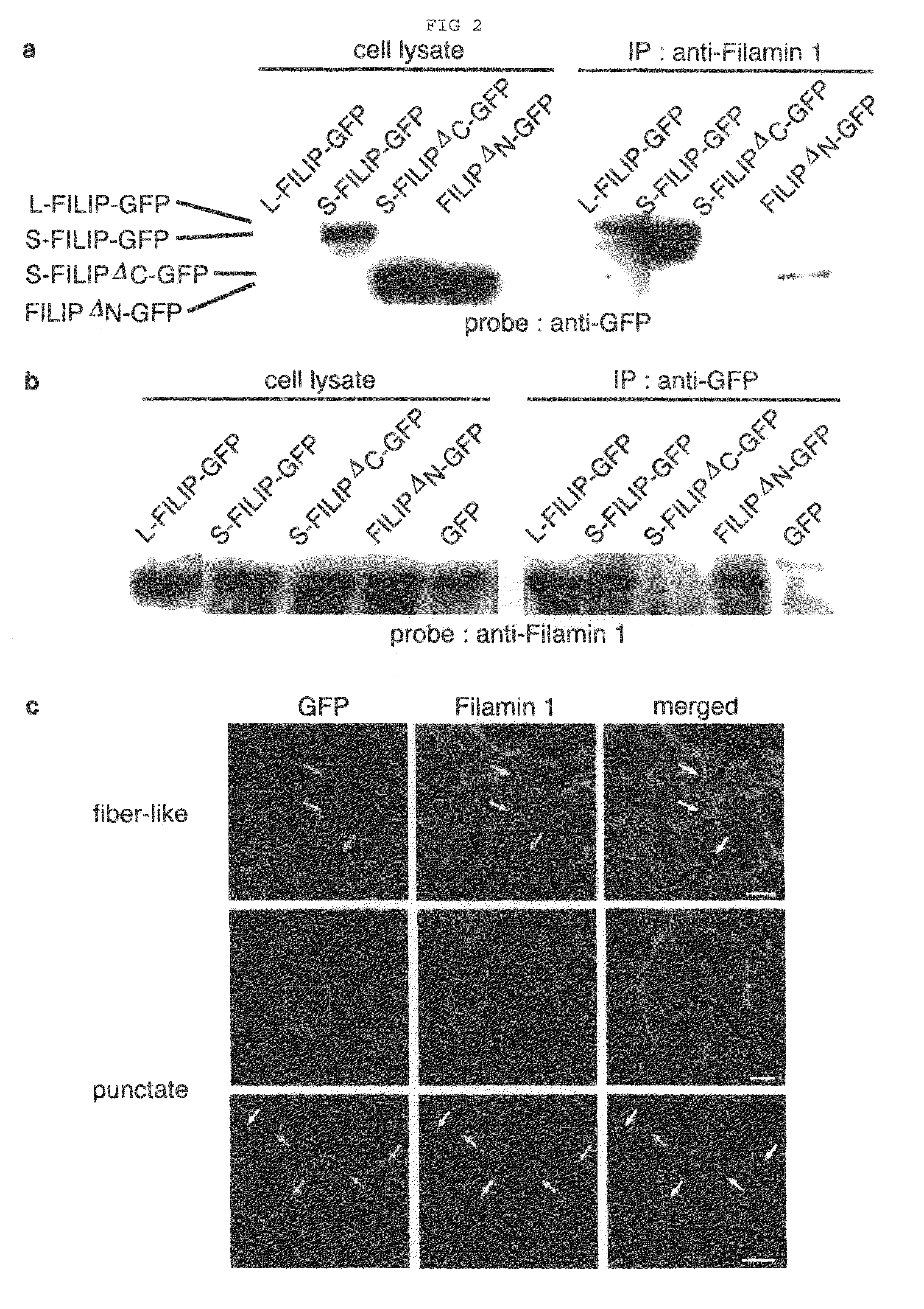
[0129]In order to further examine the unique localization of S-FILIP associated with F-actin, and elucidate the factor that might serve as a link between both molecules, a yeast two-hybrid screen was performed using the C-terminal half of S-FILIP (bait) and the whole embryo library (prey) of mouse E11. Using MATCHMAKER™ Two-Hybrid system (CLONTECH) for a yeast two-hybrid screen, the whole embryo library derived from brain of E11 mouse preintegrated into MATCHMAKER™ library (CLONTECH) was transformed with yeast strain PJ69-2A which was transformed with pAS2-1 plasmid vector carrying cDNA that encoded the common C-terminal region of FILIPs (residues 508-965 of the deduced amino acid sequence of S-FILIP), and C-terminal half of S-FILIP were mated with the whole embryo library of E11 mouse. As a result, over 8×10⁶ clones were screened and 17 clones were selected based on three selection markers. In that way, a clone encoding Fila...
example 3
Degradation of Filamin 1 by FILIPs and Decrease of Cell Motility by the Degradation
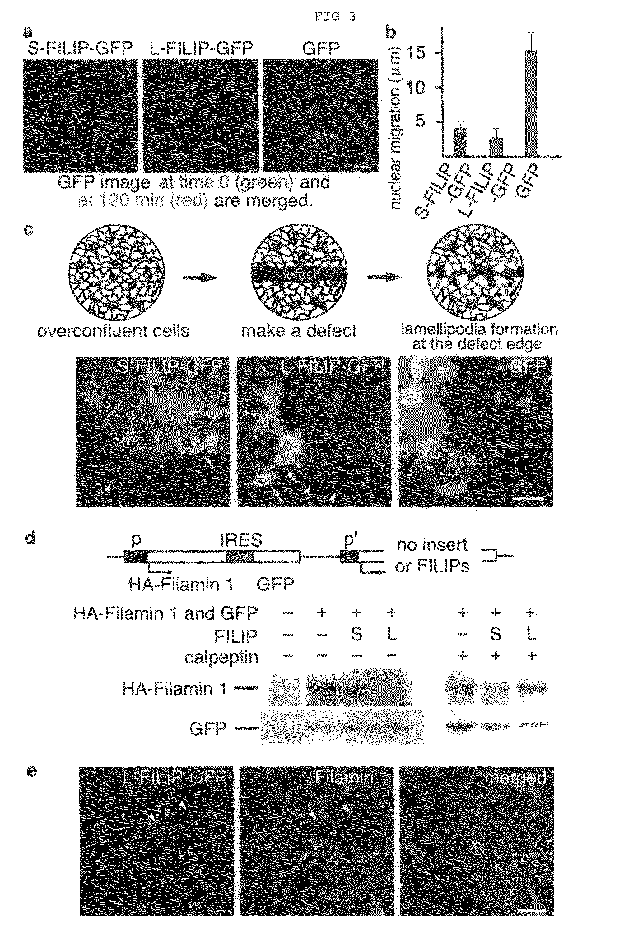
[0132]Since Filamin 1 is deeply involved in cell migration in various cells (Science 255, 325-327, 1992), it is probable that FILIPs control cell migration via Filamin 1. Thus, investigation was performed whether or not FILIPs affect cell migration rate by introducing FILIPs into COS-7 cells, which possessed Filamin 1 but not FILIPs. On the day after plating (approximately 1×10⁴ cells per 1.88 cm2 area), for analyzing the ratio of cell migration rate, COS-7 cells were transfected with expression vectors including S-FILIP-GFP (FIG. 3a left), L-FILIP-GFP (FIG. 3a center), and GFP only (FIG. 3a right). After 36 to 48 h of transfection, the cells were cultured in Dulbecco's modified Eagle medium (DMEM) containing 10% fetal bovine serum (FBS) on an IX-70 microscope equipped with n IX-IBC culturing apparatus (OLYMPUS™) under low cell density condition, image was analyzed twice at an interval of 120 min (FIG...
PUM
 Abstract
Description
Claims
Application Information
 - R&D
- Intellectual Property
- Life Sciences
- Materials
- Tech Scout
- Unparalleled Data Quality
- Higher Quality Content
- 60% Fewer Hallucinations
Browse by: Latest US Patents, China's latest patents, Technical Efficacy Thesaurus, Application Domain, Technology Topic, Popular Technical Reports.
© 2025 PatSnap. All rights reserved.Legal|Privacy policy|Modern Slavery Act Transparency Statement|Sitemap|About US| Contact US: help@patsnap.com


