Determination of clinical stress of a subject in pulse oximetry

a clinical stress and subject technology, applied in the field of clinical stress determination of subjects in connection with pulse oximetry, can solve problems such as increased blood pressure, heart rate and adrenal secretions, and too little stress, and achieve the effect of cost-effectiveness

Inactive Publication Date: 2009-12-22
GENERAL ELECTRIC CO
View PDF16 Cites 24 Cited by
  • Summary
  • Abstract
  • Description
  • Claims
  • Application Information

AI Technical Summary

Benefits of technology

[0028]The present invention seeks to provide a novel mechanism for determining the clinical stress of a patient in connection with pulse oximeters widely available in clinical set-ups. The invention further seeks to provide a cost-effective mechanism for monitoring the state of the patient in post anesthesia care areas and during stressful treatment.
[0033]As the present invention allows the functionality of a conventional pulse oximeter to be expanded by a measurement that evaluates the stress status of a patient, automatic evaluation of the stress status may be introduced in clinical set-ups in a cost-effective way. This is useful not only for finding an optimal balance between a sufficient pain relief and an undisturbed respiration, but also for monitoring patients who are not able to describe their stress status normally or whose description of the their status may not be reliable. The invention therefore provides added reliability for post anesthesia care areas and for stressful therapies. Since the measurement rests on a conventional SpO2 measurement it is also easy to perform and requires no additional measures or sensors.

Problems solved by technology

Some stress keeps us motivated and alert, while too little stress may create problems.
However, too much stress can trigger problems with mental and physical health, particularly over a prolonged period of time.
Pain, discomfort, and clinical stress may activate the sympathetical branch of the ANS and cause an increase in blood pressure, heart rate and adrenal secretions.
It may cause exposure to unwanted side effects during the recovery from the surgery.
Too light sedation causes traumatic experiences both for the patient and for the anesthesia personnel.
From economical point of view, too deep sedation may cause increased costs through extra use of drugs and time, and also extended time required for care.
Too deep a sedation may also cause complications and prolong the usage time of expensive facilities, such as the intensive care theater.
This, however, correlates poorly with sympathetical effects, i.e. with the pain and nociception, and therefore also poorly with the adequacy of analgesia.
The proposed method works reasonably well for spontaneously breathing patients, who in addition have a large RSA, such as children, but often fails in connection with artificially ventilated older patients.
As a result, a low value of clustering may suggest too deep an anesthesia, leading to a decrease in the level of hypnosis.
This may, however, lead to a worse situation, as a result of which the patient may even wake up, especially if surgical stimulations are intense.
However, the method lacks the specificity to noxious stimuli and detects also the variations caused by respiration and other increasing slopes resulting from normal sympathetical activation without noxious stimulation.
For instance, when the patient is in light anesthesia, both the sympathetical and parasympathetical branch of the ANS is active and the indices show erroneously high values suggesting insufficient analgesia.
A further drawback of the prior art technologies is that the measurement values show a considerable inter-patient variability.
This makes their interpretation, i.e. the interpretation of the adequacy of anesthesia, difficult.
The prior art technologies also focus on recording the adequacy of anesthesia during the surgery and are not applicable post-operatively on spontaneously breathing patients.
Since the method is based on detection of noxious events, i.e. short-lived changes in the signal, it cannot provide an indication of the basic level of antinociception.
Since the index is only indicative of the probability of nociception, it cannot provide quantitative information of the level of nociception or of changes in the said level.
However, prior art techniques are complicated as many parameters are required for the monitoring.
The monitoring therefore requires measuring equipment, which is not always available in clinical set-ups.

Method used


Image

  • Determination of clinical stress of a subject in pulse oximetry
  • Determination of clinical stress of a subject in pulse oximetry
  • Determination of clinical stress of a subject in pulse oximetry

Examples


Embodiment Construction

[0045]A pulse oximeter comprises a computerized measuring unit and a probe attached to the patient, typically to a finger or ear lobe. The probe includes a light source for sending an optical signal through the tissue and a photo detector for receiving the signal transmitted through of reflected from the tissue. On the basis of the transmitted and received signals, light absorption by the tissue may be determined. During each cardiac cycle, light absorption by the tissue varies cyclically. During the diastolic phase, absorption is caused by venous blood, tissue, bone, and pigments, whereas during the systolic phase there is an increase in absorption, which is caused by the influx of arterial blood into the tissue. Pulse oximeters focus the measurement on this arterial blood portion by determining the difference between the peak absorption during the systolic phase and the constant absorption during the diastolic phase. Pulse oximetry is thus based on the assumption that the pulsatil...


PUM


Abstract

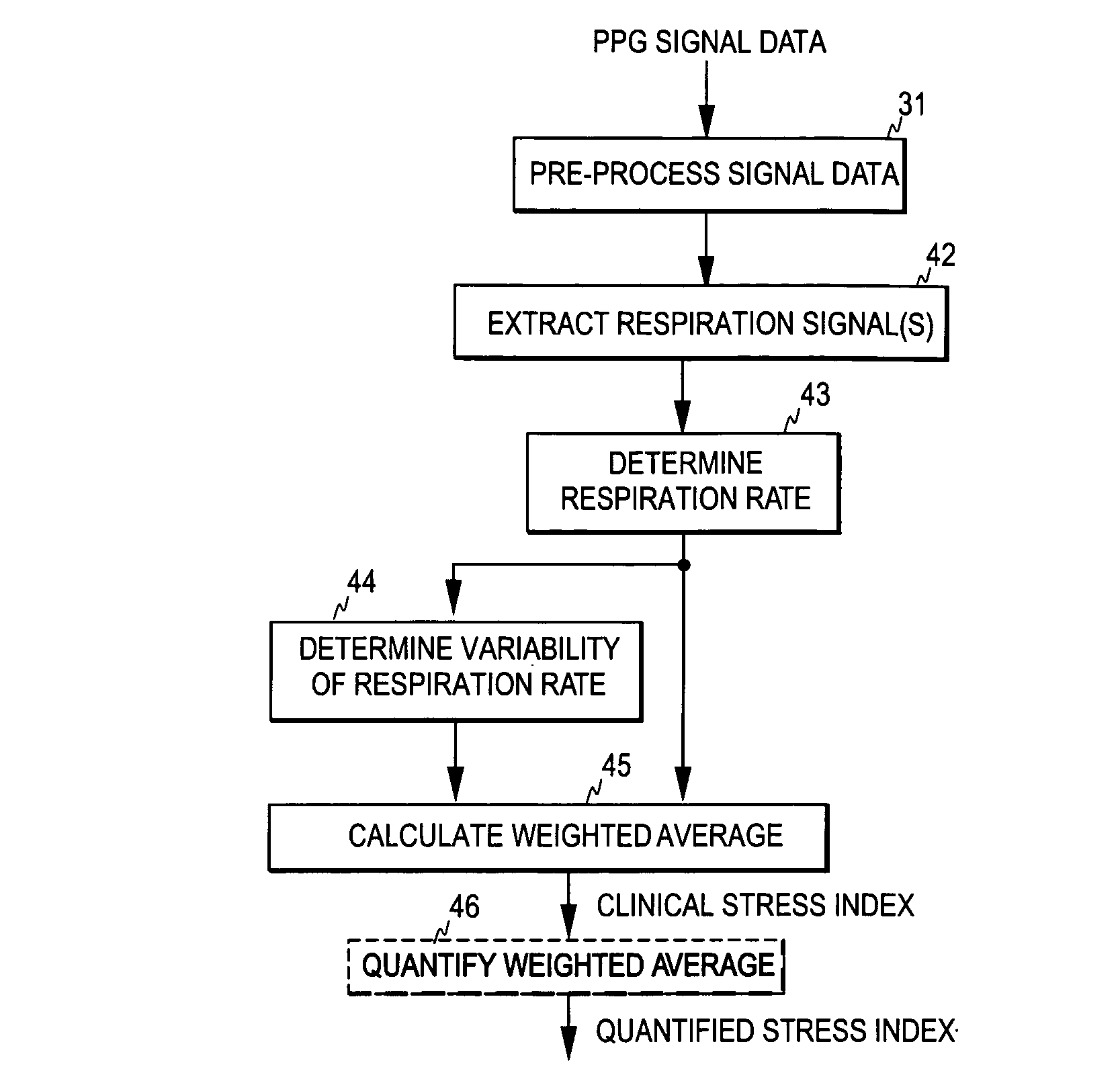
The invention relates to the determination of the clinical stress of a subject. In order to bring about an uncomplicated and cost-effective method for monitoring the stress status of a subject, plethysmographic signal data is acquired from the subject and a first measurement signal is derived, which is indicative of a predetemined feature of the respiration modulation appearing in the said signal data. An index signal is then formed based on the first measurement signal and the index signal is employed as an index indicative of the clinical stress of the subject.

Description

FIELD OF THE INVENTION[0001]The present invention relates generally to the determination of the clinical stress of a subject in connection with pulse oximetry.BACKGROUND OF THE INVENTION[0002]Pain is an unpleasant sensory or emotional experience that is associated with actual or potential tissue damaging stimuli. It is always an individual and subjective sensation, which may be acute (nociceptive), elicited by noxious stimuli, or chronic pain that has outlived its usefulness to preserve tissue integrity. The perception of pain takes mainly place at the cortex, and it may be suppressed in deep sedation and anesthesia by the general (global) inhibitory effects of sedative drugs and anesthetic agents. The responses to noxious stimulus may also be suppressed when the pain signal pathway is sufficiently suppressed at the subcortical level, often in the region of the brainstem and spinal cord. Both cortical and subcortical mechanisms play a role in pain management in modern surgical anest...

Claims


Application Information

Patent Timeline
Patent Type & Authority Patents(United States)
IPC IPC(8): A61B5/02A61B5/08
CPCA61B5/08A61B5/4824A61B5/4821A61B5/14551
Inventor HUIKU, MATTITAKALA, PANU
Owner GENERAL ELECTRIC CO
Why Eureka
Try Eureka