
Plastic implant impregnated with a degradation protector

a technology of degradation protector and plastic implants, applied in the field of plastic implants, can solve the problems of large bone loss, implant failure, and relative short functional existence, and achieve the effects of prolonging the useful life of the prosthesis, prolonging the useful life of the joint, and easy and accurate diagnosis of degradation of the prosthesis

Active Publication Date: 2009-11-10
RUSH UNIV MEDICAL CENT
View PDF6 Cites 23 Cited by
  • Summary
  • Abstract
  • Description
  • Claims
  • Application Information

AI Technical Summary

Benefits of technology

The use of a rare earth metal tracer in prosthetic implants enables early and accurate detection of wear, reducing the risk of catastrophic failure and improving patient safety by allowing for timely intervention and extending the implant's functional life.

Problems solved by technology

This relatively short functional existence is due to problems with decomposition (wear) of the prosthetic's fabrication materials and loosening of the joint due to osteolysis (bone loss) and other complications such as fatique and degeneration of the artificial joint.
Wear debris and the resulting osteolysis from inflammatory cellular responses to wear debris are the most significant factors contributing to failure of hip replacements.
Osteolysis, when undetected and untreated, can result in massive bone loss and implant failure.
Joint revision (replacement) procedures can be more difficult than the original surgery due to diseased, damaged, and decomposed bone in the area of the former prosthesis.
Often there is little original natural bone remaining to attach a new prosthesis after the damaged prosthesis is removed.
The magnitude of this problem is significant.
The function and long term survival of revision total hip arthroplasty is generally inferior to primary hip arthroplasty and leads to a worsened quality of life for many patients.
Metal on metal and ceramic on ceramic implants sometimes have less wear debris than plastic implants; however they come with other risks, including suffering adverse biological effects to increased metal ions in the body, experiencing chipping of ceramic components, and risking fracture of the implant.
Also, with metal and ceramic implants there are additional hurdles to overcome in fabrication such as the difficulty in obtaining conforming surfaces and consistently correct clearances.
Lastly, wear debris from a prosthesis can also lead to bacterial infection in the bone causing failure of the prosthesis.
Current in vivo methods for measuring wear of a prosthetic implant are limited to radiological surveying and physical examination of the patient.
However, in practice, both of these methods are truly useful only for detecting painful, catastrophic prosthetic failure, such that which happens in implant fracture or severe loosening from massive bone loss.
Lesser amounts of implant decomposition are very difficult to diagnose due to the modest degree of physical symptoms.
In addition to wear, artificial joints, particularly those made of UHMWPE, a preferred artificial joint material, also degrade and fatigue due to free radical reactions that can occur prior to implantation and also in vivo, during use.
As mentioned before, radiology often underestimates the amount of wear due to implant decomposition.
In addition, the method is imprecise, and as such, is not reliable for diagnosing less than component failure and / or catastrophic loss of bone stock.
The reason for this unreliability is the physical difficulty in measuring small incremental changes that the prosthetic components may have made by X-ray methods.
It is also difficult to standardize radiographs.
In addition, there are health risks such as cancer due to the patient repeatedly being X-rayed multiple times. Furthermore, in polyethylene prosthetics, fluid absorption and / or creep of the polyethylene samples can be in the same order of magnitude as the wear itself.

Method used


Image

  • Plastic implant impregnated with a degradation protector
  • Plastic implant impregnated with a degradation protector

Examples


example 1

Preparation of a Polyethylene Prosthesis Containing Europium Acetate Tracer

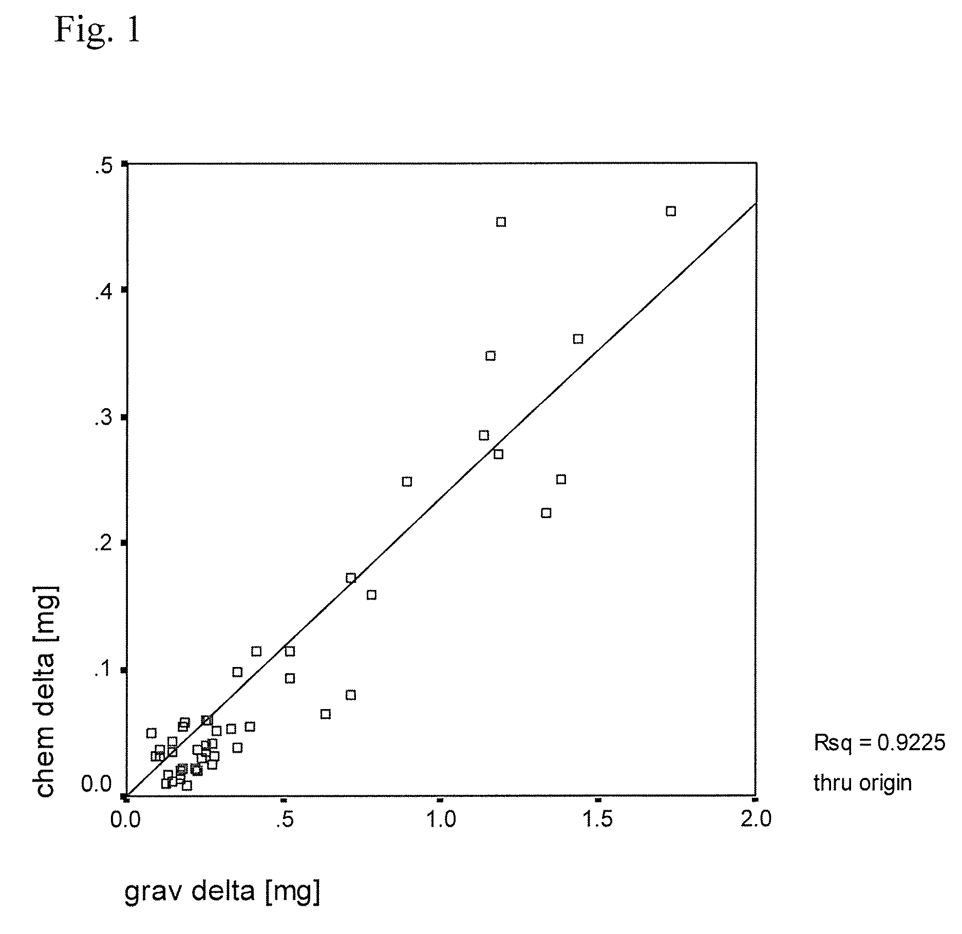
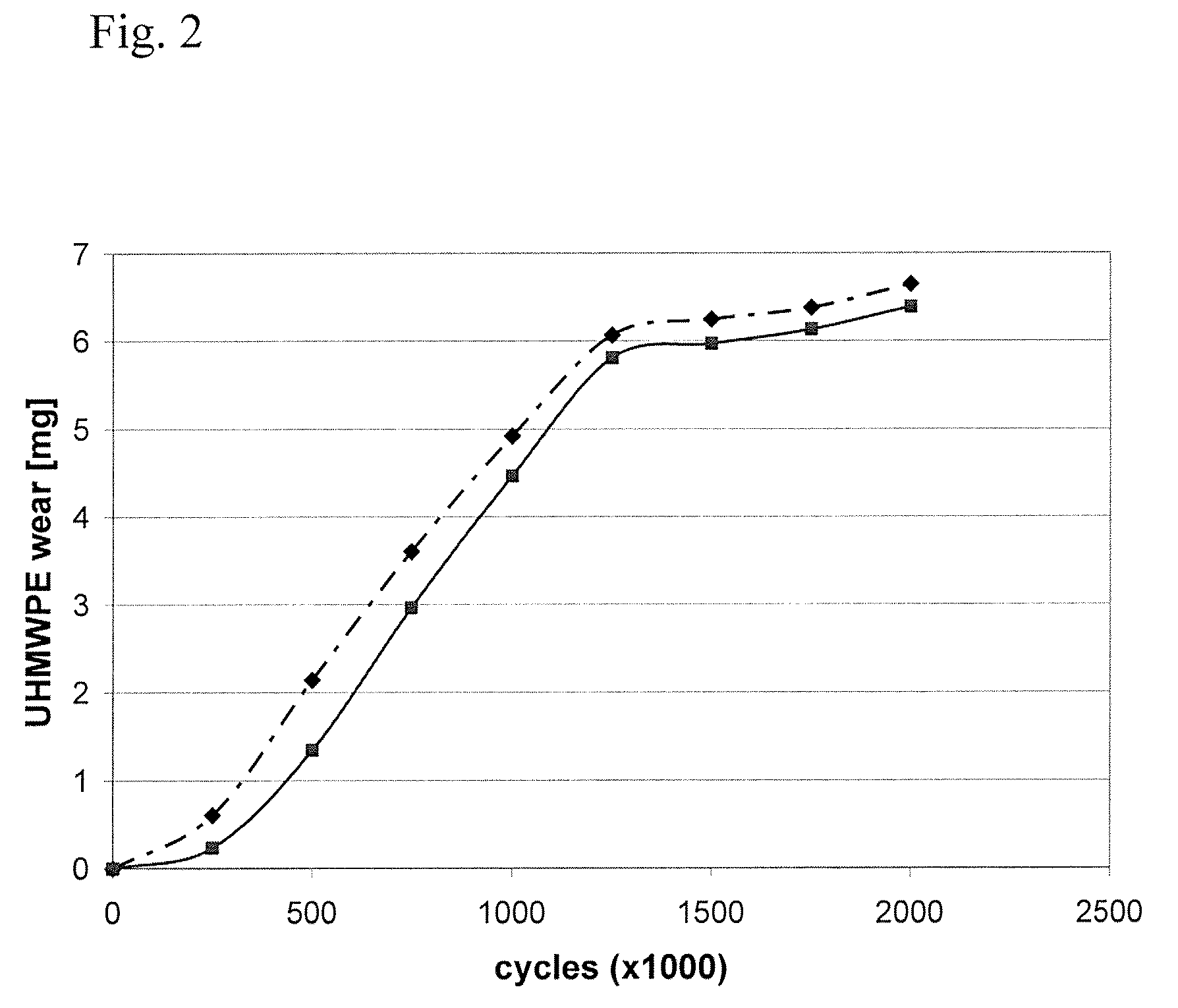
[0047]In this example, several small pins of ultra high molecular weight polyethylene (UHMWPE) containing europium acetate tracer were manufactured and their physical properties assayed. To begin, nascent ultra high molecular weight polyethylene powder was mixed with a solution of europium acetate as described in detail below. Different mixing techniques such as liquid nitrogen milling and n-hexane etching were tried to further homogenize the tracer before consolidation. Tracer homogenicity was analyzed by scanning electron microscopy and backward concentration measurement. The pins were then molded (10 mm diameter) and run on a 0.5×0.5 mm square motion pin-on-disk testing apparatus to test the effects of tracer addition on wear. The wear test was conducted in a lubricant that contained 30% bovine serum to simulate the proteins of an in vivo environment. Liquid samples were taken at regular intervals during t...


PUM

PropertyMeasurementUnit
weight percentaaaaaaaaaa
diameteraaaaaaaaaa
diameteraaaaaaaaaa

Abstract

A plastic implant device for a mammal that contains a rare earth metal compound tracer and a method for detecting degradation such as wear of the implanted device are disclosed. The tracer can also be present with a separate antioxidant or the tracer compound can be can be the salt of a C6-C22 unsaturated carboxylic acid. The rare earth metal compound tracer is released when the prosthetic is worn down or otherwise degraded in the mammalian body in which it was implanted. The presence and amount of released tracer present in a body fluid or tissue sample measured and is proportional to the degree of degradation of the implant.

Description

CROSS-REFERENCE TO RELATED APPLICATION[0001]This application claims priority from Ser. No. 60 / 733,748 filed on Nov. 4, 2005, whose disclosures are incorporated by reference.TECHNICAL FIELD[0002]The present invention relates to a plastic implant that contains an implant degradation protector. Also disclosed is a method for detecting degradation such as wear of an implant in vivo, as well as an in vitro method for assaying wear of materials to be used in an implant.BACKGROUND ART[0003]There are approximately 200,000 hip replacement surgeries each year in the U.S. alone and about 800,000 worldwide. The number of knee replacement surgeries is approximately double this amount. In the future, the number of hip, shoulder, disk and knee replacement surgeries will continue to grow exponentially as the population ages.[0004]Hip replacement surgery entails surgical intervention of two parts of the hip joint, the acetabulum (a cup-shaped bone in the pelvis) and the femoral head (the ball-shaped...

Claims


Application Information

Patent Timeline
Patent Type & Authority Patents(United States)
IPC IPC(8): A61F2/02C01F17/00
CPCA61F2/30A61L27/18A61L27/16A61L27/047C08L23/06C08L71/12A61F2/32A61F2/38A61F2/442A61F2002/30062A61F2002/488A61F2210/0004A61F2310/00035A61F2/488C08L71/00
Inventor KUNZE, ACHIMWIMMER, MARKUS
Owner RUSH UNIV MEDICAL CENT
Learn More