Eureka AIR delivers breakthrough ideas for toughest innovation challenges, trusted by R&D personnel around the world.

Method bacterial endospore quantification using lanthanide dipicolinate luminescence

a technology of lanthanide dipicolinate and bacterial endospores, which is applied in the field of method bacterial endospore quantification using lanthanide dipicolinate luminescence, can solve the problems of increasing the cost and technical difficulty of endospore production, etc., to achieve the effect of improving the sensitivity of bacterial endospor

Active Publication Date: 2007-12-11
CALIFORNIA INST OF TECH
View PDF20 Cites 16 Cited by
  • Summary
  • Abstract
  • Description
  • Claims
  • Application Information

AI Technical Summary

Benefits of technology

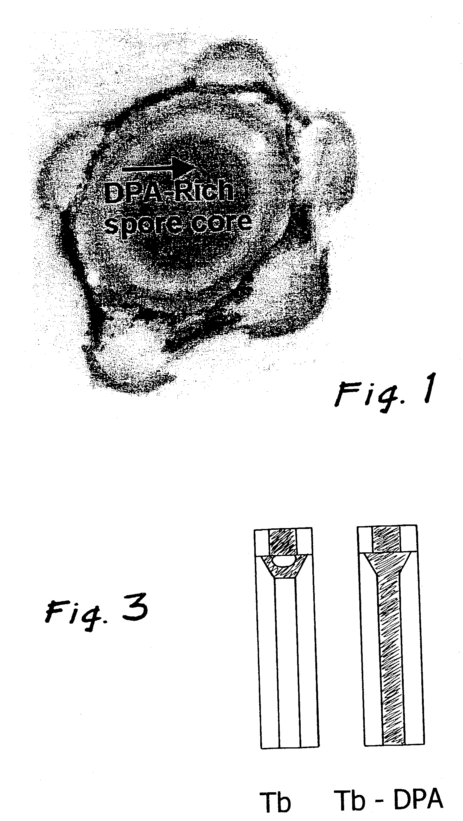
[0013]An improved sensitivity for bacterial endospore detection is realized based on dipicolinic acid triggered lanthanide luminescence. A lanthanide is combined with a medium to be tested for endospores. Dipicolinic acid is released from endospores by inducing germination or destructive lysing (e.g., microwaving, autoclaving, chemical oxidation). The released dipicolinic acid binds the lanthanides, which have distinctive emission (i.e., luminescence) spectra, and are detected using photoluminescence measurements. The concentration of spores is determined by preparing a calibration curve that relates emission intensities to spore concentrations for test samples with known spore concentrations. A lanthanide complex is used as the analysis reagent. The lanthanide complex is comprised of the lanthanide ion bound to a multidentate ligand that increase the dipicolinic acid binding constant through a cooperative binding effect with respect to lanthanide chloride (i.e, lanthanide aquo complex in solution). In addition to enhancing dipicolinic acid binding, the deleterious effects of coordinated water and multiple equilibria are also eliminated by encapsulating the lanthanide ions multidentate ligands. The combined effect of increasing the binding constant and eliminating coordinated water and multiple equilibria increase the sensitivity of the endospore assay by an estimated three orders of magnitude over prior art of endospore detection based on lanthanide luminescence.
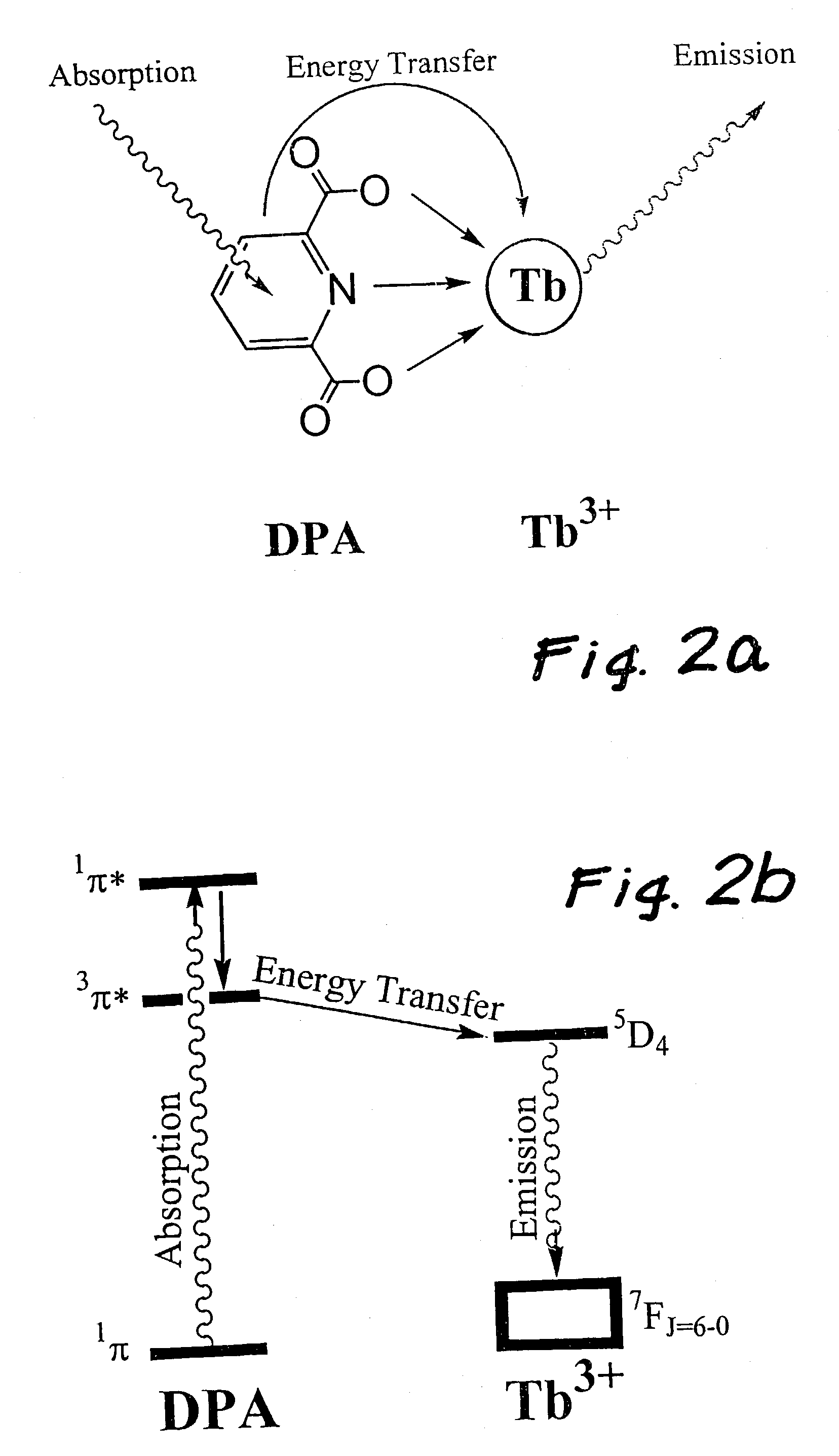
[0015]The mechanism of this method is based on the unique photophysical properties of lanthanide ions. The luminescence of lanthanide ions is characterized by long lifetimes (0.1 to 1 ms), small extinction coefficients (i.e., absorptivity ˜1 M-1 cm-1) and narrow emission bands. Narrow emission bands arise because the valence f-orbitals are shielded from the environment by the outer s and p electrons, and long lifetimes / small extinction coefficients arise because the transition between the emitting excited state and ground state is highly forbidden. Thus, direct excitation of terbium ions leads to weak luminescence due to the small absorption cross section. However, coordination of organic chromophores, like DPA, triggers intense terbium luminescence. The juxtaposition of DPA, which has an absorptivity of 5000 M-1 cm-1, serves as a light-harvesting center (i.e., antenna effect). Strong electronic coupling and downhill energetics allow the DPA centered excitation energy to be efficiently transferred to the lanthanide ion, which subsequently luminesces bright green.
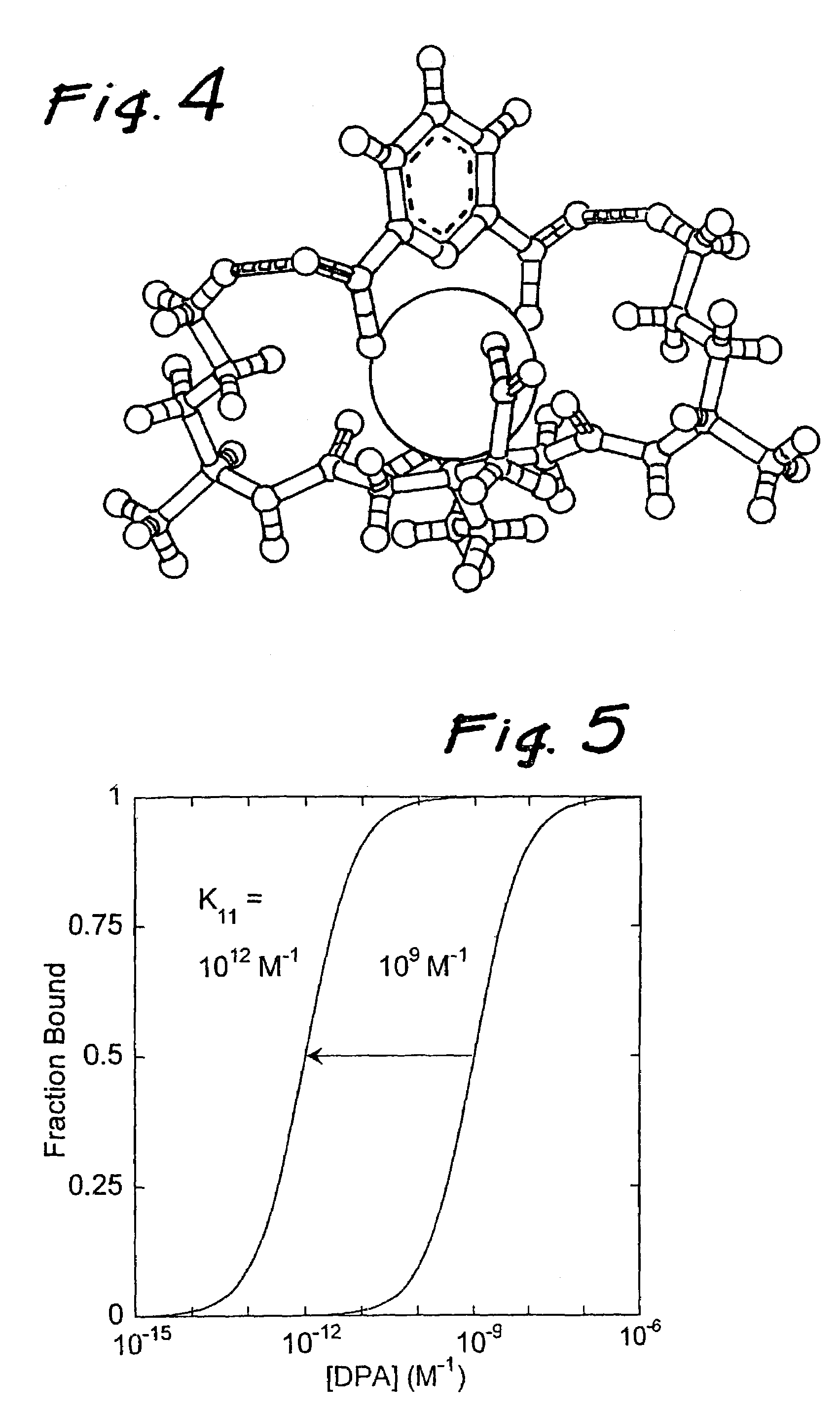
[0017]In addition to increasing the DPA to Tb binding constant, these multidentate ligands (i.e., ligands that coordinate with multiple ligating moieties) eliminate the deleterious effects of coordinated water and multiple equilibria. A Tb-ligand complex with a coordination number of 6 (i.e., the ligand coordinates to the lanthanide at six locations) allows only one DPA molecule to bind and the Tb-crown ether DPA complex contains no coordinated water, which increases the quantum yield. The detection limit is increased by approximately an order of magnitude due to elimination of coordinated water and the reduction of Tb-complex background luminescence.
[0021]In the step of combining the lanthanide ions with the medium in the form of a lanthanide complex as the analysis reagent reacting with the endospores, the ligand that enables cooperative binding is usually multidentate (i.e., a ligand defined as having a coordination number of more than one and preferably 4-6) and allows only one dipicolinic acid molecule to bind to the endospore, thereby eliminating multiple equilibria and the resulting dipicolinic acid complex contains few to no coordinated water molecules. Preferably the terbium is provided in slight molar excess over its stoichiometric ratio to reduce background luminescence of uncoordinated terbium.

Problems solved by technology

The cost and technical difficulty of endospore production has continued to decrease over time, thereby coming with the economic and technological means of a wider segment of the world population, including radical ideological groups incapable of being deterred by possible retaliation and harbored by unofficial transnational political networks unburdened any national identification or responsibility.
The current state of the art method requires lengthy and labor intensive methods.
However, because the limited field of view of a microscope at high magnifications, searching for stained bacterial cells in low concentrations can take a prohibitive amount of time.
These methods suffer prohibitive difficulties with low-concentration field samples.
First, endospores tend to agglomerate on particulates; attached endospores cannot be accurately counted with the traditional methods, even though they could easily represent the bulk of biomass in a field sample.
Second, the traditional methods take a long time and are labor-intensive.
Finally, colony-counting methods only work for cultivable bacteria, which are in the minority in field samples.
The current state-of-the-art luminescence endospore quantification scheme suffers from low sensitivity, which is limited by the binding constant (i.e., the affinity of dipicolinic acid for binding the lanthanide ion).

Method used


Image

  • Method bacterial endospore quantification using lanthanide dipicolinate luminescence
  • Method bacterial endospore quantification using lanthanide dipicolinate luminescence
  • Method bacterial endospore quantification using lanthanide dipicolinate luminescence

Examples


Embodiment Construction

[0036]The molecular design considerations of the invention enhance the detection limit by over three orders of magnitude. The design features a complex comprised of a central lanthanide ion that is caged by a multidentate ligand that gives rise to cooperative binding of dipicolinic acid through additional binding interaction to the dipicolinic acid (e.g., hydrogen bonds or electrostatic interactions). Cooperative binding is defined as a net increase in binding constant of dipicolinic acid to lanthanide complexes binding with respect to dipicolinic acid binding to lanthanide ions (i.e., the lanthanide aquo complex). An increase in binding constant results in larger fractions of dipicolinic acid bound to the lanthanide molecules at low dipicolinic acid, which consequently increases the sensitivity of the bacterial spore assay since dipicolinic acid binding triggers lanthanide luminescence. In addition, the multi-dentate ligand serves to increase the quantum yield by an order of magnit...


PUM


Abstract

A lanthanide is combined with a medium to be tested for endospores. The dipicolinic acid released from the endospores binds the lanthanides, which have distinctive emission (i.e., luminescence) spectra, and are detected using photoluminescence. The concentration of spores is determined by preparing a calibration curve generated from photoluminescence spectra of lanthanide complex mixed with spores of a known concentration. A lanthanide complex is used as the analysis reagent, and is comprised of lanthanide ions bound to multidentate ligands that increase the dipicolinic acid binding constant through a cooperative binding effect with respect to lanthanide chloride. The resulting combined effect of increasing the binding constant and eliminating coordinated water and multiple equilibria increase the sensitivity of the endospore assay by an estimated three to four orders of magnitude over prior art of endospore detection based on lanthanide luminescence.

Description

RELATED APPLICATIONS[0001]The present application is related to U.S. Provisional Patent Application, Ser. No. 60 / 334,463, filed Nov. 30, 2001 which is incorporated herein by reference and to which priority is claimed pursuant to 35 USC 119.GOVERNMENT RIGHTS[0002]The U.S. Government has certain rights in this invention pursuant to Grant No. NAS7-1407 awarded by NASA.BACKGROUND OF THE INVENTION[0003]1. Field of the Invention[0004]The invention is directed to improving the sensitivity of technology for the real-time quantification of bacterial endospore levels based on lanthanide dipicolinate luminescence.[0005]2. Description of the Prior Art[0006]An endospore (i.e., bacterial spore or bacterial endospore) is a generalized term for a type of spore that is produced within certain species of bacteria. The primary function of most endospores is to ensure survival through periods of environmental stress. Endospores are drought, heat, and starvation tolerant. They are protected by a hardene...

Claims


Application Information

Patent Timeline
IPC IPC(8): C12Q1/04
CPCC12Q1/04
Inventor PONCE, ADRIANVENKATESWARAN, KASTHURI J.KIRBY, JAMES PATRICK
Owner CALIFORNIA INST OF TECH
Why Eureka
Eureka Blog
Learn More