
Rapid and automated electrochemical method for detection of viable microbial pathogens

a technology of pathogen detection and electrochemical method, which is applied in the direction of liquid/fluent solid measurement, material electrochemical variables, instruments, etc., can solve the problem of sharp decline of curves

Inactive Publication Date: 2007-07-03
THE BOARD OF TRUSTEES OF THE UNIV OF ARKANSAS
View PDF10 Cites 7 Cited by
  • Summary
  • Abstract
  • Description
  • Claims
  • Application Information

AI Technical Summary

Benefits of technology

This method is more sensitive than antibody-based and nucleic acid-based methods, with a detection limit similar to conventional culture methods, and is more rapid, achieving detection times significantly lower than conventional methods while maintaining reproducibility and selectivity.

Problems solved by technology

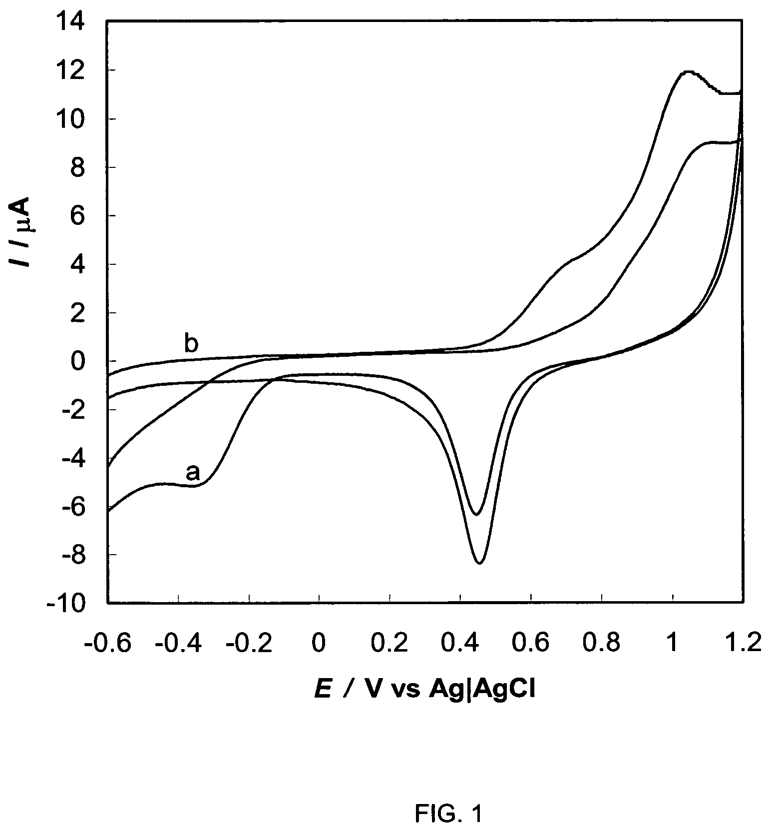
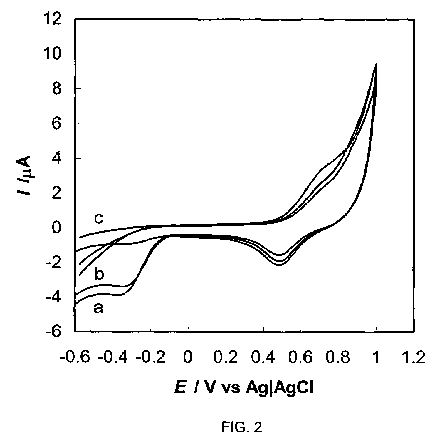
The rapid oxygen consumption at a time during the growth of bacteria resulted in a sharp decline of the curves.

Method used


Image

  • Rapid and automated electrochemical method for detection of viable microbial pathogens
  • Rapid and automated electrochemical method for detection of viable microbial pathogens
  • Rapid and automated electrochemical method for detection of viable microbial pathogens

Examples


example 1

[0040]Salmonella typhimurium (ATCC 14028) as a target pathogen was obtained from Dr Amy Waldroup (Centers of Excellence for Poultry Science, University of Arkansas). Escherichia coli O157:H7 (ATCC 43888) and Listeria monocytogenes (FDA 10143) as competing bacteria were obtained from American Type Culture Collection (Rockville, Md.). Fluid selenite cystine Broth, mannitol, trimethylamine oxide and disodium phosphate were purchased from Sigma-Aldrich (St. Louis, Mo.). Deionized water from Millipore (Milli-Q, 18.2 MΩ / cm) (Millipore, Bedford, Mass.). Buffered peptone water (BPW) was purchased from Remel (Lenexa, Kans.). Gold disk electrodes consisting of a 1.6 mm diameter disk embedded in the end of a 6 mm diameter Kel-F cylinder, platinum auxiliary electrodes, reference electrodes (Ag|AgCl) and polishing materials were from Bioanalytical Systems, Inc. (West Lafayette, Ind.).

example 2

[0041]A BAS CV-50W electrochemical analyzer and IM-6 impedance analyzer (Bioanalytical Systems, Inc., West Lafayette, Ind.) were equipped with a water-jacketed glass vial (15 ml) as an incubator containing three electrodes (Au working electrode, platinum auxiliary electrode and an (Ag|AgCl) reference electrode). The temperature in the incubator was controlled at 37±0.5° C. by a thermostatic bath.

example 3

and Conventional Plating Method

[0042]A pure culture of S. typhimurium was grown in 5 ml BPW broth at 37° C. for approximately 17˜18 h before use. The culture was serially diluted from 1 to 10−8 with selenite cystine / mannitol medium (SC / M). SC / M medium was prepared by adding 17 g fluid selenite cystine and 5 g mannitol into 1 L deionized water. SC / M medium was sterilized by boiling. The prepared medium was stored at 2–8° C. Plate counting was performed by plating 0.1 ml of dilutions on xylose lysine tergitol agar (XLT4) (Difco, Detroit, Mich.). After incubation at 37° C. for 24 h, S. typhimurium colonies on the plate were counted to determine the number of colony forming units of S. typhimurium per ml (CFU / ml) in the culture. A pure culture of E. coli or L. monocytogenes was grown in 5 ml brain heart infusion (BHI) at 37° C. for approximately 17˜18 h before use. The number of E. coli or L. monocytogenes in pure culture was determined by plate counting methods. Colonies of L. monocyto...


PUM

PropertyMeasurementUnit
timeaaaaaaaaaa
timeaaaaaaaaaa
diameteraaaaaaaaaa

Abstract

A method for in situ detection of viable pathogenic bacteria in a selective medium by measuring cathodic peak current of oxygen on cyclic voltammograms during bacterial proliferation with an electrochemical voltammetric analyzer. The rapid oxygen consumption at a time during the growth of bacteria resulted in a sharp decline of the cathodic peak current curves. The detection times (threshold values) obtained from the cathodic peak current curve were inversely related to the concentrations of the pathogenic bacteria in the medium. This method for detection of pathogenic bacteria is more sensitive than nucleic acid-based polymerase chain reaction (PCR) methods and any of antibody-based methods such as enzyme-linked immunosorbent assay (ELISA) technology, electrochemical immunoassays, immunosensors, and it has a sensitivity similar to conventional culture methods and impedimetric methods but is more rapid than both of them. A calibration curve was obtained by plotting initial cell concentrations (CFU / ml) determined by conventional plate counting, as a function of the detection time.

Description

CROSS-REFERENCE TO RELATED APPLICATIONS[0001]The present application claims the benefit of U.S. Provisional Application No. 60 / 401,489 filed Aug. 6, 2002, which is incorporated herein by reference.STATEMENT REGARDING FEDERALLY SPONSORED RESEARCH OR DEVELOPMENT[0002]The invention was made with Government support under the terms of Grant No. 99-34211-7563 awarded by USDA / CSREES. The Government has certain rights in the invention.BACKGROUND OF THE INVENTION[0003]1. Field of the Invention[0004]This invention pertains to a method for the detection of viable pathogenic bacteria in a food or water sample.[0005]2. Brief Description of the Related Art[0006]Various pathogenic bacteria are known to be borne by food products, water or the like. These pathogenic bacteria can include Salmonella typhimurium, Listeria monocytogenes, Escherichia coli O157:H7 and the like. Salmonella typhimurium is considered to be one of the most virulent foodborne pathogens. The ingestion of these organisms via con...

Claims


Application Information

Patent Timeline
Patent Type & Authority Patents(United States)
IPC IPC(8): C12Q1/04
CPCC12Q1/04
Inventor LI, YANBINYANG, LIJURUAN, CHUANMIN
Owner THE BOARD OF TRUSTEES OF THE UNIV OF ARKANSAS
Learn More