
Oncogenes and methods for their detection

a technology of oncogenes and methods, applied in the field of molecular biology, to achieve the effect of illiciting an immune respons

Inactive Publication Date: 2004-03-30
MASSACHUSETTS INST OF TECH
View PDF13 Cites 6 Cited by
  • Summary
  • Abstract
  • Description
  • Claims
  • Application Information

AI Technical Summary

Benefits of technology

As described previously, alteration of the Gly.sub.12 codon in the EJ oncogene makes possible a simple diagnostic assay for carcinogenesis or transformation caused by alteration of this codon in the oncogene. Any mutation of the Gly.sub.12 codon which occurs during carcinogenesis or related processes alters the cleavage recognition site for the NaeI and HpaI endonucleases, and render the altered DNA of the oncogene resistant to cleavage of this site. Thus, any test of the cleavability of the DNA at this site by these endonucleases constitutes a diagnostic test for the mutational alteration of this region of the proto-oncogene.
In practice, a peptide segment can be synthesized by a standard organic synthetic technique, the sequence of this peptide corresponding precisely with the amino sequence of the region of interest of the protein to be studied. This peptide can then be coupled to a carrier protein and injected into a suitable host (e.g., mouse) to illicit an immune response. The serum of the animal immunized in this fashion is then used to immune-precipitate both the immunizing peptide, and more importantly, the protein carrying this amino acid sequence in one of its domains. Consequently, a serum can be made against an oligopeptide sequence (e.g., decapeptide) spanning the amino acid residue site that is altered during the conversion of the normal proto-oncogene to the oncogene. Such serum can be made against the normal peptide sequence, or alternatively, against the altered sequence. The specificity of the immunoglobulin-antigen interaction will insure that the serum reacting with one oligopeptide will only react with the protein bearing the same, corresponding sequence in one of its domains and not cross-react with a protein bearing an altered version of this sequence in one of its domains.

Problems solved by technology

It is generally thought that transformation begins with damage to DNA, although the exact nature of the damage is in dispute.

Method used


Image

  • Oncogenes and methods for their detection
  • Oncogenes and methods for their detection
  • Oncogenes and methods for their detection

Examples


example 2

Definition of DNA Fragment Containing Transforming Sequences

Transforming DNAs, obtained from the EJ cell line as described in Example 1, were exposed to various restriction endonucleases prior to transfection and tested to see whether the biological activity of the DNA survived enzyme treatment. Survival of biological activity would indicate that the functionally essential sequences were carried entirely within one of the many DNA fragments generated after enzyme treatment.

KpnI and PvuII were found to inactivate the transforming potency of DNAs prepared from EJ tumor cells. EcoRI was found to spare the biological activity. Identical results were obtained when cultures were grown up from transfected foci and their DNAs tested by enzyme treatment prior to a second round of transfection. It was concluded, therefore, that the transforming sequences were contained entirely within one EcoRI-generated fragment of EJ bladder carcinoma DNA.

example 3

Detection and Identification of the Transforming DNA Segment for Sequence Hybridization

That fraction of human DNA which self-anneals at a rate of Cot=1 or less was employed as a probe. This probe was prepared from total cellular DNA from a human bladder carcinoma cell line A1663. The DNA concentration was adjusted to 125 ug per ml in 0.01 M PIPES buffer (pH 6.8), and sonicated to ca. 400 bp size fragments. Denaturation was done by boiling at 100.degree. C. for ten min. The NaCl concentration was adjusted to 0.3 M and reannealing performed at 68.degree. C. for 40 min. The solution was quenched on ice, equal volumes of 2.times.S1 buffer (0.50 M NaCl, 0.06 M NaOAc, 2 mM ZnSo.sub.4, 10% glycerol, pH 4.5) was added, followed by addition of 20 ul S1 enzyme (Boehringer, 10.sup.3 units per ul). This was incubated at 37.degree. C. for one hour. 1 M Na-phosphate buffer (equimolar mixture of Na.sub.2 HPO.sub.4 and NaH.sub.2 PO.sub.4) was added to 0.12 M Na-phosphate final concentration and the...

example 4

Molecular Cloning of a Transforming Segment

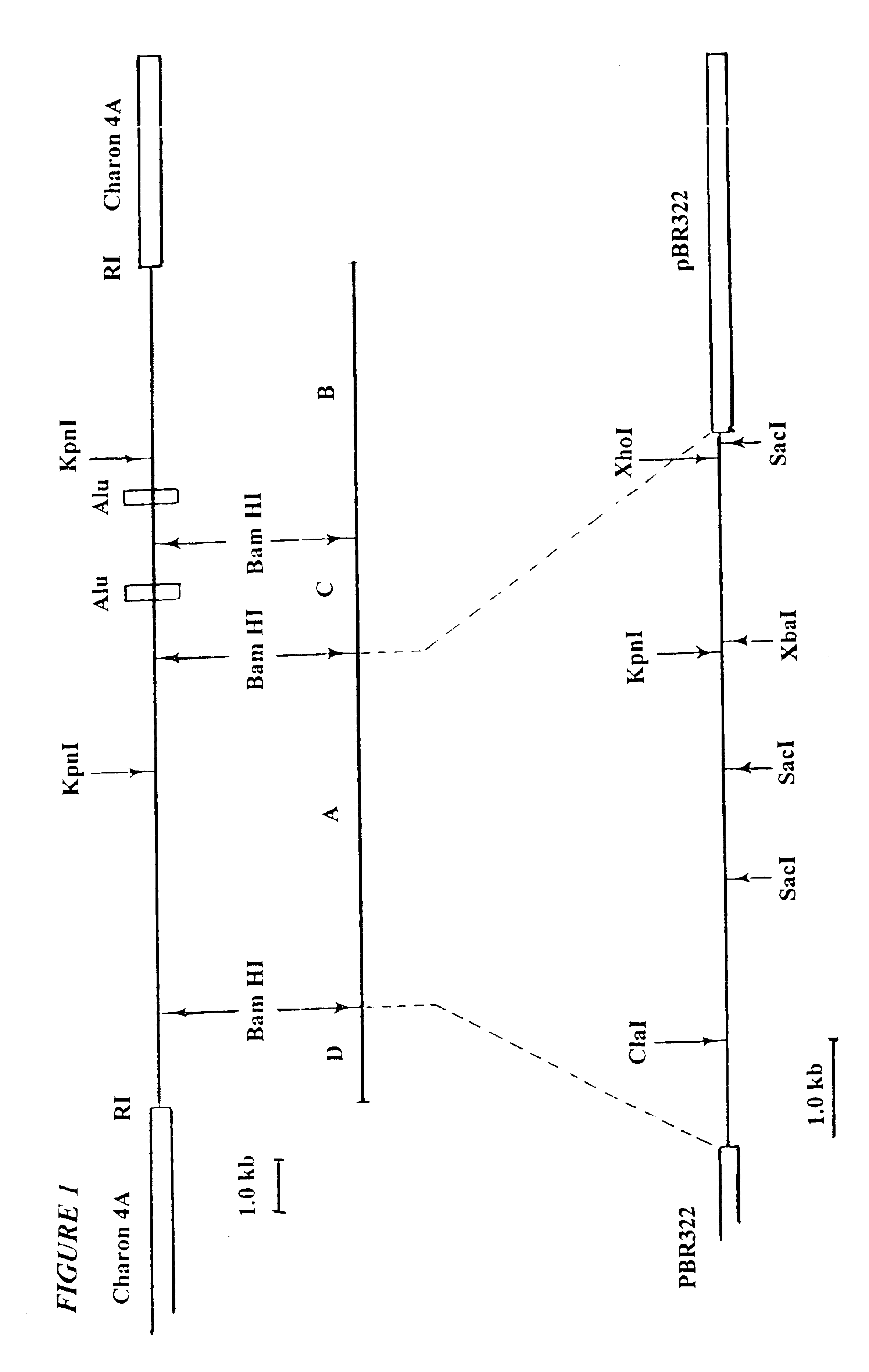
The strategy for molecular cloning of the large EcoRI fragment and associated oncogene began with insertion of EcoRI fragments of a 2.degree. transfectant into lambdaphage vectors. The plan was to screen for components of the resulting library that exhibited sequences reactive with the Cot=1 probe. However, the EcoRI fragment containing the oncogene seen in the Southern blot had a size of about 25 kb, which exceeded the carrying capacity of available lambdaphage vectors. Nevertheless, in occasional 2.degree. transfectants, smaller versions of the large Alu-containing EcoRI fragments had been observed. Such smaller fragments appeared to have resulted from mechanical breakage of the DNAs prior to transfection. Thus, truncated DNA fragments carrying the transforming and Alu sequences appear to have been become fused after transfection with new arrays of EcoRI sites present in co-transfected DNA.

An example of such DNA of the 2.degree. focus lin...


PUM


Abstract

Experiments designed to define the differences between the 21 oncogene of DNA isolated from human bladder cancer cells and its corresponding proto-oncogene are described herein. Also described is the determination of the difference between the rat neu oncogene and its corresponding proto-oncogene. Also described are nucleic acid probes reactive with regions of the proto-oncogene or oncogene, as are methods for their use in detecting the occurrence of the two types of genes. Antibodies specific for gene products encoded by the neu genes are also described, as are methods for their use.

Description

GOVERNMENT SUPPORTThe work described herein was supported by grants from the National Institutes of Health, the National Cancer Institute and the American Business Cancer Research Foundation.This invention is in the field of molecular biology and more specifically relates to isolating discrete genes from mammalian DNA and to defining differences between mutant alleles and their corresponding wild type alleles, particularly oncogenes and proto-oncogenes, and to assays which take advantage of such differences.Previous work relating to chemical carcinogenesis has demonstrated that carcinogenic potency of a compound often correlates with its mutagenic power. See McCann, J., Choi, E., Yamasaki, E. and Ames, B. N. Proc. Natl. Acad. Sci. USA 72: 5135-5139 (1975); McCann, J. and Ames, B. N. Proc. Natl. Acad. Sci. USA 73: 950-954 (1976); Bridges, B. A. Nature 261: 195-200 (1976); and, Bouck, N. and diMayorca, G. Nature 264: 722-727 (1976). This suggests that DNA is the ultimate target of car...

Claims


Application Information

Patent Timeline
IPC IPC(8): C03C8/18C03C8/00C07K14/435C07K14/71C07K16/18C07K16/30C07K16/32C12Q1/68G01N33/531H01B3/12A61K38/00H01G4/008H01G4/232
CPCC03C8/18C07K14/71C07K16/3038C07K16/32C12Q1/683C12Q1/6886G01N33/531H01B3/12H01G4/0085H01G4/2325A61K38/00C12Q2600/156C12Q2600/158
Inventor WEINBERG, ROBERT A.TABIN, CLIFFORD J.BRADLEY, SCOTT M.
Owner MASSACHUSETTS INST OF TECH
Learn More