
Method and apparatus for simultaneous quantification of different radionuclides in a large number of regions on the surface of a biological microarray or similar test objects

a technology of biological microarrays and test objects, applied in the field of methods and apparatus for simultaneous quantification of different radionuclides in a large number of regions on the surface of biological microarrays or similar test objects, can solve the problems of limited dynamic range, high amount of material excluded from the use of standard technology, and degree of bias of starting material

Inactive Publication Date: 2003-08-19
BIOMOLEX
View PDF12 Cites 9 Cited by
  • Summary
  • Abstract
  • Description
  • Claims
  • Application Information

AI Technical Summary

Benefits of technology

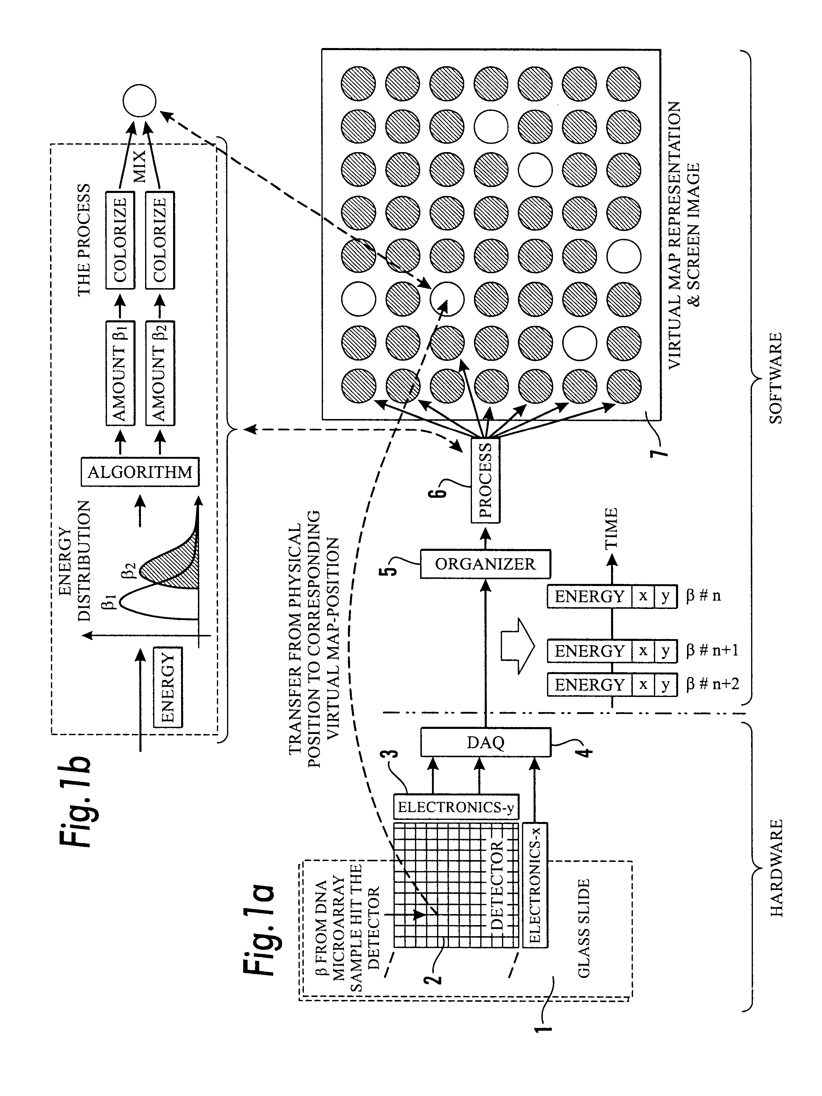
The main objective of this invention is to provide a method and apparatus for performing simultaneous quantifications and / or comparisons of one or more target biological molecules that are distinctively marked with radioactive isotopes and then adsorbed onto a probe array of biological molecules deposited on a substrate, and which substantially eliminates or reduces the above mentioned problems.
By employing this specific combination of solid state technology and highly integrated read-out electronics, one achieves a highly sensitive accurate and reliable instrument which records the position and energy of each radioactive particle that penetrates the sensor plate of the instrument. Thus by placing the sensor plate in close contact with the biological microarray, one can measure the radioactivity from each test spot on the biological microarray. In order to make the instrument versatile and suitable for different types of radiation, it is envisioned that the instrument is equipped with replaceable sensor plates which have equal dimension but are made of different monolithic semiconductor materials, and which can easily be inserted into the integrated read-out electronics. Thus as an example, if one employs a sensor plate made of silicon which responds well to .beta.-radiation, and then wants to investigate a biological microarray tagged with .gamma.-emitters, the instrument can momentarily be made ready for this by substituting the silicon sensor plate with a sensor plate made of cadmium-zinc-tellurium which responds well to .gamma.-radiation.
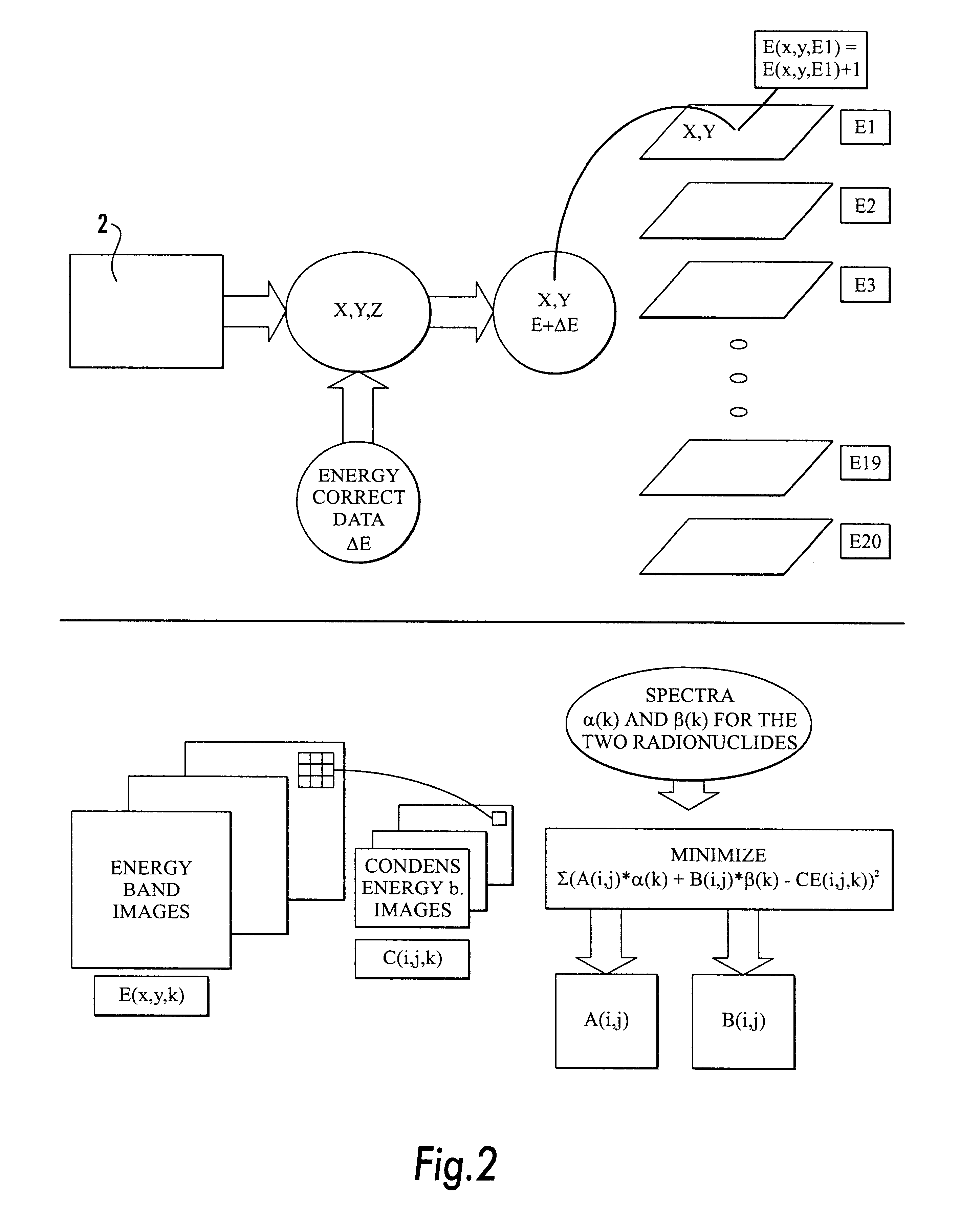
In order to obtain a flexible instrument for a wide range of applications, the instrument is equipped with a very high resolution sensor plate containing a large number of pixels. Thus, for many applications the resolution of the biological microarray will be coarser than the resolution of the sensor plate. In these cases, considerable computing time can be saved if the registered data from neighbouring pixels can be condensed by adding them to the centre pixel. That is, by adding e.g. the registered events in one pixel and all of its nearest surrounding neighbours (a total of 9 pixels) into the centre pixel, the resolution and thus number of computations are decreased by a factor 9. If even coarser resolution is wanted one can include all neighbour pixels in two surrounding rows (a total of 25 pixels) and so forth. In this way we simulate a coarser sensor resolution and thus reduces the necessary computing steps accordingly. The spatial condensing into regions does also give an important increase in number of registered events for each element E.sub.rk, in the resulting condensed energy band image, thus leading to an improved statistical accuracy of the estimated contributions.
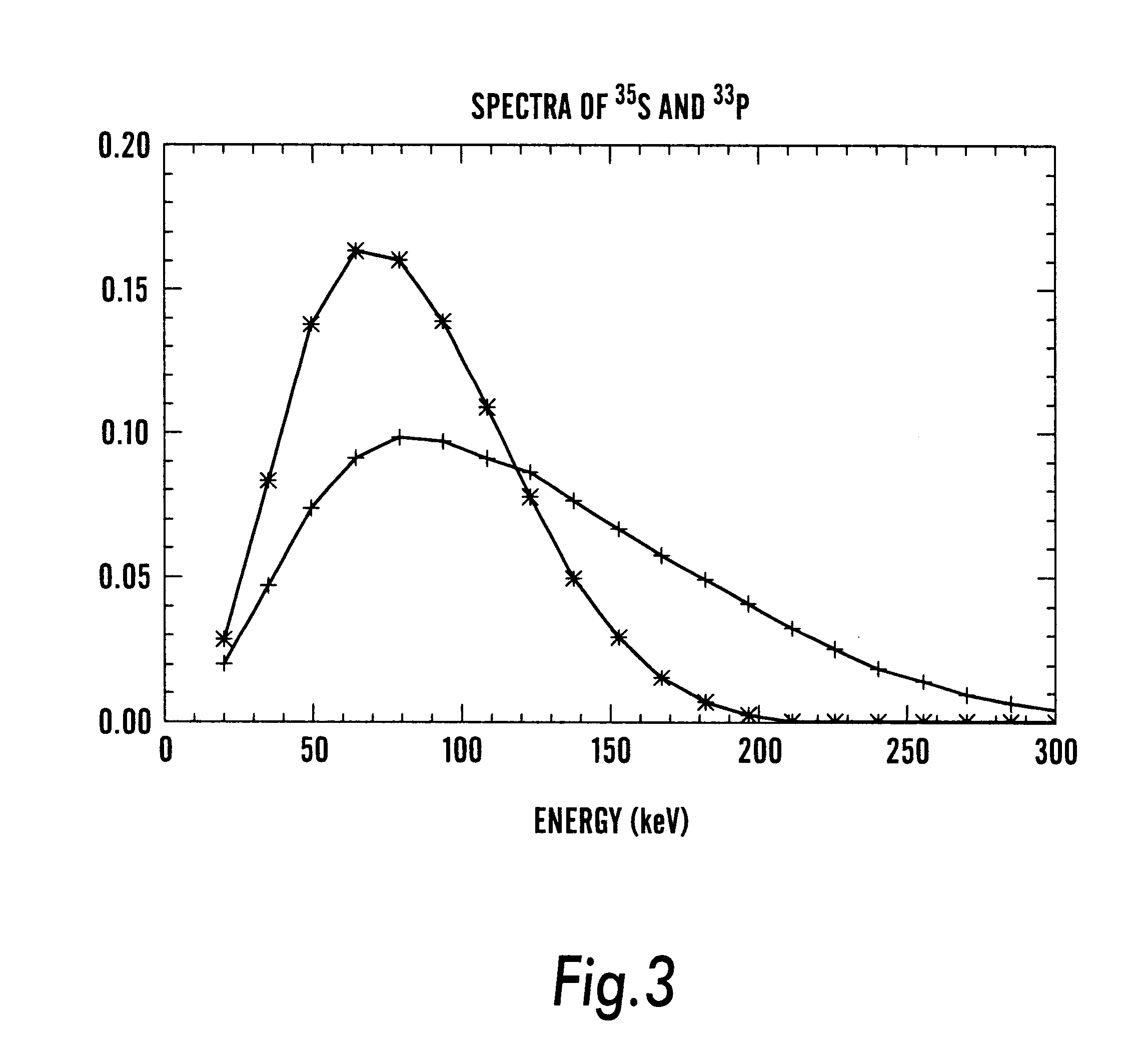
Also, the method requires advance knowledge of the expected energy spectrum of each actual radionuclide. This can be obtained by calibration of the instrument with surface distributed sources of each of the actual radionuclides before determination of the unknown sample. The calibration procedure is incorporated as an automatic procedure in the instrument, where measured spectra are obtained for each of one side (the x-side) strips of the sensor plate. By determining the first and second moment of these spectra, their zero offset and gain with reference to a reference strip can be determined (note that for the calibration procedure, neighbour strip deposited energies are not taken into account). These calibration data can then be used in real time to correct the energy of each detected radioactive event. Also, this calibration procedure avoids problems encountered with external photon (gamma) sources where scattered radiation gives rise to changes of the spectra.
Since the glass plate containing the biological microarray may be slightly rotated with respect to the principal axes of the instrument, the rotation angle must be determined so that the test spots on the microarray form exactly vertical and horizontal rows and columns in the image. The determination of the optimal angle may be carried out as follows: A sum image is formed by adding all bands in the energy band images, E.sub.ijk, i.e. summation over the index k. This image is then rotated in steps of 0.1 degrees, and for each new angle, the projection of the image on the x-and y-axes, respectively, are calculated. The optimal rotation angle corresponds to the maximum of the variance (i.e. the maximal contrast variation). The projections can easily be used to determine the upper, lower, leftmost, and rightmost position of a test spot. Thus, identification of a test spot is easy, and the associated region occupied by that test spot can be identified. It is thus possible to create a new energy band image where each point corresponds to the region occupied by one test spot. This will significantly strengthen the statistical accuracy of the final results.
This method and apparatus have been described as a general approach suitable for simultaneous determinations of the contribution from more than one radioactive isotope of a registered energy spectrum, which is a superimposition of unknown quantities from each isotope. The method is obviously necessary in the case of .beta.-emitters since these have overlapping energies, but may seem unnecessary for other types of radiation (.alpha., .gamma.) which has a monoenergetic nature and thus gives very distinct energy spectra which in general do not overlap each other. But the method according to this invention may be beneficial even for these cases since the method allows to employ radioactive isotopes which emit photons or particles with small energy differences because the method will separate possible overlapping energy contributions. Also, the method will automatically treat the problem with secondary registrations due to background radiation and unavoidable scattering, and thus give determinations with excellent precision. This feature is an obvious benefit for all types of radiation.

Problems solved by technology

This relatively high amount of material excludes the use of standard technology from a number of very relevant applications, including clinical diagnostics.
It is likely that amplification techniques will lead to degrees of bias of the starting material, due to the enzymatic nature of the process combined with the large variation in mRNA length for different transcripts.
The large amount of test material necessary to achieve adequate signal strength, and the problem that the available fluorophores are accepted only with difficulty by the reverse transcriptase enzyme represents thus considerable disadvantages in the prior art.
However, it suffers from a limited dynamic range (only 1.5-3 orders of magnitude) and low sensitivity, is laborious and gives inaccurate determinations.
However, areas having activities below the minimum threshold are not quantified accurately, and inevitably imposes a limitation to the accuracy in measurements of activity distributions.
A major practical problem of the two latter radiography modalities is the need to know the exposure time before acquiring the picture so that underexposure or overexposure may be avoided.
Especially with film, where exposure times may be months, a bad estimate of the exposure time may cause waste of a considerable time.
Even though the geometrical resolution of the available systems cannot compete with that of the film technique, it is comparable with phosphor screen imaging systems.
However, these techniques can not separate the contributions from different non-monoenergetic nuclides, and thus cannot be applied for simultaneous quantification of two or more different radioactive emitters at the same spot on a biological microarray.
Obviously, this impedes (hinders) every application where one wants to simultaneously compare the activity from more than one specimen, such as for instance a healthy cell and a tumour cell.
Also, according to our knowledge, the known methods and apparatuses for performing the conventional radiography methods are all limited to labelling all molecules on the surface with only one radio nuclide at time and a limited number of radio isotopes.
However, this camera cannot resolve structures with spatial extent on the sub millimeter scale.

Method used


Image

  • Method and apparatus for simultaneous quantification of different radionuclides in a large number of regions on the surface of a biological microarray or similar test objects
  • Method and apparatus for simultaneous quantification of different radionuclides in a large number of regions on the surface of a biological microarray or similar test objects
  • Method and apparatus for simultaneous quantification of different radionuclides in a large number of regions on the surface of a biological microarray or similar test objects

Examples


example 2

In order to verify the applicability of the method and apparatus according to the invention for microarray applications, an experiment using two cell line RNAs in reduced amounts were performed. Otherwise standard microarray procedures were employed. By using RNA from two different cell lines, OHS osteosarcoma cell line and MCF-7 breast cancer cell line, a demonstration of a successful separation of the nuclides are performed, and given as a fluorophore plot in form of an overlay pseudo-colour image of the two isotopes in FIG. 5. The figure demonstrates clearly that the method and apparatus according to the invention achieves the dynamic variation of signals that may be expected from similar experiments using fluorophores, but with significantly lower starting RNA-material.

As mentioned, radioactively labelled biological substances are easily incorporated into living cells as opposed to most fluorescent substances. This may be done by adding medium substituents, such as nucleotides, ...

example 3

The amount of any protein in a cell is partly regulated by the stability of it's corresponding RNA. This is generally considered to be related to the size of the poly-A tail, and partly other sequence-specific factors. To date, examination of individual mRNA species have been the method of choice. By the incorporation of two radioactive labels, it becomes possible to monitor all mRNA species in a population by comparison with a standard. This permits a genome wide scanning of RNA half life, when combined with DNA microarrays.

Instead of using cDNA with radioactivity in a cell free system enzymatically, a35S-UTP and a33P-UTP are added to the culture medium of parallel cell cultures in appropriate concentrations at time 0. Then the cells are allowed to grow until time 1, when the radioactive nucleotides are sufficiently incorporated to allow detection of RNA produced in the cells from time 0. At time 1, one parallel is treated chemically to block RNA synthesis. At time 2, both cultures...

example 4

In the emerging field of proteomics, similar methods to the one given in example 3 may be applied when using amino acid tags. For instance, the monitoring of various protein degrees of phosphorylation, a major regulatory phenomenon of all cells, may be performed by incorporating radioactivity labelled tags that are added to the medium. By printing antibody arrays, the incorporation of phosphor tags in a number of different proteins may be monitored simultaneously.

Although the invention has been described as a case of examples of comparison tests of two DNA / RNA-substances tagged with a distinctive S-emitting radionuclide each, it should be emphasised that the method and apparatus according to the invention is a general approach for determining the amounts of radioactivity tagged biological molecules that are adsorbed onto a biological microarray. This applies for all types of radioactivity including .alpha., .beta., .gamma., and positron radiation and all radioactive nuclides, especi...


PUM

PropertyMeasurementUnit
diametersaaaaaaaaaa
diametersaaaaaaaaaa
radioactivityaaaaaaaaaa

Abstract

The present invention relates to a method and apparatus for simultaneous quantification of the amounts of one or more radioactive nuclides within arbitrary regions on a surface where these nuclides have been deposited, adsorbed or fixed. These radioactive nuclides serve as markers on compounds that typically have been incorporated into tissue sections or into larger biological molecules that by various mechanisms have been bound to chemical substances on this surface. The method is especially well suited for DNA microarray deductions through the use of nucleotides labelled with different beta-emitting radionuclides.

Description

The present invention relates to a method and apparatus for simultaneous quantification of the amounts of one or more radioactive nuclides within arbitrary regions on a surface where these nuclides have been deposited, adsorbed or fixed. These radioactive nuclides serve as markers on compounds that typically have been incorporated into tissue sections or into larger biological molecules that by various mechanisms have been bound to chemical substances on this surface. The method is especially well suited for DNA microarray deductions through the use of nucleotides labelled with different beta-emitting radionuclides.It is widely believed that thousands of genes and their products, i.e. RNA and proteins, in a living organism function in a complicated and orchestrated way that creates the mystery of life. However, traditional methods in molecular biology generally work on a "one gene in one experiment" basis, which makes it hard to achieve the overall picture of the gene function. Thus...

Claims


Application Information

Patent Timeline
Patent Type & Authority Patents(United States)
IPC IPC(8): C12Q1/68G06F19/00G16B25/00
CPCC12Q1/6809G06F19/20C12Q2565/102C12Q2563/101G16B25/00
Inventor HOVIG, EIVINDSKRETTING, ARNENYGARD, EINARKVINNSLAND, YNGVEBREISTOL, KNUTYOSHIOKA, KOKI
Owner BIOMOLEX
Learn More