Spatiotemporal resolution enhancement of biomedical images

a biomedical image and spatiotemporal resolution technology, applied in the field of deep learning methods, can solve the problems of image detail loss, algorithm clearly outperforms traditional image interpolation strategies (i.e., cubic interpolation), and cannot incorporate knowledge across time domains, so as to improve anatomic detail, reduce scan time, and preserve anatomic detail

Pending Publication Date: 2022-04-14
RGT UNIV OF CALIFORNIA
View PDF0 Cites 3 Cited by
  • Summary
  • Abstract
  • Description
  • Claims
  • Application Information

AI Technical Summary

Benefits of technology

The invention is a method and system for improving the resolution of small heart MRIs using convolutional neural networks. These networks can learn the patterns of the images and then use that information to increase the resolution of the images. This approach outperformed traditional methods and can be used with both MRIs and CTIs. It can help to capture important details in the images faster, which can be useful in challenging situations like arrh context and motion. Overall, the invention can accelerate image acquisition and improve the accuracy of diagnostic imaging.

Problems solved by technology

Even with only 1 / 64 of the original image data, some image detail can be lost, but the algorithm clearly outperforms traditional image interpolation strategies (i.e., cubic interpolation) that are not able to incorporate knowledge across the time domain.

Method used


Image

  • Spatiotemporal resolution enhancement of biomedical images
  • Spatiotemporal resolution enhancement of biomedical images
  • Spatiotemporal resolution enhancement of biomedical images

Examples


Embodiment Construction

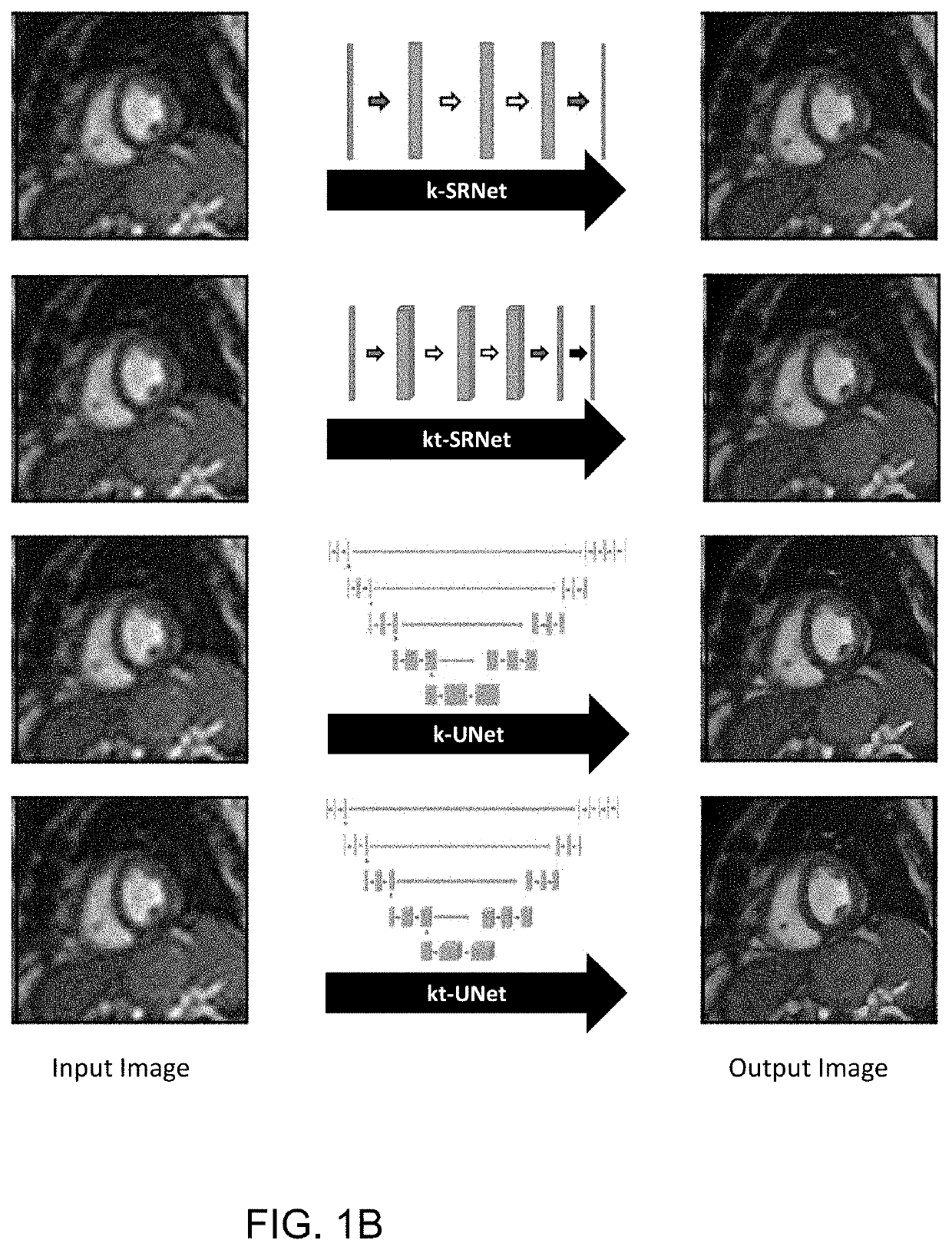
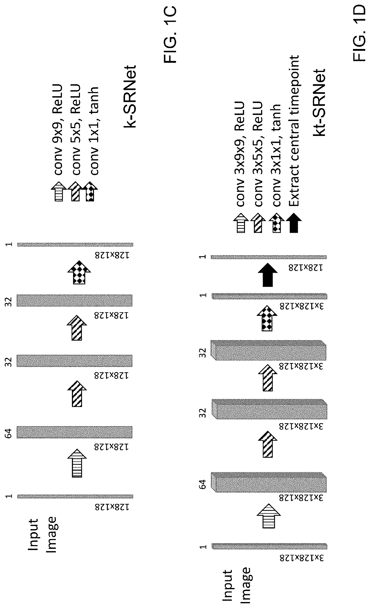
[0029]According to embodiments of the invention, convolutional neural networks (CNNs) are used for super-resolution to recover high-resolution images from low-resolution observations in MR and CT images. In the example implementations described herein, the inventive method provides high-frequency spatial information from short-axis cardiac MR images with superior performance compared to conventional upscaling methods. Additionally, improvement can be obtained through the use of adjacent temporal frames for multi-frame super-resolution.

[0030]Four neural networks were developed and evaluated for their ability to perform single and multi-frame super-resolution. Two general neural network architectures were explored for feasibility in performing this task. The first, a relatively shallow network is a SRCNN-inspired neural network with a custom hybrid loss function, which is referred to as “k-SRNet”, based on the Shallow Wide ResNet that was described by R. Yasrab (“SRNET: A Shallow Skip...


PUM


Abstract

A method for improving resolution of a lower resolution image includes inputting at least one digital image into a convolutional neural network (CNN) comprising one of a SRNet and a UNet to output a two-dimensional image having a higher resolution. In some embodiments, multiple image frames may be processed using a 3D CNN to generate a two-dimensional output image.

Description

RELATED APPLICATIONS[0001]This application claims the benefit of the priority of U.S. Provisional Application No. 63 / 090,154, filed Oct. 9, 2020, which is incorporated herein by benefit in its entirety.FIELD OF THE INVENTION[0002]The present invention relates to the use of deep learning methods to enhance spatial detail from a small matrix MRI acquisition.BACKGROUND[0003]Cardiac MRI is essential for the evaluation of cardiac size and function and valuable in the diagnosis of cardiac disease but is limited by relatively long acquisition times. Acquisition of smaller matrix images can be performed more quickly, but at the cost of spatial detail. The advent of deep learning has reinvigorated interest in the possibility of recovering spatial detail from low resolution images, known as “super-resolution”, which may enable faster acquisition of diagnostic images.[0004]Computed tomography (CT) and magnetic resonance (MR) imaging lie at the crux of modern diagnosis and management of a wide ...

Claims


Application Information

Patent Timeline
IPC IPC(8): G06T3/40G06N3/02G16H30/40
CPCG06T3/4053G06T3/4046G06T2200/04G16H30/40G06N3/02G06N3/08G06N3/045
Inventor HSIAO, ALBERTMASUTANI, EVAN
Owner RGT UNIV OF CALIFORNIA
Why Eureka
Try Eureka