
Cell surface coupling of nanoparticles

a nanoparticle and cell surface technology, applied in the field of cell surface coupling of nanoparticles, to achieve the effect of efficient and stable coupling of nanostructures

Active Publication Date: 2021-09-02
MASSACHUSETTS INST OF TECH
View PDF0 Cites 3 Cited by
  • Summary
  • Abstract
  • Description
  • Claims
  • Application Information

AI Technical Summary

Benefits of technology

This patent provides a method to attach nanostructures to the surface of T cells, which can deliver therapeutic agents and other substances in vivo without being internalized. The method uses a ligand that binds to a protein called CD45, which is found on T cells, to maintain the nanostructures on the surface. The patent also describes a polycation that increases the efficiency of attachment between the nanostructures and T cells. This approach can lead to more effective and safe use of T cells for therapeutic purposes. The compositions provided by this patent can improve the efficiency and stability of attaching nanostructures to carrier cells for various biological and pharmaceutical applications.

Problems solved by technology

Such results were unexpected given the cellular toxicity known to result from delivery of DNA coated and condensed using a polycationic coatings.

Method used


Image

  • Cell surface coupling of nanoparticles
  • Cell surface coupling of nanoparticles
  • Cell surface coupling of nanoparticles

Examples


example 1

ation of CD45 as Stable Cell Surface Anchor for Maximizing Liposome Loading

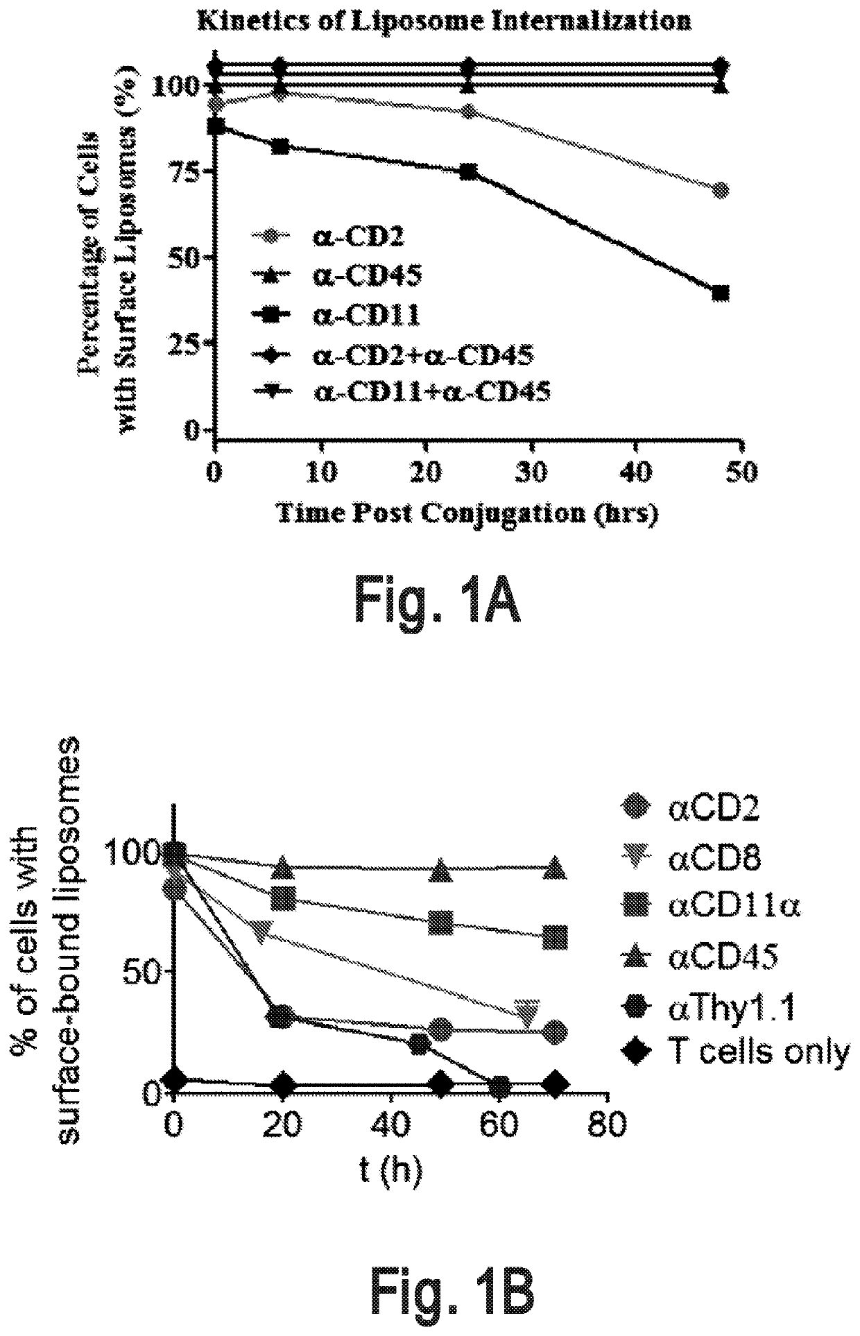
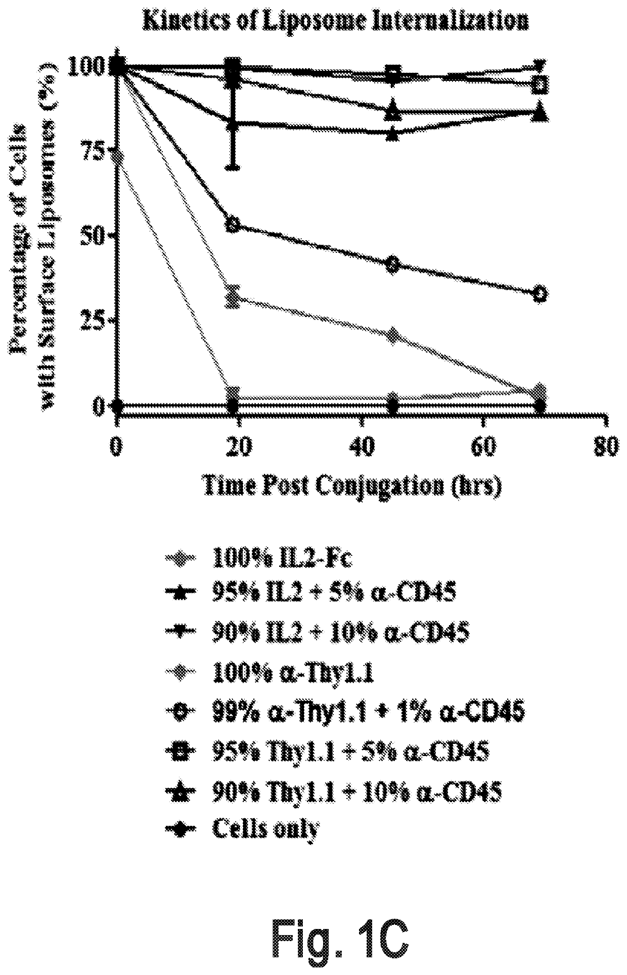
[0309]An interbilayer-crosslinked multilamellar vesicle (ICMV) (Nature Mater. 2011, 10, 243-251) is one type of particle that can remain on the cell surface (e.g., T cell surface) for more than four days. A candidate pool of slowly-internalizing receptors was obtained by mass spectrometry analysis of the total surface proteins ICMVs bound most abundantly. The screening for the main contributors for ICMVs' long surface retention was conducted by using liposomes which are surface coupled with an antibody against top candidate surface receptors such as CD2, CD11 and CD45. Antibody-conjugated liposomes coupled to carrier cells.

[0310]Liposome Synthesis

[0311]Antibody-conjugated liposomes were produced by hydrating dried high-TM phospholipid films containing 2.5% PEG-maleimide and 1% biotin head groups. Specifically, vacuum dried lipid films composed of 1,2-distearoyl-sn-glycero-3-phospho ethanolamine-N-[maleimide(p...

example 2

on and Surface Modification of Cytokine Nanogels for Efficient and Stable T Cell Surface Coupling

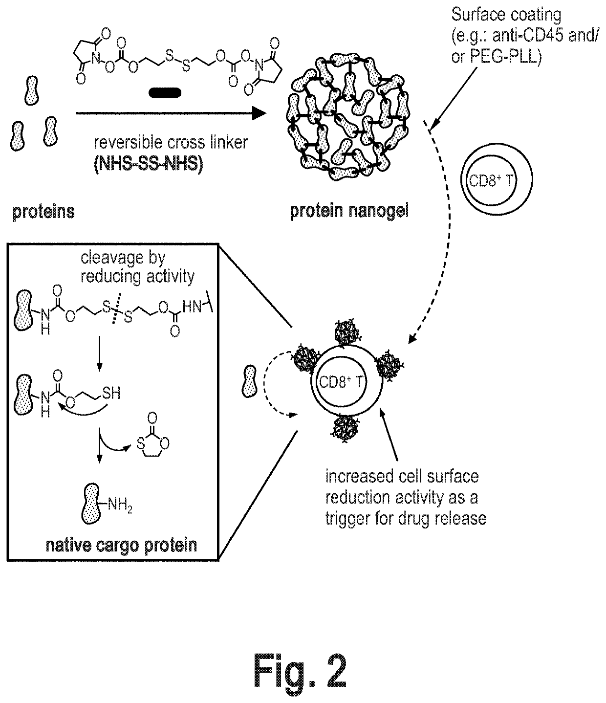
[0318]Cytokine nanogels (NGs) were prepared as described in International Publication Number WO 2015 / 048498 A2, published on 2 Apr. 2015, incorporated by reference herein. Further modifications we made to the cytokine nanogels as described herein. Generally, the cytokines were chemically crosslinked with a reversible linker (NHS-SS-NHS) to form crosslinked nano-structure of proteins, named protein nanogel (NG, FIG. 2). After completion the crosslinking reaction of cytokines, anti-CD45 antibody and / or polycations are added in situ for modifying the NG surface (FIG. 2). Following synthesis, the nanogels were characterized and coupled to carrier cells as described further below.

[0319]Synthesis of Nanogels

[0320]Synthesis of NHS-SS-NHS crosslinker: In a 125 mL round-bottom flask, 2-hydroxyethyl disulfide (1.54 g, 10 mmol) was dissolved in tetrahydrofuran (THF, 30 mL, anhydrous) and added drop...

example 3

Nanogels Promote Effector T Cell Expansion In Vitro and In Vivo

[0336]In vitro proliferation assays of T cells were conducted to determine the effect of anti-CD45 on T cell expansion. Naïve pmel-1 CD8+ T-cells were labelled with carboxyfluorescein succinimidyl ester (CFSE) and then conjugated with aCD45 / IL-15Sa-NG, IL-15Sa-NG, or aCD45 / IL-15Sa-NG (non-deg.) respectively as described above. After removing unbound NGs, T-cells were resuspended in RPMI with 10% FCS (5.0×10⁵ / mL) and added to anti-CD3 / CD28 coated beads at a 1:2 bead:T-cell ratio. Free IL-15Sa was added to the cells in control groups at equivalent dose (pulsed or continuous). Cell media were replaced every 3 days and free IL-15Sa was replenished in the continuous treatment group. At selected time points, replicates of T-cells were added with counting beads and washed with flow cytometry buffer (PBS with 2% FCS) followed by aqua live / dead staining. Cells were stained for surface markers (CD8, CD122) with antibodies followed...


PUM


Abstract

The present disclosure is directed, in some embodiments, to methods and compositions of comprising a cell having a non-internalizing receptor, and a nanoparticle surface-modified with a ligand that binds to the non-internalizing receptor.

Description

CROSS-REFERENCE TO RELATED APPLICATIONS[0001]This application is a division of U.S. patent application Ser. No. 15 / 236,186, filed on Aug. 12, 2016, pending, which claims priority to and the benefit of U.S. Provisional Patent Application No. 62 / 204,337, filed Aug. 12, 2015, the entire contents of which is herein incorporated by reference.FEDERALLY SPONSORED RESEARCH[0002]This invention was made with Government support under Grant No. R01 CA172164 awarded by the National Institutes of Health. The Government has certain rights in the invention.SEQUENCE LISTING[0003]The instant application contains a Sequence Listing which has been submitted electronically in ASCII format via EFS-Web and is hereby incorporated by reference in its entirety. Said ASCII copy, created May 12, 2021, is named “MITN1-004DV_Sequence-Listing.txt” and is 14005 bytes in size.BACKGROUND OF THE INVENTION[0004]Combining nanostructure (e.g.: nanoparticle-and liposome) based drug delivery strategies with cell-based the...

Claims


Application Information

Patent Timeline
Patent Type & Authority Applications(United States)
IPC IPC(8): C07K14/725C07K16/28C07K14/705C07K14/715C07K19/00A61K47/60A61K47/69C07K14/54C07K16/30A61K39/00C07K14/55A61K47/68A61K9/127C12N5/00C12N5/0783A61P35/00
CPCC07K14/7051A61K2039/5156C07K14/70589C07K14/7155C07K19/00A61K47/60A61K47/6913C07K14/70503C07K16/2845C07K14/70517C07K14/70578C07K14/5443C07K16/30A61K39/0011A61K47/6903C07K14/55A61K47/6849A61K9/127C07K16/289A61K47/6889C07K2319/03C07K2319/00C07K2319/33A61K2039/55527C12N5/0006A61K2039/5158C12N5/0636C07K2319/30A61K2039/55533A61K2039/585A61K39/001104A61P35/00C12N2501/2315A61K2039/505C12N5/0638A61K2039/57C07K2317/622C07K2319/70C07K16/2806A61K9/06A61K47/6901A61K47/645A61K38/2013A61K38/2086A61K39/395A61K39/4631A61K39/4611A61K39/464404A61K39/464492A61K2239/39A61K2239/57
Inventor IRVINE, DARRELL J.ZHENG, YIRANTANG, LI
Owner MASSACHUSETTS INST OF TECH
Learn More