Method of isolating and detecting cancer stem cells

a cancer stem cell and isolating technology, applied in the field of isolating and detecting cancer stem cells, can solve the problems of its implementation being faster than existing methods, and achieve the effects of facilitating detection, confirming the effectiveness of enrichment, and being more reliabl

Inactive Publication Date: 2021-03-18
CARCIDIAG BIOTECH
View PDF1 Cites 1 Cited by
  • Summary
  • Abstract
  • Description
  • Claims
  • Application Information

AI Technical Summary

Benefits of technology

[0302]This detection is facilitated and made more reliable by the enrichment of the sample with cancer stem cells.
[0303]Detection by these methods also validates the effectiveness of the enrichment of the sample with cancer stem cells of organs involved in respiration by the method according to the invention, i.e. the effectiveness of the method in isolating cancer stem cells of organs involved in respiration.
[0304]In the diagnostic methods of the present invention, the higher the intensity of detection in the biological sample is, compared to a healthy sample adjacent to the biological sample, the higher the risk of recurrence of cancer of organs involved in respiration is and more aggressive the cancer is.
[0305]Similarly, the higher the quantity of cancer stem cells of organs involved in respiration in the biological sample is, compared to a healthy sample adjacent to the biological sample, the higher the risk of recurrence of cancer of organs involved in respiration is and more aggressive the cancer is.
[0306]The detection and quantification of cancer stem cells of organs involved in respiration in a biological sample thus makes it possible to determine the aggressiveness of the cancer of organs involved in respiration and its ability to develop.
[0307]The detection and quantification of cancer stem cells of organs involved in respiration is also part of a personalized medical approach. Indeed, the detection of cancer stem cells f organs involved in respiration in the biological sample allowing to assess the prognostic value of the treatment, thus allows the treatment to be adapted.

Problems solved by technology

In addition, its implementation is faster than existing methods, as the latter are not generalizable due to their non-reproducibility and include both cancerous stem and non-stem cells.

Method used


Image

  • Method of isolating and detecting cancer stem cells
  • Method of isolating and detecting cancer stem cells
  • Method of isolating and detecting cancer stem cells

Examples


example 1

for the Isolation of Pulmonary Cancer Stem Cells, a Key Example of Respiratory Tract Cancers

[0476]I. Materials Required

[0477]Reagents and Equipment[0478]Individual biotinylated lectin or mixture of biotinylated lectins specifically labelling Pulmonary Cancer Stem Cells (prepared from individual lectins Vector Laboratories)[0479]Kit CELLection Biotin Binder (Invitrogen) containing magnetic beads coupled to streptavidin by a DNA bound[0480]Magnet

[0481]Buffers[0482]Versene (Invitrogen) comprising saline phosphate buffer (PBS) and EDTA[0483]Buffer 1: PBS (Saline phosphate buffer without Ca2+ and Mg2+) with 0.1% BSA (bovine serum albumin), pH 7.4[0484]Buffer 2: PBS (Phosphate Buffer Saline without Ca2+ and Mg2+) with 0.1% BSA (Bovine Serum Albumin) and 0.6% sodium citrate[0485]Buffer 3: RPMI 1640 with 1% FCS (Fetal Calf Serum), 1 mM CaCl2 and 5 mM MgCl2, pH 7.0-7.4.

[0486]II. Duration of the Experiment[0487]20 min to prepare the cells[0488]20 min to label the cells[0489]20 min to incubate...

example 2

city Test

[0521]The objective of a clonogenicity test is to observe the ability of cells to reassemble spheres (corresponding in the patient to the reassembly of a tumor mass) and therefore their proliferative capacity.

[0522]The clonogenicity test is used in this example to confirm the presence of pulmonary cancer stem cells and to quantify said cells in a sample after isolation of pulmonary cancer stem cells by the isolation method described in the present invention. It thus makes it possible to demonstrate the effectiveness of the isolation method according to the invention compared to a control sample not subjected to this method (unsorted cells).

[0523]Clonogenicity tests were performed in a 6-well plate at a density of 500 cells / cm2 in a DMEM composition medium (Gibco) supplemented with 50 units / mL penicillin, 50 units / mL streptomycin (Gibco) and 2.4 g / L sodium bicarbonate, 1 M HEPES buffer (Sigma Aldrich, Saint-Quentin-Fallavier, France), 1× progesterone (Sigma Aldrich), 1× putr...

example 3

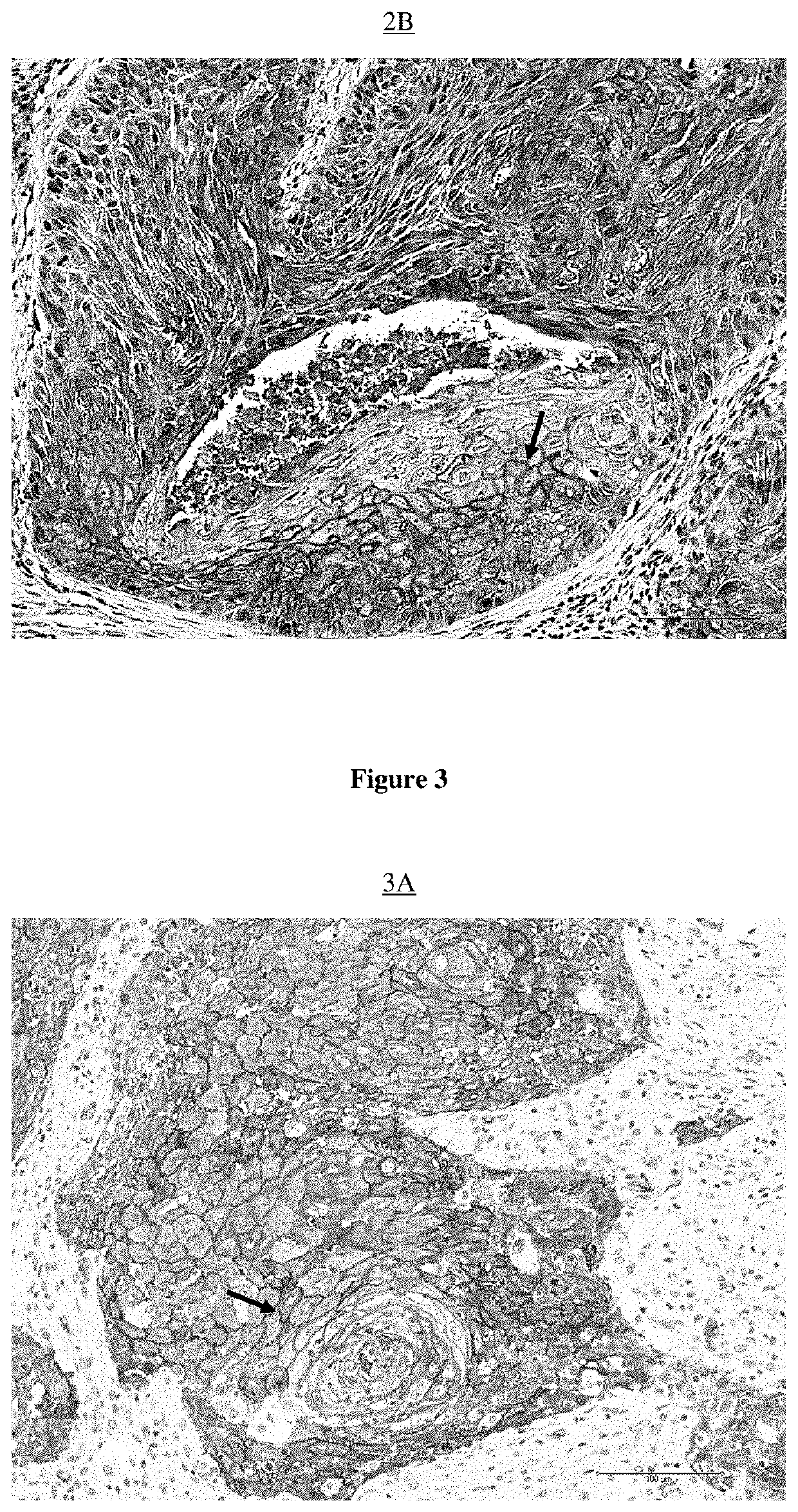
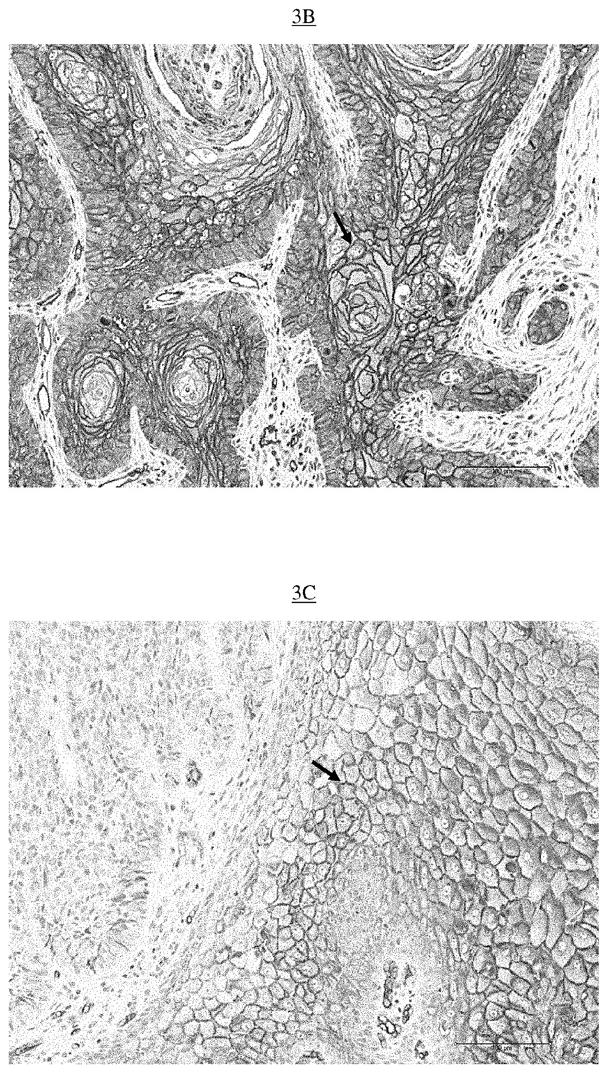
abelling of Lectins on Waxed Histological Section: Example of Lung Cancer

[0526]Equipment used: Paraffin blocks, Ice, Microtome, Microtome, Superfrost® blades, Bond Max Automate (Leica Microsystems) with computer, Leica consumables (alcohol, wash buffer, ER1 buffer, dewax buffer, labels, coverslips, tubes), PBS-BSA buffer 5%, Biotinylated Lectins (UEA-1, Jacaline, ABA, ACA, GSL-I and GSL-II) (Vector Lab), Kit Bond Intense R detection (Leica), Leica mounting medium, slides and microscope.

[0527]The paraffin blocks containing lung cancer samples from each of the patients identified by their number (given by the pathological anatomy department) were placed in the ice for about 1 hour to be cooled, in order to facilitate their microtome section to a thickness of 5 μm.

[0528]The so-called “superfrost” slides, this for maximum adhesion of the sectioned tissue, were identified by the same numbers as those present on the blocks. A drop of water was placed in the center of each of these slides....


PUM

PropertyMeasurementUnit
weight ratioaaaaaaaaaa

Abstract

Disclosed is the in vitro use of at least one lectin which recognises the fucose alpha(1-2) galactose unit for labelling cancer stem cells of organs involved in respiration, in order to obtain labelled cancer stem cells of organs involved in respiration, in a biological sample. In one particular embodiment, the at least one lectin is chosen from the lectins Ulex Europaeus agglutinin 1 (UEA-1) or the homologue thereof, Trichosanthes japonica agglutinin II (TJA-II), Agaricus Bisporus agglutinin (ABA), Amaranthus Caudatus agglutinin (ACA), jacalin, Griffonia Simplicifolia lectin I (GSL-I) and Griffonia Simplicifolia lectin II (GSL-II). In one particular embodiment, the organ involved in respiration is chosen from the lungs, the larynx, the pharynx, the mouth, the nose, the throat, the tongue, the sinuses, the trachea and the saliva glands including the tonsils and the parotid gland.

Description

BACKGROUND OF THE INVENTIONField of the Invention[0001]This invention concerns a method of isolating and detecting cancer stem cells (CSCs) of organs involved in respiration.Description of the Related Art[0002]Lung cancer is the leading cause of cancer deaths in France for men and the second for women, the first being breast cancer. It affects 37,000 people per year. The 5-year survival rate is very low, hence the notion of early diagnosis that occurs in this pathology where the present invention makes sense.[0003]Lung cancer, like other types of cancer, has many factors leading to a high mortality rate in the concerned population, with in particular a late diagnosis, which results in both a more advanced cancer called metastatic and a high recurrence rate. Indeed, this rate directly depends on the stage at which the cancer is detected. Nevertheless, it should be noted that even when the detection stage is early, the recurrence rate remains high because some parameters are not yet t...

Claims


Application Information

Patent Timeline
Patent Type & Authority Applications(United States)
IPC IPC(8): G01N33/574
CPCG01N33/57423G01N2333/42G01N33/56966G01N33/57469G01N2800/54C12N5/0688C12N5/0693
Inventor CARRE, VINCENTLACROIX, AURÉLIE
Owner CARCIDIAG BIOTECH
Why Eureka
Try Eureka