
Collagen scaffold modified by covalent grafting of adhesion molecules, associated methods and use thereof for cardiovascular and thoracic cell therapy and contractile tissue engineering

a collagen scaffold and covalent grafting technology, applied in the field of collagen scaffolds, can solve the problems of lack of differentiation of transplanted cells, difficult production of cell therapy scaffolds, and substantial cell mortality

Inactive Publication Date: 2016-07-14
SCHUSSLER OLIVIER
View PDF1 Cites 7 Cited by
  • Summary
  • Abstract
  • Description
  • Claims
  • Application Information

AI Technical Summary

Benefits of technology

The patent text discusses the use of a support fixation system, called RGD peptides, to improve the differentiation and survival of endothelial cells and cardiac myocytes in collagen scaffolds. The RGD peptides were tested in the presence of contractile cells or endothelial cells and were found to have beneficial effects on cell differentiation and survival. The text also mentions the use of various growth factors, chemokines, and other biological agents that can be associated with the collagen scaffold to further enhance cell fate. The functionalized collagen scaffold with RGD peptides also showed anti-apoptotic effects on cells associated with it. Overall, the patent text provides a technical solution for improving the regenerative potential of collagen scaffolds and promoting cell fate in cardiovascular tissue engineering.

Problems solved by technology

Producing cell therapy scaffolds is difficult because such supports enable the free diffusion of nutrients and oxygen, possess the mechanical properties but also the specific biological ligands to be able to interact with the associated cells of interest so as to promote their survival and their differentiation as well.
To this day, techniques for transplanting isolated cells suffer from causing a very substantial cell mortality and often a lack of differentiation of the transplanted cells.
The lack of production of new myocardial fibers in sufficient numbers for a positive effect to be clinically transposed has been attributed to the substantial cell death occurring after the graft (approx.
The destruction of the native extracellular matrix (ECM) essentially composed of type I and type III collagen in the myocardium and its replacement with a less vascularized tissue having altered mechanical properties (less compliant), which, in addition, due to a change in its composition, does not allow an optimal interaction anymore, could explain the relatively poor results of the free cell transplantation.
However the cell survival in this type of support is still low and the differentiation remains partial, with especially a lack of terminal differentiation for cardiac myocytes5 and for endothelial cells36-39.
In the present state of our knowledge, the use of contractile tissues obtained using MATRIGEL™ is not allowed for human clinical medicine due to the origin of such compound.
Moreover, the presence of MATRIGEL™ may generate a number of side effects.
The diffusion deficiencies limit the thickness of the expected tissue and make it unsuitable for replacing a cardiac muscle.
Such accelerated matrix degradation subsequent to the strong inflammatory response is responsible for the local release of great amounts of enzymes, free radicals and various degradation products which may also compromise the survival of the cells associated with those supports.
The collagen degradation products themselves have been reported as being toxic in nature, especially for contractile cells48.
Moreover, the mechanical properties of the supports are impaired by this degradation even before the associated cells could form their own matrice49.
Moreover, the poor angiogenesis in the implant limits the functionality thereof.
However Levenberg and al. underlined that MATRIGEL physical properties are not stable in vivo and that cells, after their first differentiation, do rapidly de-differentiate in this type of gel.
Moreover, the limitation to the MATRIGEL™ use for such applications is explained by the fact that MATRIGEL™ is above all a tumor extract and therefore cannot be used in human medicine.
In addition, MATRIGEL™ induces the formation of a secondary cicatricial and poorly vascularized fibrosis which on the long run compromises the cell survival.
However glutaraldehyde may auto-polymerize, then slowly depolymerize within the biological preparation and, thus, release free glutaraldehyde which is toxic to the cell and therefore not compatible with cell therapy.
It should be noted nevertheless that in these experiments, the RGD is not covalently fixed to the support and that fibrin gels, in addition, tend to get compact as time goes, their mechanical properties being impaired and the support not allowing angiogenesis to develop anymore74.
Using this type of gel with stem cells also create a number of problems because if the fibrinogen concentration is too low, the gel does liquefy and get lost within a couple of weeks.
This method results in the formation of numerous by-products, it is not selective and does not allow to use spacers which maintain the grafted peptide sequence that may bind to the integrin receptor reasonably spaced apart from the protein collagen.
The technique described by J L. Myles and al.82 is limited to the use of adhesion peptide sequences bound to a thiol-containing residue, which generally requires to modify the peptide by coupling it with a cysteine molecule.
In addition, the purification of the reaction products carried out in a homogeneous phase is difficult and the by-product separation makes it necessary to use several chromatography purification steps.
Lastly, the technique of Myles J L and al.82 does not allow to monitor the coupling reaction progress and leaves aside available amino groups that might create peptide sequences duplication reactions.

Method used


Image

  • Collagen scaffold modified by covalent grafting of adhesion molecules, associated methods and use thereof for cardiovascular and thoracic cell therapy and contractile tissue engineering
  • Collagen scaffold modified by covalent grafting of adhesion molecules, associated methods and use thereof for cardiovascular and thoracic cell therapy and contractile tissue engineering
  • Collagen scaffold modified by covalent grafting of adhesion molecules, associated methods and use thereof for cardiovascular and thoracic cell therapy and contractile tissue engineering

Examples


example 1

[0085]Covalent coupling of adhesion molecules such as RGD / RGE peptides to the collagen scaffold or biological agents such as proteoglycans, growth factors or cytokines if the scaffold is made of collagen and / or contains thiol, amine or carboxyl accessible groups.

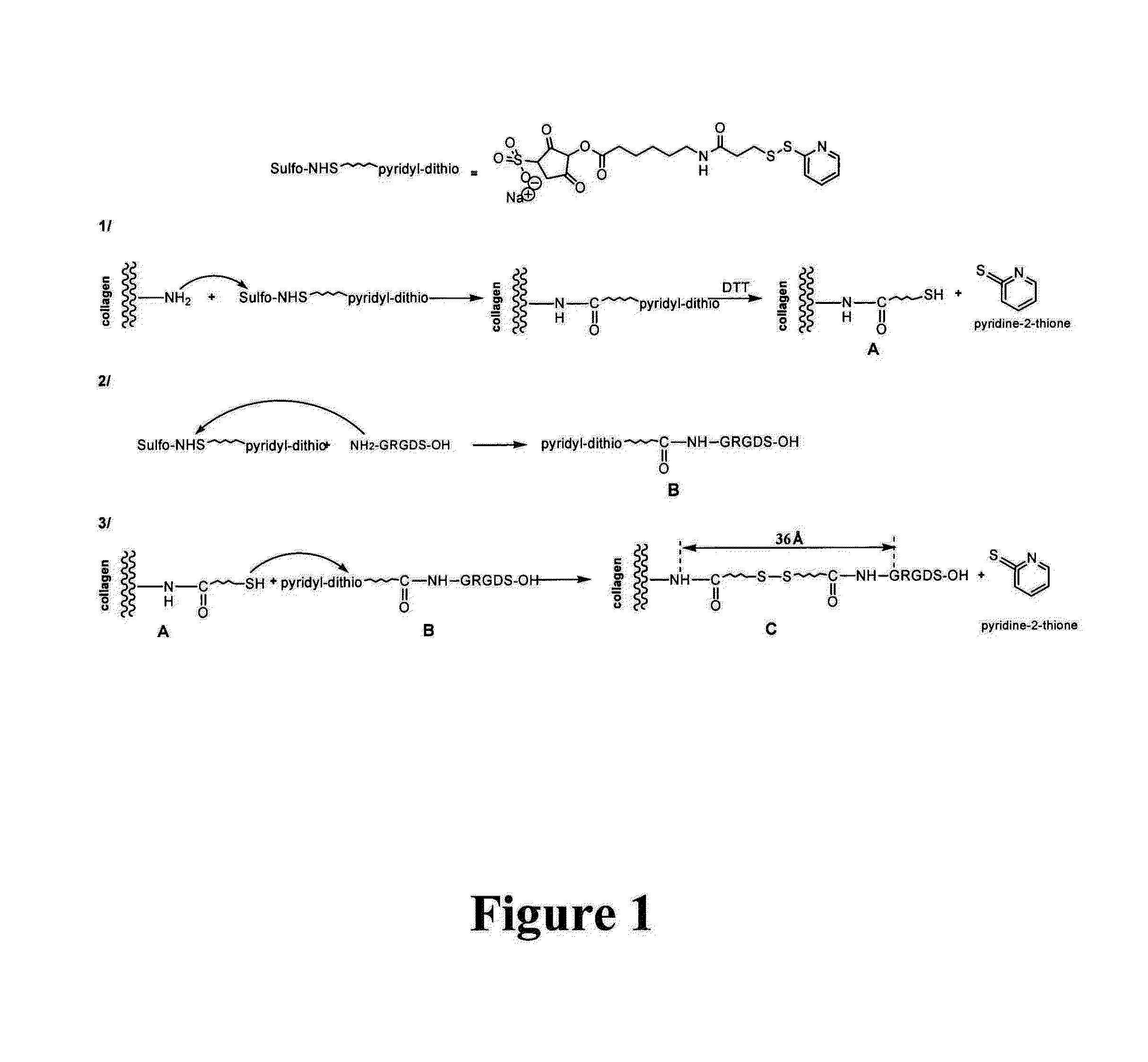
[0086]The coupling chemical reaction principle is illustrated in FIG. 1. The coupling described here makes it possible to illustrate the RGD moiety at a medium distance from the support, here a 30-40 Angstrom collagen matrix, elongation that is favorable to the maximum interaction of the peptide sequence with the integrin receptor site (cf. Beer J H. and al. 1992, Craig W S. and al. 1995)23,24.

[0087]Collagen scaffolds are used, and advantageously DHT-cross-linked porous supports like with ULTRAFOAM®, materials that are already used in clinical medicine as hemostatics: 5 mm-thick ULTRAFOAM® sheets (2.5 mm after rehydration in PBS) (Davol Inc., Cranston, R.I.) come from beef, DHT-cross-linked type I and type III collagen fiber...

example 2

[0089]Using collagen scaffolds functionalized with adhesion molecules for making a contractile tissue

[0090]Contractile Cells

[0091]Different cell types have a contractile activity prior to or after a complete differentiation as for example smooth muscle cells, skeletal muscle cells or cardiac myocytes. Embryonic stem cells, pre-differentiated or not (where the differentiating agent may be for example a growth factor or a combination of growth factors. The removal or the inhibition of some factors from the culture medium as for example growth factors of the FGF, TGF beta, BMP-2, SDF1 type, physical factors such as hypoxia, electrostimulation, freezing, mechanical stress, etc.) also has shown their ability to promote the differentiation towards contractile cells. Marrow bone cells (hematopoietic cells or mesenchymal cells), cells isolated from the circulating blood (also including cells isolated from the umbilical cord blood). Contractile potential cells were also isolated from differe...

example 3

[0105]Angiogenesis induction in vitro by associating endothelial cells in a functionalized matrix with adhesion molecules:

[0106]In another type of preparation, cells of interest such as endothelial cells (mature or progenitor cells) may be transplanted into the functionalized 3D-collagen matrix with adhesion molecules, advantageously the RGD moiety, in the presence of or in the absence of contractile cells or other types of cells as for example fibroblasts, keratinocytes, contractile cells, genetically modified cells, etc. which may also be used independently. This support will promote the survival and the differentiation of the associated cell population as would do endothelial cells. This cell population may be used in association with contractile cells or independently. Such support may sometimes be also used without any initially associated cell population, the cells colonizing subsequently the support. However it has been demonstrated that associating endothelial cells does pro...


PUM

PropertyMeasurementUnit
Pore sizeaaaaaaaaaa
Dielectric strengthaaaaaaaaaa
Thicknessaaaaaaaaaa

Abstract

Three-dimensional solid contractile collagen scaffolds having improved biological properties and electromechanical performance are provided. The scaffolds can be used for cell transplantation, including the fixation of biocompatible reagents and adhesion molecules which control cell adhesion, apoptosis, survival and / or differentiation simultaneously. Grafting adhesion molecules to the collagen matrices renders the scaffold suitable for use in vascular and cardiothoracic surgery / medicine, as well as in cell therapy for the heart and in artificial heart muscle engineering. Also, a method for grafting and optimizing the presentation of adhesion peptides or biological agents in the scaffold is provided.

Description

FIELD OF THE INVENTION[0001]The present invention relates to collagen scaffolds characterized in that they are chemically modified by covalent grafting of adhesion molecules which facilitate the cell implantation, their survival and their differentiation. The collagen scaffold (hereunder referred to as “the support” or “the scaffold”) may advantageously be used with contractile potential cells but other cell types may also be suitably used, or combined, such as angiogenic potential cells.[0002]The present invention also relates to the covalent grafting method and to the uses of such substrates for tissue engineering, cell therapy or for modifying the collagen content of devices used in the thoracic and cardiovascular medical and surgical field.[0003]The present invention relates to coated collagen substrates, especially for use in medicine, for tissue repair and regeneration.[0004]Collagen takes part in many devices used in cell therapy, for tissue engineering and in the thoracic an...

Claims


Application Information

Patent Timeline
IPC IPC(8): A61L27/24A61L27/54A61L27/38A61L27/56
CPCA61L27/24A61L27/56A61L27/54A61L2300/412A61L2430/20A61L2300/25A61L27/38A61L31/044A61L31/16A61L2300/00A61L2300/80A61L2400/06
Inventor SCHUSSLER, OLIVIERMICHELOT, ROBERT
Owner SCHUSSLER OLIVIER
Learn More