
Mussel-inspired bioactive surface coating composition generating silica nanoparticles

a bioactive surface coating and mussel technology, applied in the direction of peptide/protein ingredients, prosthesis, peptide sources, etc., can solve the problems of many restrictions in the industrial application of the naturally extracted mussel adhesive protein, potent toxic risk, and inability to meet the requirements of in vivo environments, and achieve excellent adhesion strength of the mussel adhesive. , the effect of promoting cell proliferation or differentiation

Active Publication Date: 2016-04-07
POSTECH ACAD IND FOUND
View PDF0 Cites 8 Cited by
  • Summary
  • Abstract
  • Description
  • Claims
  • Application Information

AI Technical Summary

Benefits of technology

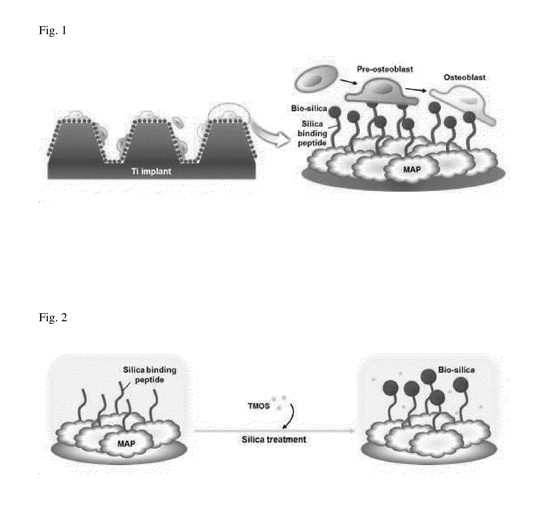
The present invention is about using silica nanoparticles to create a coating that can promote cell growth and differentiation using the mussel adhesive protein. This coating can be applied to a variety of surfaces without limitation. The advantage is that the mussel adhesive protein provides strong adhesion to the surface.

Problems solved by technology

However, the silica nanoparticles have disadvantages that they are easily detached from the material surface when a widely used physical / chemical deposition method is used, and they require acidic or basic conditions and various chemical additives and thus are accompanied by a potential risk of toxicity when a sol-gel precipitation method is used.
Further, a lithography method is problematic in that this method is limited to the target surface on which silica is formed, and is not suitable for in vivo environments (Caruso, R A, Antonietti, M, 2001.
Further, mussel adhesive proteins are able to adhere to various surfaces such as plastics, glass, metal, Teflon and biomaterials, and the like, and allow adhesion on wet surface within a few seconds, which remains an unsolved problem in development of chemical adhesives (Lee, B P, Messersmith, P B, Israelachivili, J N, Waite, J H, 2011.
Therefore, despite very excellent physical properties of the mussel adhesive protein, there are many restrictions in industrial applications of the naturally extracted mussel adhesive proteins.
However, there have been no reports about a mussel-inspired adhesive material, which is prepared by incorporating the recombinant mussel adhesive proteins having adhesion strength to various surfaces while being produced in a large amount with the silica nanoparticles.

Method used


Image

  • Mussel-inspired bioactive surface coating composition generating silica nanoparticles
  • Mussel-inspired bioactive surface coating composition generating silica nanoparticles
  • Mussel-inspired bioactive surface coating composition generating silica nanoparticles

Examples


example 1

Preparation of Fusion Protein

[0066]Primers (Table 3) for a silica-binding peptide sequence derived from the diatoms C. fusiformis were constructed. These primers were used to perform polymerase chain reaction, thereby preparing a fusion protein, in which the silica-binding peptide was linked to a mussel adhesive protein, fp-1 (SEQ ID NO. 1) or fp-151 (SEQ ID NO. 3).

TABLE 3 SEQ IDNO.PrimerNucleotide sequence (5′→3′)8ForwardGCGCCATATGAGCAGCAAAAAATCTGGCTCCTATTfor R5CAGGCTCGAAAGGTTCTAAACGTCGCATTCTGGGTGGCGGAGGGGCGAAACCGAGCTATCCGCCGACC9ReverseGCGCCTCGAGCTTGTACGTTGGAGGATAAGAAGGfor MAP

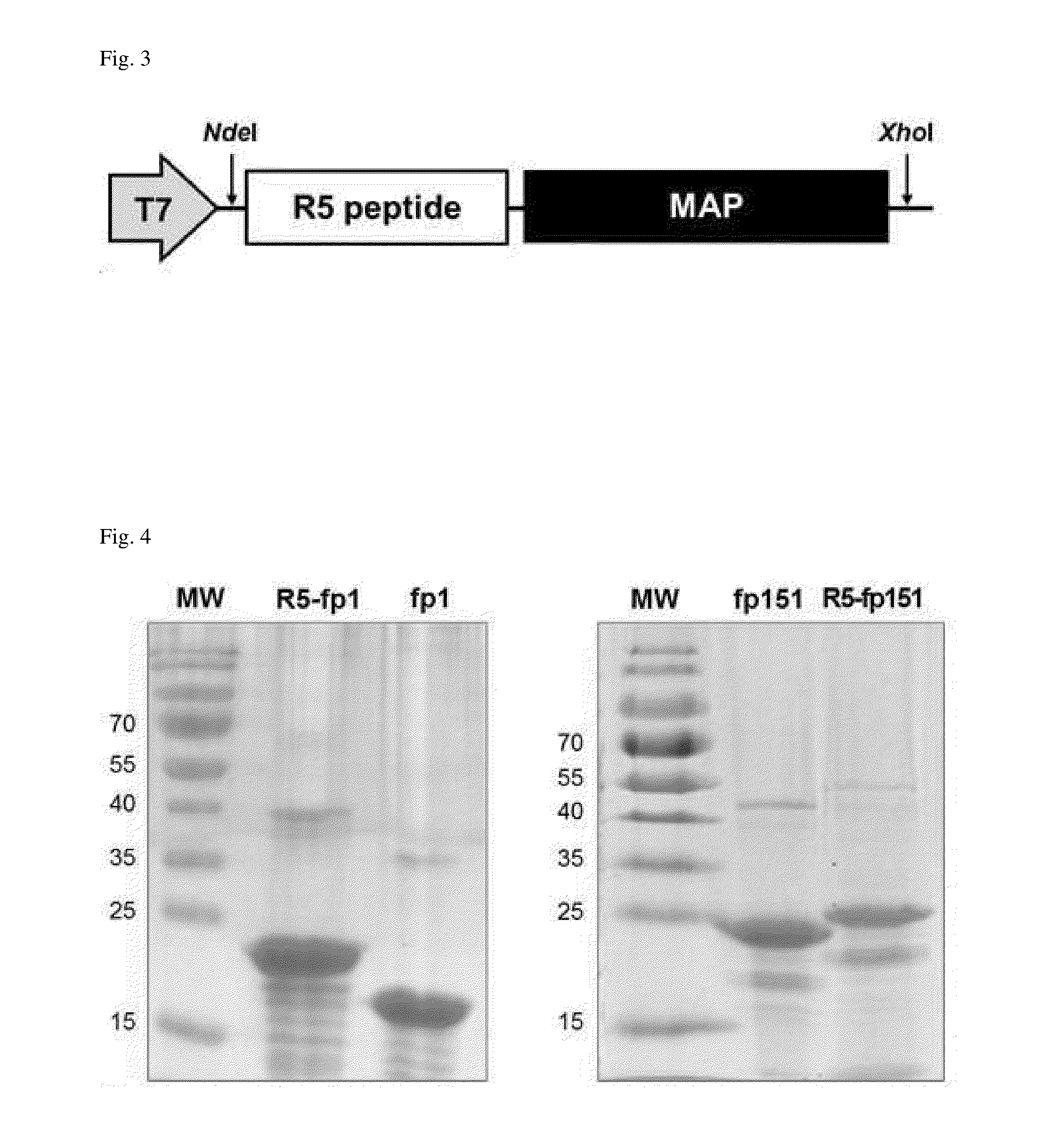
[0067]As in FIG. 3 showing the schematic illustration of fusion protein (R5-MAP) construction, an R5 peptide (SEQ ID NO. 5; SSKKSGSYSGSKGSKRRIL) was linked to a mussel adhesive protein (MAP), such as mussel adhesive protein fp-1 or fp-151. A pET-22b(+) vector containing T7 promoter was used as a plasmid vector, and transformed into an E. coli TOP10 strain. Further, for expression of the fusion protein, the clo...

example 2

Preparation of Fusion Protein-Silica Nanoparticle Complex

[0070]2-1: Use of Polymer Substrate Surface

[0071]To coat the surface of a coverslip made of polystyrene with the fusion protein-silica nanoparticle complex, 5% acetic acid solution containing 5 mg / ml of the fusion protein prepared in Example 1 was applied to the substrate surface, and left at room temperature for 12 hours to perform protein deposition. In this regard, to remove the fusion proteins which were not properly adhered to the surface, the substrate surface was washed with distilled water so as to obtain a fusion protein(R5-MAP)-coated substrate surface.

[0072]The fusion protein-coated substrate surface was immersed in 1 M trimethylorthosilicate (TMOS) solution for 2 minutes so as to prepare a complex, in which the silica nanoparticles were linked to the fusion protein. To remove silica which was not properly adhered to the substrate surface, the substrate surface was washed with distilled water.

[0073]To confirm format...

example 3

In Vitro Cell Test of Surface Coating Composition

[0082]3-1: Cell Culture by Use of Surface Coating Composition

[0083]A cell function-improving ability of the bioactive surface coating composition including the fusion protein-silica nanoparticle complex of Example 2 was examined in vitro.

[0084]In the same manner as in Example 2-1, four types of the coated substrate surfaces were prepared by coating the surface of the polystyrene coverslip. In detail, the four types of the coated substrate surfaces include 1) the surface of polystyrene coverslip (NC) which was coated with none of the fusion protein and TMOS, 2) the surface (Si—NC) which was coated with TMOS, but without R5-MAP fusion protein, 3) the surface (R5-MAP) which was coated without TMOS, but with R5-MAP fusion protein, and 4) the surface (Si-R5-MAP) which was coated with R5-MAP fusion protein-silica nanoparticle complex formed after treatment of TMOS solution.

[0085]5×10⁴ mouse osteoblast MC3T3-E1 cells were cultured on the fou...


PUM

PropertyMeasurementUnit
Adhesivityaaaaaaaaaa
Cell proliferation rateaaaaaaaaaa

Abstract

The present invention relates to a fusion protein comprising a mussel adhesive protein and a silica-binding peptide linked to the mussel adhesive protein, a silica nanoparticle a silica connected to the fusion protein, a fusion protein-silica nanoparticle complex comprising the silica nanoparticle having bioactivity and adhesiveness for cell proliferation and accelerating the differentiation, a surface coating composition including the complex, its use, and a method of coating a surface using the surface coating composition.

Description

TECHNICAL FIELD[0001]The present invention relates to a mussel-inspired bioactive surface coating composition generating silica nanoparticles, and use thereof, and more particularly, to a bioactive protein prepared by fusion of a recombinant mussel adhesive protein with a silica-binding peptide generating silica nanoparticles, a fusion protein-silica nanoparticle adhesive complex of the fusion protein and silica, and a method thereof, and a bioactive surface coating composition including the complex and a surface coating method.BACKGROUND ART[0002]Bone loss occurs in surgical treatment procedures such as orthopedic and dental surgery, traumatic injuries, or tumor removal, and implants and substitutes are widely used to restore areas of bone loss. In this regard, for successful in vivo repair, it is important to use a material capable of bearing loads of hard tissues and stimulating osteoprogenitor cells to induce bone regeneration (Kokubo, T, Kim, H-M, Kawashita, M, 2003. Biomateria...

Claims


Application Information

Patent Timeline
IPC IPC(8): C07K14/435
CPCC07K14/43504C07K2319/00A61K38/00A61L27/025A61L27/30A61L27/3604C07K14/43509
Inventor CHA, HYUNG JOONJO, YUN KEECHEONG, HOGYUNKIM, CHANGSUPCHOI, BONG-HYUK
Owner POSTECH ACAD IND FOUND
Learn More