
Method and apparatus of aiding detection of surface abnormality in the oesophagus

a technology of oesophagus and surface abnormality, which is applied in the direction of nucleotide libraries, instruments, library screening, etc., can solve the problems of small work on the precise ordering of these alterations in large cohorts, associated clinical follow-up data, and striking and unexpected findings

Inactive Publication Date: 2016-01-07
UK RES & INNOVATION LTD +1
View PDF1 Cites 0 Cited by
  • Summary
  • Abstract
  • Description
  • Claims
  • Application Information

AI Technical Summary

Benefits of technology

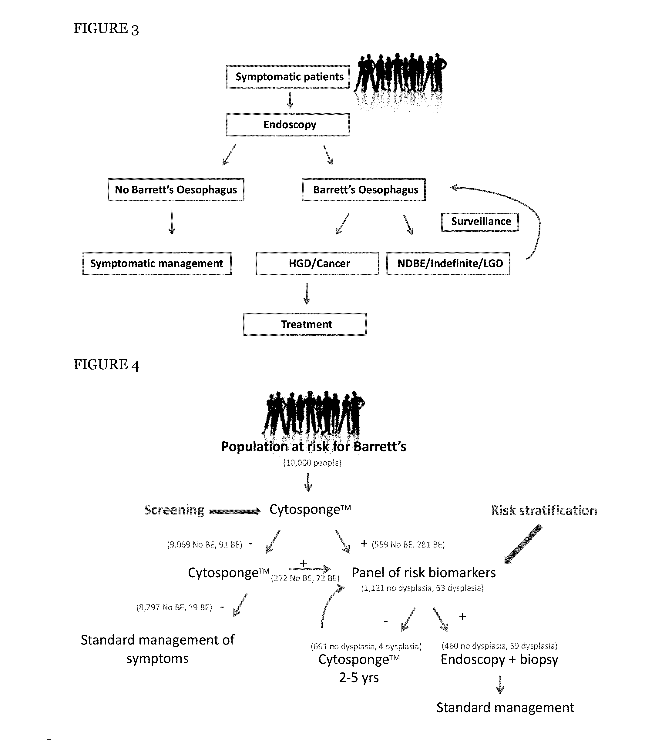
The invention provides a method to sample the surface of the oesophagus more effectively than prior art techniques such as endoscopic biopsy or brushing. The method involves introducing a swallowable device with abrasive material into the subject's stomach or oesophagus, and then retrieving the device after it has collected cells from the surface. The method can result in more accurate and reliable samples, and can also sample the entire oesophagus.

Problems solved by technology

However, little work has yet focused on the precise ordering of these alterations in large cohorts of patients with pre-malignant disease and associated clinical follow-up data.
The findings were striking and unexpected.
This approach is highly controversial due to the inherent difficulties in accurate identification of dysplastic lesions, and recent data suggest that endoscopic surveillance of BE is not effective13,23.
The difficulties involved in endoscopic surveillance for BE include sampling bias inherent in random biopsies protocols and the subjective and time-consuming histopathological diagnosis of dysplasia.
Though their recurrent nature suggests a role in clonal expansion at the pre-malignant stage they do not seem to provide any long term increase in the likelihood of malignant progression.

Method used


Image

  • Method and apparatus of aiding detection of  surface abnormality in the oesophagus
  • Method and apparatus of aiding detection of  surface abnormality in the oesophagus
  • Method and apparatus of aiding detection of  surface abnormality in the oesophagus

Examples


example 1

Selection of Markers

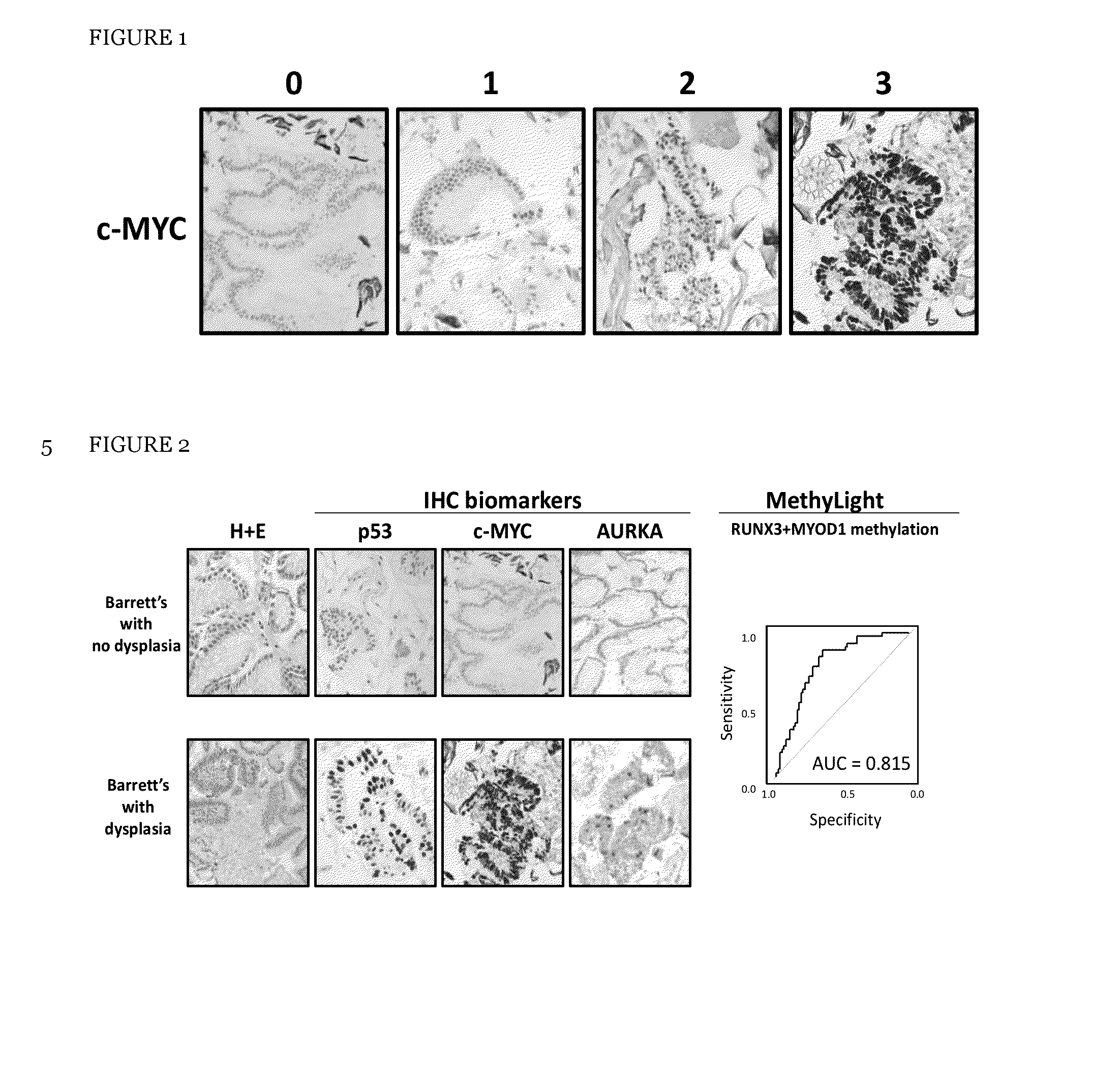
[0313]There were a number of reasons for selecting the four molecular risk stratification biomarkers, (optionally plus a fifth marker atypia), examples of which are set out below:

[0314]p53 protein accumulation was selected as a biomarker as p53 is one of the best characterised tumour suppressor proteins and has been shown to be associated with dysplasia in Barrett's oesophagus (Bian et al., 2001) as well as with increased risk of progression to OAC (Kastelein et al., 2012; Sikkema et al., 2009).

[0315]c-MYC, a well characterised oncogene, was included as it is recurrently amplified in OAC (Miller et al., 2003; Rygiel et al., 2008) and displays increased gene expression in Barrett's with high grade dysplasia in our in-house gene expression arrays.

[0316]Aurora kinase A (AURKA) was selected as a surrogate marker of aneuploidy as AURKA overexpression, centrosome amplification and aneuploidy have been shown to be associated. AURKA is a key regulator of mitotic entry, c...

example 2

Exclusion of Markers

[0318]There are robust reasons for excluding other potential biomarkers for use on the Cytosponge™. Some examples of markers excluded from the design of the panel are discussed below:

[0319]Eleven other potential biomarkers were evaluated to determine whether these could be used in conjunction with the Cytosponge™ to detect Barrett's with dysplasia. The 11 biomarkers were EGFR, CDNK2A, FGFR2, CCNA1, DDX21, MSLN, PLK1, HER2, DNMT1, MYHFD2 and VNN2. EGFR, HER2, CDNK2A, CCNA1 and FGFR2 were selected from published literature and DDX21, MSLN, PLK1, DNMT1, MTHFD2 and VNN2 were selected from in-house gene expression array data. VNN2 was eliminated as there are no antibodies available for staining formalin fixed paraffin embedded (FFPE) slides for this protein. FGFR2 and CDKN2A were eliminated as expression of both these proteins was detected in gastric glandular tissue which would also be sampled by the Cytosponge™. MTHFD2 was excluded as the staining was only cytoplasm...

example 3

Sample Processing and Preparation

Processing of the Capsule Sponge Specimens

[0325]Cytosponge™ capsules were swallowed by patients and then placed directly into preservative solution at 4° C. until processed further. The samples were vortexed extensively and shaken vigorously to remove any cells from the sponge material. The preservative liquid containing the cells was centrifuged at 1000 RPM for 5 minutes to pellet the cells. The resulting pellet was re-suspended in 500 μL of plasma and thrombin (Diagnostic reagents, Oxford, UK) was then added in 10 μL increments until a clot formed. The clot was then placed in formalin for 24 h prior to processing into a paraffin block. The sample was cut into 3.5 μm sections to provide 15 slides, named slides 1 to 15, with two sections placed on slide 1 and 2.


PUM

PropertyMeasurementUnit
morphologyaaaaaaaaaa
affinityaaaaaaaaaa
nucleic acidaaaaaaaaaa

Abstract

The invention relates to a method of aiding detection of a surface abnormality in the oesophagus of a subject, wherein said surface abnormality is selected from the group consisting of low-grade dysplasia (LGD), high-grade dysplasia (HGD), asymptomatic oesophageal adenocarcinoma (OAC) and intra-mucosal cancer (IMC), the method comprising:a) providing a sample of cells from said subject, wherein said sample comprises cells collected from the surface of the subject's oesophagus;b) assaying said cells for at least two markers selected from(i) p53;(ii) c-Myc;(iii) AURKA or PLK1, preferably AURKA; and(iv) methylation of MyoD and Runx3;wherein detection of abnormal levels of at least two of said markers infers that the subject has an increased likelihood of a surface abnormality in the oesophagus. The invention also relates to certain kits, apparatus and uses.

Description

FIELD OF THE INVENTION[0001]The invention is in the field of testing for, or aiding the detection of, surface abnormality in the oesophagus.BACKGROUND[0002]Oesophageal cancer (OAC) is currently the eighth most common cancer type worldwide and its incidence has risen almost 5-fold over the past three decades.[0003]Barrett's oesophagus is the first step in the pathway towards OAC and meta-analyses have demonstrated that Barrett's oesophagus confers a 0.12-0.5% increased risk of progression to adenocarcinoma per year. Barrett's oesophagus occurs when the normal oesophageal cells are replaced by glandular cells and this, with time, can progress to low-grade dysplasia (LGD), high-grade dysplasia (HGD) and then finally to adenocarcinoma.[0004]Early diagnosis of OAC and / or its pre-malignant precursor Barrett's oesophagus can improve patient management and prognosis of OAC. In one known approach, the Cytosponge™ cell collection device has been developed, for example as published in WO2011 / 0...

Claims


Application Information

Patent Timeline
Patent Type & Authority Applications(United States)
IPC IPC(8): G01N33/574C12Q1/68
CPCG01N33/57407C12Q1/6886C12Q2600/154G01N2333/4703G01N2333/912G01N2333/4748
Inventor FITZGERALD, REBECCAROSS-INNES, CARYN
Owner UK RES & INNOVATION LTD
Learn More