Oral cancer point of care diagnostics
a point of care and diagnosis technology, applied in the field of oral cancer point of care diagnosis, can solve the problems of primitive initial experimnents
- Summary
- Abstract
- Description
- Claims
- Application Information
AI Technical Summary
Benefits of technology
Problems solved by technology
Method used
Image
Examples
example 1
Proof of Principle
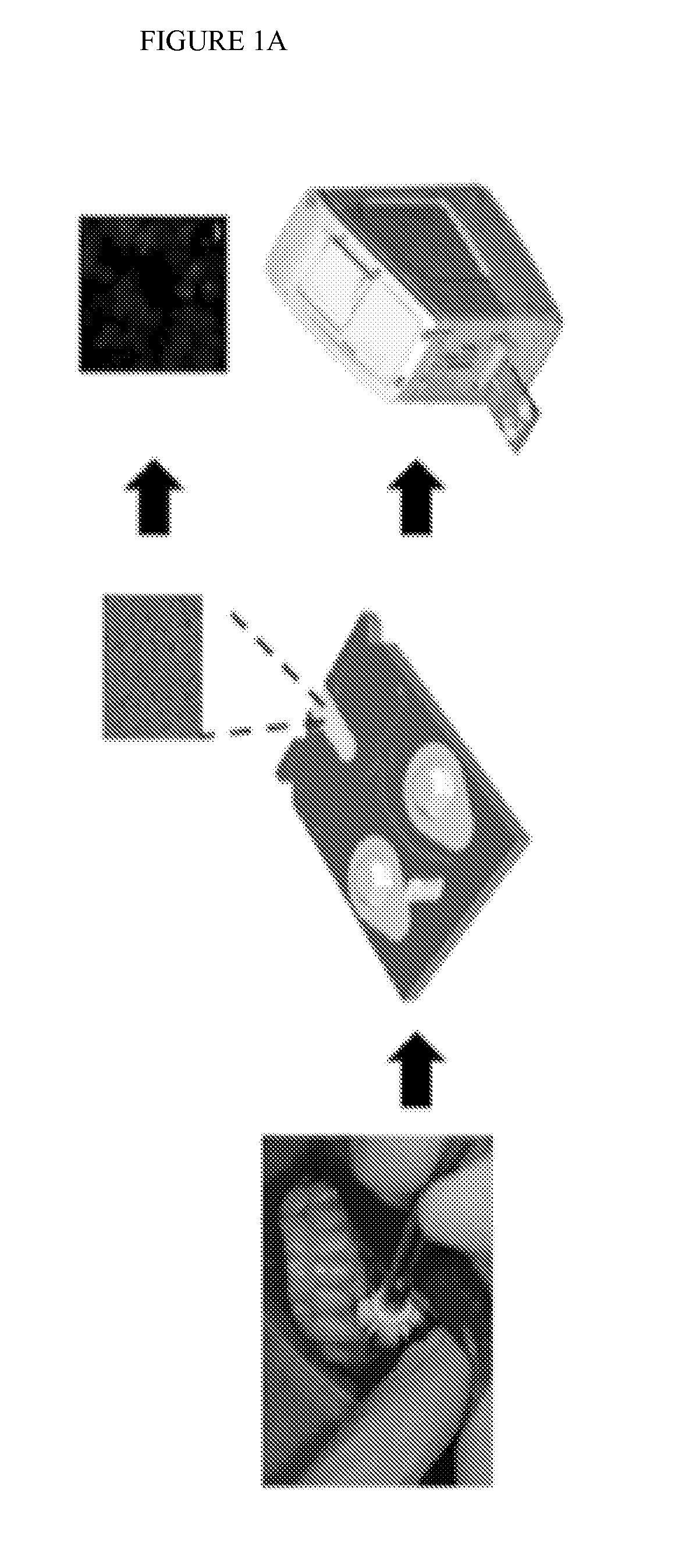
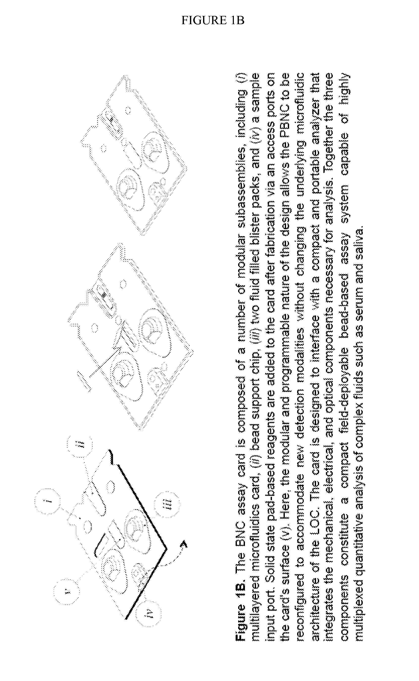
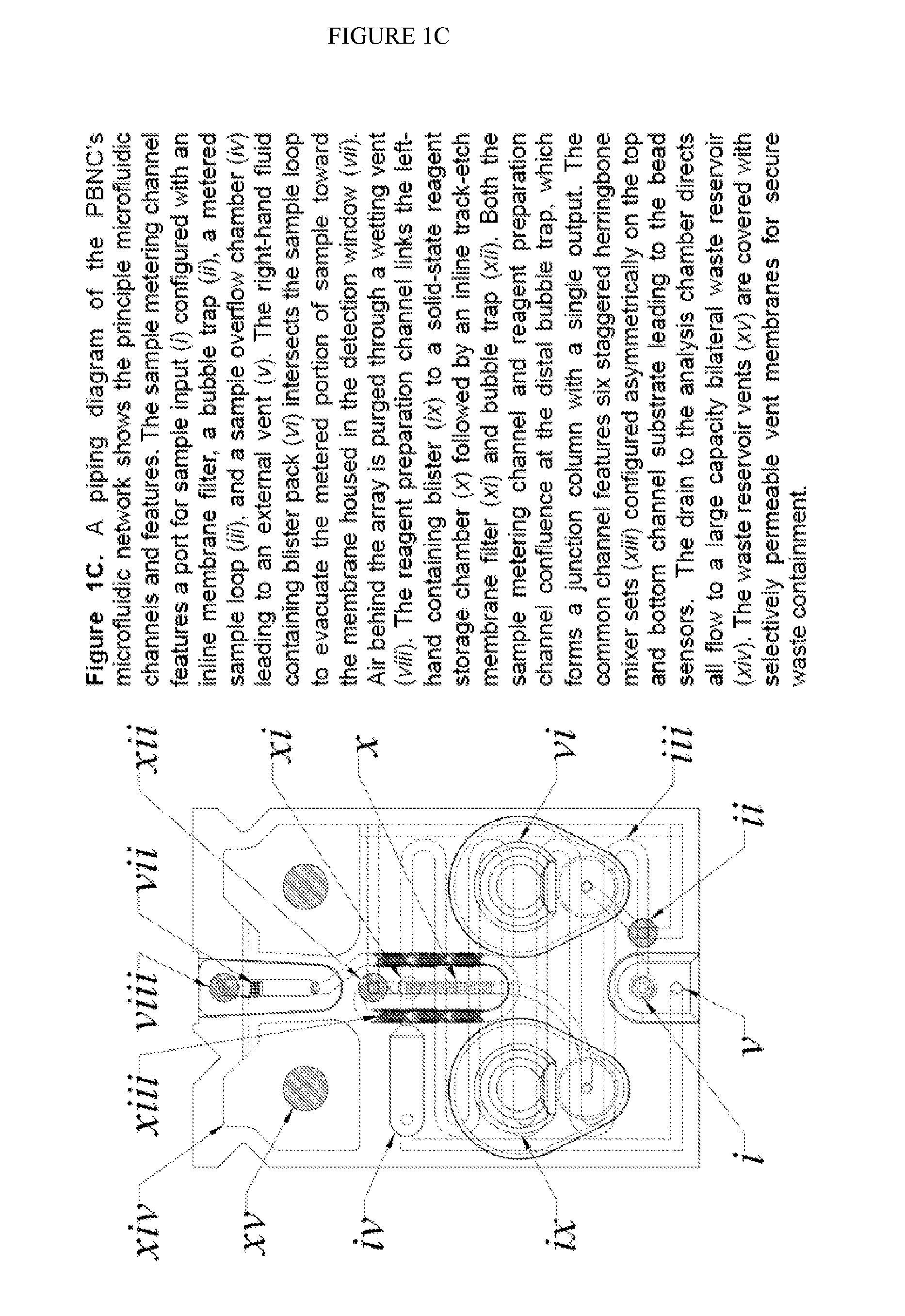
[0056]Using some of the same concepts borrowed from our HIV immune function test, we adapted the flow cell for use with brush biopsy samples to explore the cytomorphometric features and biomarker signatures for their capacity to distinguish between oral cancer and benign lesions. The PNBC sensor system for assessing OSCC integrates multiple laboratory processes onto a microfluidic platform in three primary steps: 1) cell separation / capture on the membrane filter, 2) biomarker immunolabeling and cytochemical staining, and 3) fluorescent imaging and analysis. No human intervention is needed once the sample is applied to the device, until the results are obtained, and the method is fully automated.
[0057]During cell capture, an oral cytology suspension is delivered to the PNBC sensor, using pressure-driven flow, whereupon any particles or cells larger than the membrane pore size are retained on the micro-membrane surface. Once captured, the cells are serially fluoresce...
example 2
Additional Markers
[0064]According to the Oral Cancer Foundation, HPV is now the leading cause of oral cancers in the US. Indeed, while tobacco continues to be an important risk factor, an increasing number of never-smoker patients have been diagnosed with oral cancer, developed by HPV16 infection. The system demonstrated here allows for future implementation of additional markers already discovered, emerging, or that will be discovered, that could specify HPV-related oral cancers.
[0065]The Speight and Thornhill team's long track record of research into the mechanisms associated with the malignant conversion of oral keratinocytes has enabled identification of specific markers of malignant conversion. Integrins are a family of molecules that are important in cell-cell and cell-matrix interactions for which the importance of cell surface integrin expression, particularly the expression of αvβ6 by oral keratinocytes, in the development of a malignant phenotype and invasion of the connec...
example 3
Applications
[0068]The innovative aspect to the validation of multiple biomarkers (separately or in a multiplex manner) for differentiating potentially malignant lesions is the application of a minimally-invasive brush biopsy test that can be performed in clinics or dentist's offices. Results will be available in a matter of minutes, versus hours to days for scalpel biopsies or OraScan analyses of brush biopsy samples.
[0069]The new device is intended to serve as a diagnostic device and not simply a screening aid for patients that have potentially malignant lesions (PMLs). In this context the new device is meant to replace (or augment) the current scalpel biopsy and pathology exam that now takes more than three days to complete.
[0070]Unlike the highly invasive scalpel biopsy, the new bio-nano-chip device will collect samples with a noninvasive, soft cytology brush and in doing so will be compatible with sampling of multiple areas.
[0071]These new devices have potential for broad usage ...
PUM
| Property | Measurement | Unit |
|---|---|---|
| transparent | aaaaa | aaaaa |
| cell aspect ratio | aaaaa | aaaaa |
| fluorescent | aaaaa | aaaaa |
Abstract
Description
Claims
Application Information
 - R&D
- Intellectual Property
- Life Sciences
- Materials
- Tech Scout
- Unparalleled Data Quality
- Higher Quality Content
- 60% Fewer Hallucinations
Browse by: Latest US Patents, China's latest patents, Technical Efficacy Thesaurus, Application Domain, Technology Topic, Popular Technical Reports.
© 2025 PatSnap. All rights reserved.Legal|Privacy policy|Modern Slavery Act Transparency Statement|Sitemap|About US| Contact US: help@patsnap.com


