
Biological detection device and detecting method

a detection device and detection method technology, applied in the field of detection devices and detection methods, can solve the problems of a large amount of test samples, a long waiting time in hospitals, and a higher voltage (1000v) and absolute temperature, so as to improve the long process of analyzing blood quality, obtain physiological information easily, and test a large quantity of test samples quickly

Active Publication Date: 2013-07-04
NAT CHIAO TUNG UNIV +1
View PDF1 Cites 18 Cited by
  • Summary
  • Abstract
  • Description
  • Claims
  • Application Information

AI Technical Summary

Benefits of technology

The present invention provides a simple and quick method to detect the concentration of triglyceride and high density liposome in serum, which can indicate the user's health information. This method can be easily performed using an electrically controlled surface polarity of a polymer thin film and is faster than the traditional blood analysis method. Additionally, the invention improves the long process of analyzing blood quality and allows users to know about preliminary information of their blood in a timely manner. The invention also provides an easy testing apparatus for LCD manufacturers, allowing designers to test a large quantity of test samples quickly.

Problems solved by technology

Medically, many factors must be taken into consideration for determining hyperlipidemia, and these factors include a comprehensive comparison of concentrations of triglyceride (TG), high density lipoprotein cholesterol (HDL), and low density lipoprotein cholesterol (LDL), and different chemical methods are used for testing each item, and thus the detection device and detecting method adopted require complicated and time-consuming processes.
At present, there is still a long waiting time in hospitals for patients requiring to have a physical examination to check whether or not they are a risk group of hyperlipidemia.
Although the primer can be used for protecting the metal surface, problems of a higher voltage (˜1000V) and absolute temperature and unstable acid-alkali sensitivity still exist.
During the chemical analysis process, the nature of the triglyceride may be damaged.
This process is also complicated and time-consuming.
As disclosed in U.S. Pat. No. 7,811,780B2, HDL test sample solution and testing solution are used to observe the concentration of the resultants or the residual of reactants to determine the concentration of HDL, but this method requires special solutions and complicated testing procedures, and the test can be used for the testing of HDL concentrations only.
As disclosed in U.S. Pat. No. 7,838,631B2, HDL, VLDL, LDL and chylomicron with different physical properties can be used for manufacturing a simple filter device to separate HDL and other molecules, but this method cannot give the concentration levels of the HDL.
Although the aforementioned PCT and U.S. patents have provided various different detection devices and detecting methods to determine hyperlipidemia, these devices and methods still have many problems.
The prior art still lacks of a hypelipidemia detection device capable of detecting the concentration of triglyceride and high density liposome by a simple, easy and quick detecting method.

Method used


Image

  • Biological detection device and detecting method
  • Biological detection device and detecting method
  • Biological detection device and detecting method

Examples


first preferred embodiment

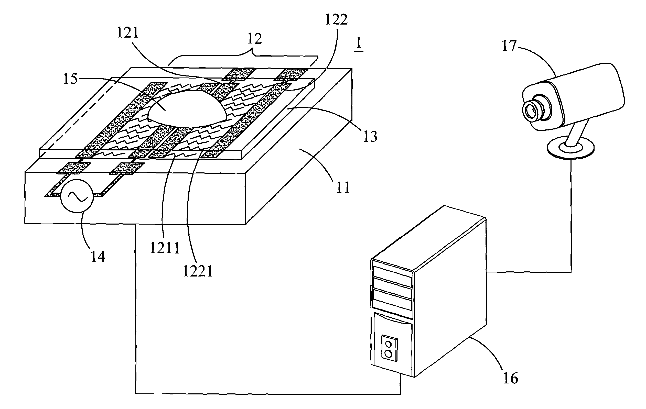
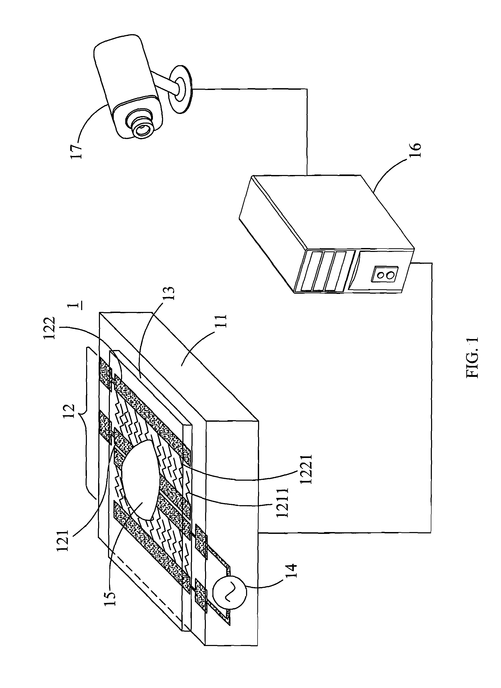
[0036]With reference to FIG. 1 for a schematic view of a biological detection device in accordance with the first preferred embodiment of the present invention, the biological detection device 1 comprises a substrate 11, an electric field unit 12, a liquid crystal / polymer composite film 13, a power supply 14, a processing unit 16 and an image sensor 17.

[0037]The substrate 11 includes but is not limited to a glass substrate. The electric field unit 12 comprises a plurality of electrode pairs, and each electrode pair is installed in parallel with each other on the substrate 11, wherein each electrode pair is consisted of a first strip electrode 121 and a second strip electrode 122, and the first strip electrode 121 has a plurality of first extensions 1211, and each first extension 1211 is arranged with a gap from the other. The second strip electrode 122 has a plurality of second extensions 1221, and each second extension 1221 is arranged with a gap from the other, and each second ext...

second preferred embodiment

[0062]In this preferred embodiment, the test sample is a serum solution obtained from a hospital. The biological detection device and the detecting method of the present invention perform a detection and analysis when a voltage is applied or when no voltage is applied.

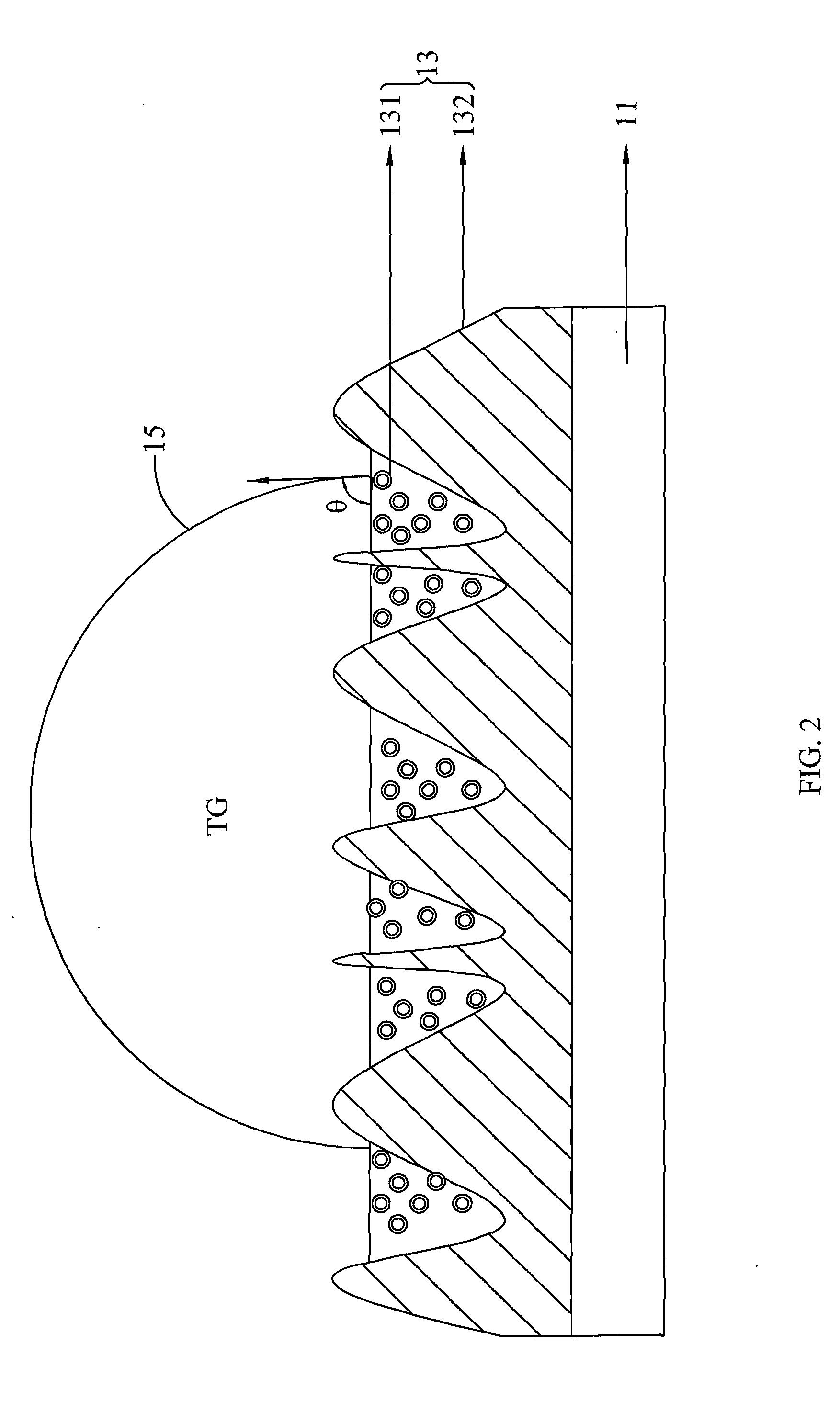
[0063]Firstly, a serum solution is obtained from a hospital, and the serum solution is set aside to cool to room temperature, and then a test tube containing the serum solution is shaken slightly and uniformly, and a dropper is used to place the serum solution on a liquid crystal / polymer composite film (LCPCF), and the liquid crystal / polymer composite film is put onto an observation table. A high-speed CCD camera is installed to the side to record the condition of the serum droplet and transmit the data to a computer, and a computer program FTA32 is provided for identification and analysis. The high-speed CCD camera (Model No. CVM30, CCD, by Pentad) takes the photo of a contact angle of the observed serum droplet on th...


PUM

PropertyMeasurementUnit
voltageaaaaaaaaaa
lengthaaaaaaaaaa
widthaaaaaaaaaa

Abstract

The present invention discloses a biological detection device and a detecting method. The biological detection device comprises a substrate, an electric field unit, a liquid crystal / polymer composite film (LCPCF), a power supply, a processing unit, and an image sensor. Because of the electrically tunable orientations of the liquid crystal (LC) director anchored among the polymer grains, the wettability of the LCPCF changes with an applied electric field. As a result, we can manipulate a blood droplet on the LCPCF by a wettability gradient owing to the distribution of LC directors on the LCPCF. The motion states of the blood droplet can be related to the various qualities of the blood, and finally determines the health of the test sample. The change of contact angle of blood on LCPCF and the blood droplet motion on LCPCF indicate the concentration of TG and the concentration of HDL.

Description

CROSS-REFERENCE TO RELATED APPLICATION[0001]This application claims the benefit of Taiwan Patent Application No. 100149671, filed on Dec. 29, 2011, in the Taiwan Intellectual Property Office, the disclosure of which is incorporated herein in its entirety by reference.BACKGROUND OF THE INVENTION[0002]1. Field of the Invention[0003]The present invention relates to a detection device and a detecting method, and more particularly to a biological detection device that changes the hydrophilic property of a liquid crystal / polymer composite film to produce a serum droplet motion, and combines with an image sensor to detect biological information of the blood droplet to determine the health condition of the blood.[0004]2. Description of Related Art[0005]Conventional methods of testing triglyceride in blood is to take out serum composition from the blood by cell separation and add a specific testing agent after purification and a series of chemical conversions, and the extent of a color chang...

Claims


Application Information

Patent Timeline
Patent Type & Authority Applications(United States)
IPC IPC(8): G01N33/00
CPCG01N33/49B01L2400/0427B01L3/502792
Inventor LIN, YI-HSINCHU, WEI-LINWU, LI-CHING
Owner NAT CHIAO TUNG UNIV
Learn More