
Specimen supporting member for x-ray microscope image observation, specimen containing cell for x-ray microscope image observation, and x-ray microscope

a technology of microscope image and supporting member, which is applied in the field of observation technique of x-ray microscope image, can solve the problems of specimen fixing agent itself, lack of durability, and significant reduction of the contrast of the resulting observation image, and achieve the effect of reducing damage to the specimen and convenient high-resolution x-ray observation

Inactive Publication Date: 2012-12-20
NAT INST OF ADVANCED IND SCI & TECH
View PDF4 Cites 15 Cited by
  • Summary
  • Abstract
  • Description
  • Claims
  • Application Information

AI Technical Summary

Benefits of technology

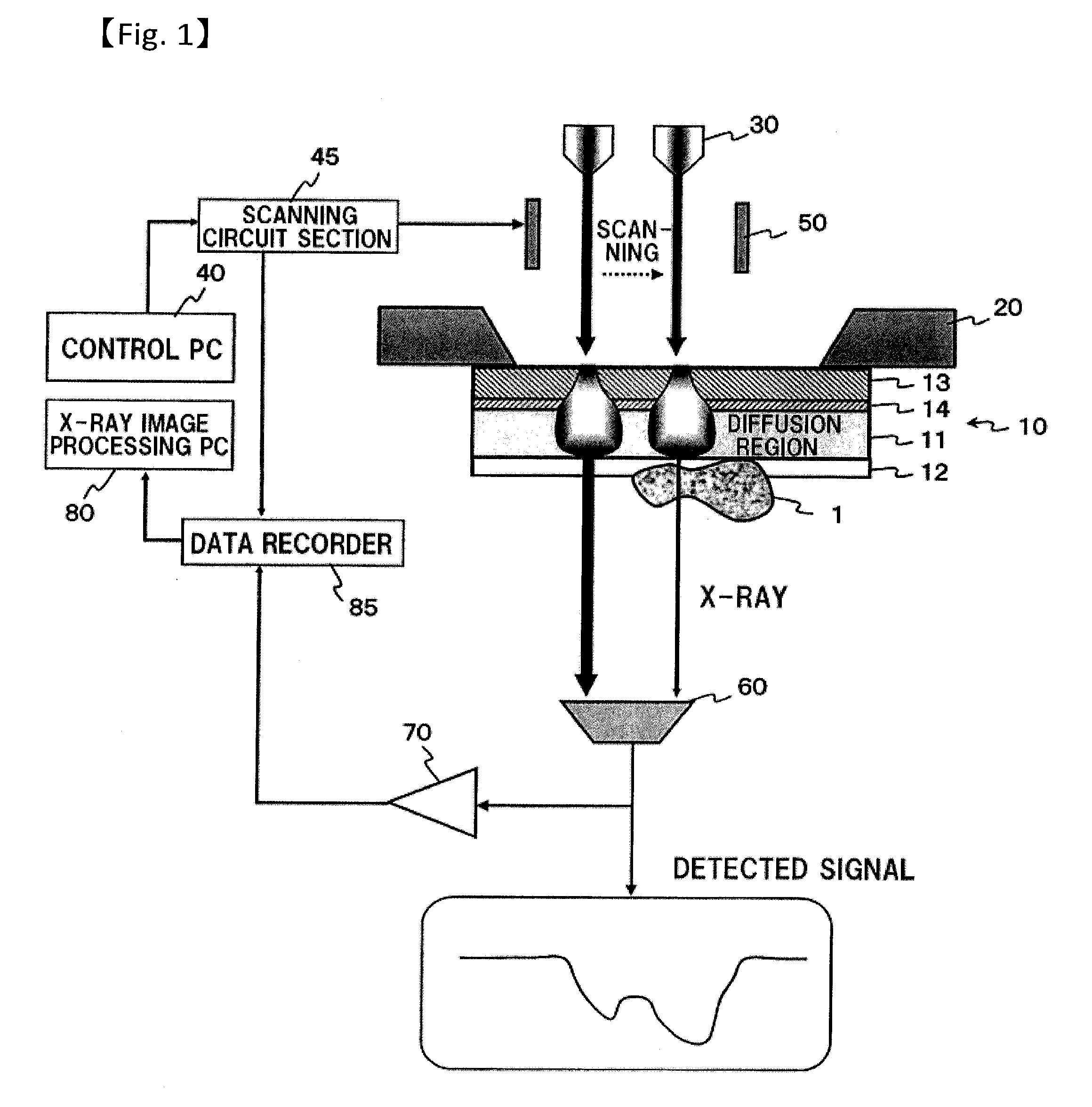
The present invention relates to an observation technique of X-ray microscope images of biological specimens in aqueous solution. The invention is about a technique that uses soft X-rays in the "water window" region (10 to 45 nm) which has lower absorption by carbon and nitrogen compared to soft X-rays in the "carbon window" region (5 to 45 nm) and the "nucleic acid window" region (0.6 to 2.3 nm). This technique allows for higher resolution observation of biological specimens in aqueous solution compared to optical microscopy. The invention also discusses the use of a thin and durable supporting film for fixing the specimen to the microscope, which is transparent to X-rays and minimizes deflections caused by Brownian motion. The invention addresses the problem of reducing the contrast of the observation image caused by the absorption of soft X-rays by organic matters commonly used as fixing agents for biological specimens.

Problems solved by technology

However, since such organic matters contain a large amount of carbon and nitrogen, soft X-ray in the above described water window region is apt to be absorbed, thereby significantly reducing the contrast of a resulting observation image.
Moreover, since the molecular size is as large as several tens of nm, a problem also exists in that the structure of such specimen fixing agent itself is observed at the time of high resolution observation.
Further, they also have drawbacks that they are susceptible to external environments such as heat and ultra violet ray or humidity, and are lack of durability.

Method used


Image

  • Specimen supporting member for x-ray microscope image observation, specimen containing cell for x-ray microscope image observation, and x-ray microscope
  • Specimen supporting member for x-ray microscope image observation, specimen containing cell for x-ray microscope image observation, and x-ray microscope
  • Specimen supporting member for x-ray microscope image observation, specimen containing cell for x-ray microscope image observation, and x-ray microscope

Examples


example 1

[0130]FIG. 13A is an X-ray image obtained by vapor depositing a nickel film of 120 nm thickness as the specimen adsorption film on a specimen supporting film made up of a silicon nitride film of 50 nm thickness, and fixing and observing yeast with this specimen adsorption film. Further, FIGS. 13B to 13D are respectively a diagram showing the result of a simulation of the case where an electron beam is made to irradiate the above described specimen supporting member; a diagram showing the result of a simulation of the radiant quantity of characteristic X-ray of nitrogen emitted from the specimen supporting film of silicon nitride at the time of electron beam irradiation shown in FIG. 13B; and a diagram showing the result of a simulation of the X-ray transmittance rate of a nickel film of 120 nm thickness.

[0131]The simulation shown in FIG. 13B shows an electron beam scattering state in the specimen supporting film and the specimen adsorption film obtained by a Monte Carlo simulation, ...

example 2

[0133]FIG. 14A is an X-ray image obtained by vapor depositing a nickel film of 60 nm thickness as the specimen adsorption film on the specimen supporting film made up of a carbon film of 40 nm thickness, and making bacteria as an observation specimen absorb to the nickel film and observing it at a magnification of 2000, and it can be confirmed that the bacteria are quite uniformly adsorbed in an appropriate manner.

[0134]FIG. 14B is an image when the above described observation magnification of bacteria is raised to 15,000, at which magnification the internal structure of bacterium is observed in detail.

[0135]FIG. 14C is a diagram showing the result of a simulation of the case where an electron beam is made to irradiate the above described specimen supporting member, in which the simulation determined the scattering state of an electron beam in the specimen supporting film and the specimen adsorption film by the Monte Carlo simulation, where the acceleration voltage of electron beam ...

example 3

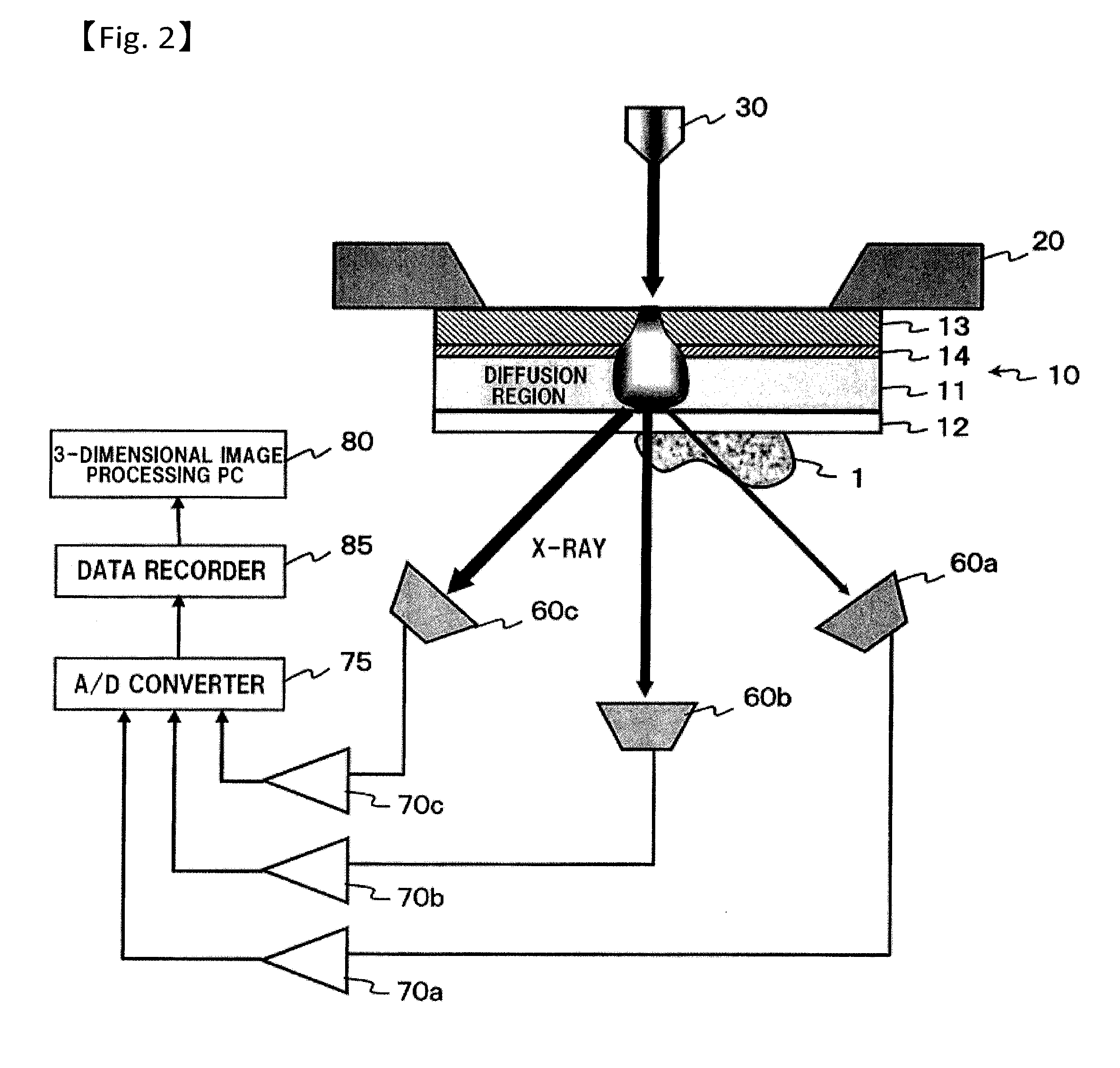
[0137]FIG. 15A is a diagram showing a scattering state of electron beam determined by a Monte Carlo simulation when plural kinds of X-ray radiation films are laminated, wherein the lamination is made from the electron beam incidence side in an order from a carbon film (20 nm), an aluminum film (20 nm), and a titanium film (20 nm), and the acceleration voltage of electron beam is 5 kV. Laminating thin films made up of plural kinds of metal allows a plurality of characteristic X-rays to be radiated, enabling the measurement of elementary composition within a specimen from those absorption ratios and the like. Moreover, forming thin film layers by laminating from the electron beam incidence side in an order from a lighter element to a heavier element allows the scattering range of electron beam to be suppressed.

[0138]FIG. 15B is a diagram showing the radiation range (the extent of spreading of characteristic X-ray intensity) of characteristic X-rays from each of a carbon film (20 nm), ...


PUM


Abstract

A specimen supporting member (10) includes: a specimen supporting film (11) such as a silicon nitride film, a carbon film, and a polyimide film; an X-ray radiation film (13) provided on one principal surface of the specimen supporting film, and for radiating a characteristic X-ray in a soft X-ray region upon irradiation with charged particles; and a specimen adsorption film (12) which is a metal film provided on another principal surface of the specimen supporting film (11), and which fixes by adsorption a specimen (1) to be observed. Since a protein which is a constitutive substance of a biological specimen has a characteristic to easily adsorb to a metallic ion, a specimen adsorption film (12) is formed on one principal surface of the specimen supporting film (11) so that an observation specimen adsorbs thereto.

Description

TECHNICAL FIELD[0001]The present invention relates to an observation technique of X-ray microscope image, and particularly to a technique suitable for the observation of a high-resolution X-ray microscope image of a biological specimen in an aqueous solution.BACKGROUND ART[0002]An X-ray microscope can observe a specimen in an aqueous solution at a higher resolution compared with an optical microscope. Particularly, soft X-ray having a wavelength in a range of 2.3 to 4.4 nm (corresponding to 284 to 540 eV), which is generally called as the “water window”, has a characteristic that its absorption coefficient significantly differs among materials constituting a living body, and that while it passes through water, it hardly passes through protein etc. due to large absorption by carbon and nitrogen.[0003]Using such soft X-ray in the “water window” region will allow an object containing water (a living body specimen and a specimen in a solution) to be observed as it is, and in addition to...

Claims


Application Information

Patent Timeline
Patent Type & Authority Applications(United States)
IPC IPC(8): G21K7/00
CPCG01N23/2251G21K7/00H01J2237/28H01J2237/2445H01J2237/20
Inventor OGURA, TOSHIHIKO
Owner NAT INST OF ADVANCED IND SCI & TECH
Learn More