
Cationic polymer coated mesoporous silica nanoparticles and uses thereof

a technology of mesoporous silica and nanoparticles, which is applied in the field of submicron structures, can solve the problems of encapsulated drugs being lost from the carrier or degraded, overblown claims, and agglomeration in the circulation, so as to reduce the translation of p-glycoproteins, and reduce the translation of proteins

Inactive Publication Date: 2012-08-16
RGT UNIV OF CALIFORNIA
View PDF1 Cites 64 Cited by
  • Summary
  • Abstract
  • Description
  • Claims
  • Application Information

AI Technical Summary

Benefits of technology

[0013]In some embodiments, the submicron structure also includes a cationic therapeutic compound. The cationic therapeutic compound may be, for example, in the interior or in the pores of the submicron structure. In some embodiments, the submicron structure also includes an oligonucleotide electrostatically bound to the cationic polymer. Some embodiments include both a cationic therapeutic compound and an oligonucleotide. In some embodiments, the oligonucleotide is an siRNA that reduces translation of a protein that causes resistance in a cell. In some embodiments, the siRNA reduces translation of a protein that causes resistance to the therapeutic compound in the cell. In some embodiments, the siRNA reduces translation of p-glycoprotein. In some embodiments, the therapeutic compound is doxirubicin.

Problems solved by technology

The key challenge now is to optimize the design features for efficient and safe in vivo drug delivery (He et al., Small, vol.
While the availability of nanocarrier drug delivery systems is an exciting development that holds the promise of a fundamental change in cancer chemotherapy, it remains at a relatively early stage of the implementation of this technology that often contains overblown claims of drug delivery nanoparticles acting as magic bullets.
Moreover, there is also a possibility that the encapsulated drugs could be lost from the carrier or degraded, as well as the fact that the colloidal instability of the carrier could lead to agglomeration in the circulation and may therefore be excluded from the intended “target site”.
It is also possible that the nanocarrier may reach the target site but that the drug is not released from the particle or that the carrier is not taken up effectively in the tumor cells.
Both effects will conspire to insufficient intracellular drug delivery.
Given these constraints, it is not a surprise that drug delivery to the tumor site seldom achieves more than 10% of the total administered dose.
The potential downside of surface coating is that PEG may also interfere in particle uptake by the tumor cells and that the longer circulation time may increase drug leakage from the carrier (Xia et al., ACS Nano, vol.

Method used


Image

  • Cationic polymer coated mesoporous silica nanoparticles and uses thereof
  • Cationic polymer coated mesoporous silica nanoparticles and uses thereof
  • Cationic polymer coated mesoporous silica nanoparticles and uses thereof

Examples


example 1

Synthesis and Surface Modification of Mesoporous Silica Nanoparticles MSNP

[0236]The basic synthesis of MSNP was conducted by mixing the silicate source tetraethylorthosilicate (TEOS) with the templating surfactants cetyltrimethylammonium bromide (CTAB) in basic aqueous solution (pH 11). In a round bottom flask, 100 mg CTAB was dissolved in a round-bottom flask containing solution of 48 ml distilled water and 1.75 ml sodium hydroxide (2 M). The solution was heated to 80° C. and stirred vigorously. After the temperature had stabilized, 0.5 ml TEOS was added slowly into the heated CTAB solution. After 15 min, 0.23 mmol of the organosilane solution was added into the mixture. 3-trihydroxysilylpropyl methylphosphonate was used for phosphonate surface modification, and aminopropyltriethoxy silane (APTS) was used for amine surface modification. After 2 hr, the solution was cooled to room temperature and the materials were washed with methanol using centrifugation. In order to incorporate f...

example 2

Physicochemical Characterization of the NP

[0239]MSNP were synthesized according to a modified procedure previously described (Radu et al., J. Am. Chem. Soc., vol. 126, pp. 13216-13217, 2004; Cai et al., Chem. Mater., vol. 13, no. 2, pp. 258-263, 2001). All MSNP were characterized for size, size distribution, shape, and charge (Table 1).

TABLE 1Size distribution of MSNPs in aqueous solutions.ZetaSize (nm)Potential (mV)DMEMBEGMH2O / DMEM +10%2 mg / mlserum / H2OserumBEGMBSABEGM + BSAMSNP-OH19664081096416−10.5 / −6.8 / −7.2MSNP-Phosphate1975306867439−8.9 / −6.5 / −5.8MSNP-PEG26754051215542−10.4 / −5.9 / −4.5MSNP-PEI 0.616894151243474+29.5 / −7.8 / −6.5KDMSNP-PEI 1.216844521298502+38.7 / −6.5 / −7.1KDMSNP-PEI 1.810535101087550+36.9 / −5.4 / −3.2KDMSNP-PEI 10614702917684+34.1 / −7.5 / −6.9KDMSNP-PEI 25147310431544825+30.8 / −5.9 / −4.0KDParticle size and zeta potential in solution were measured by a ZetaSizer Nano (Malvern).DMEM = Complete Dulbecco's Modified Eagle Media, which contains 10% fetal calf serum (FCS).BEGM = Bronc...

example 3

Differences in the Cytotoxic Potential of Anionic Vs Cationic MSNP

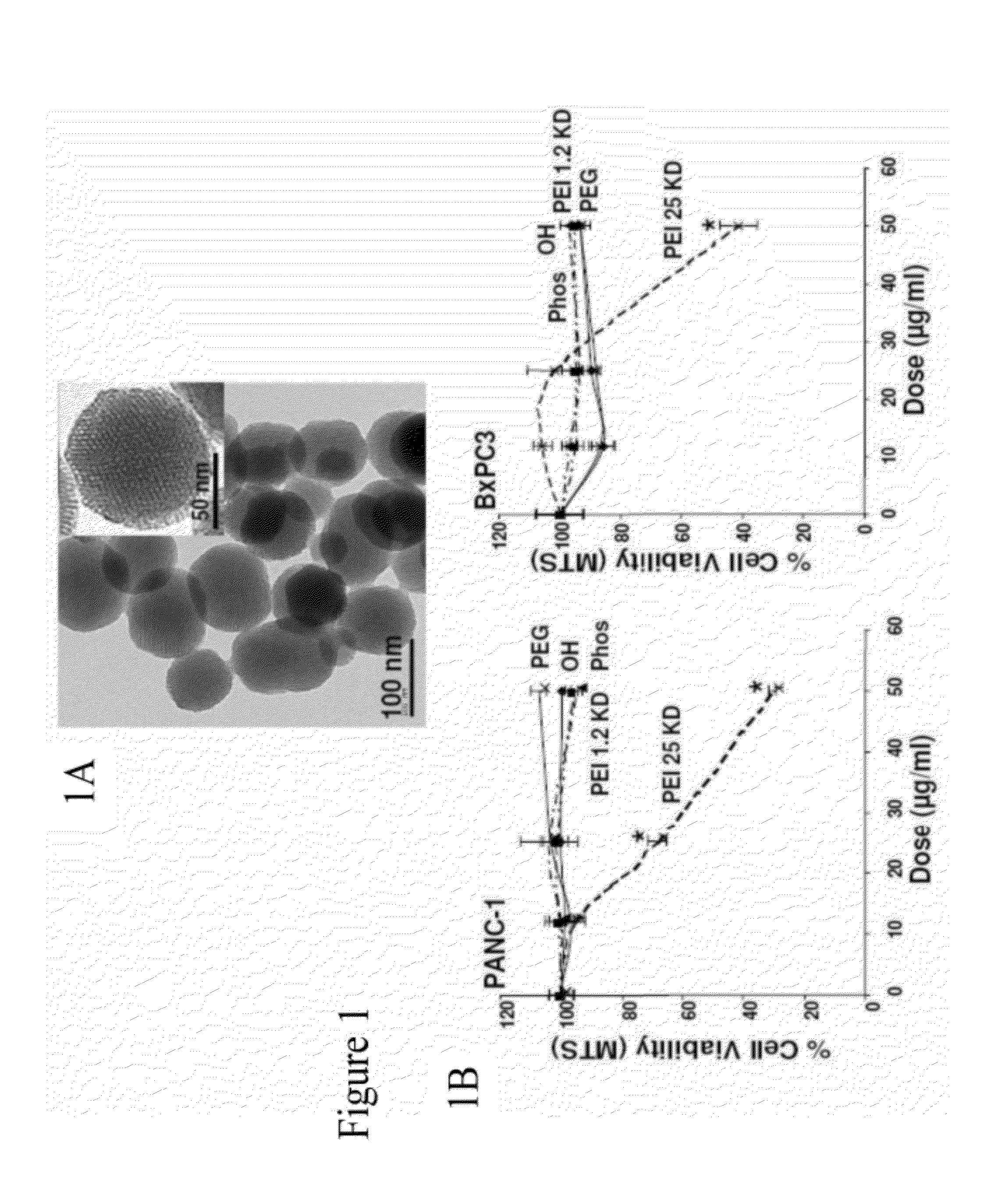
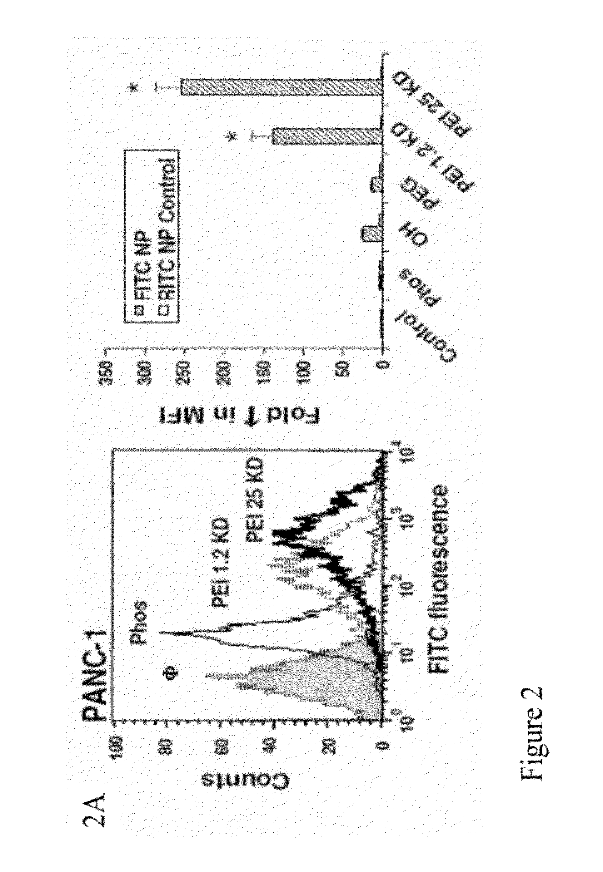
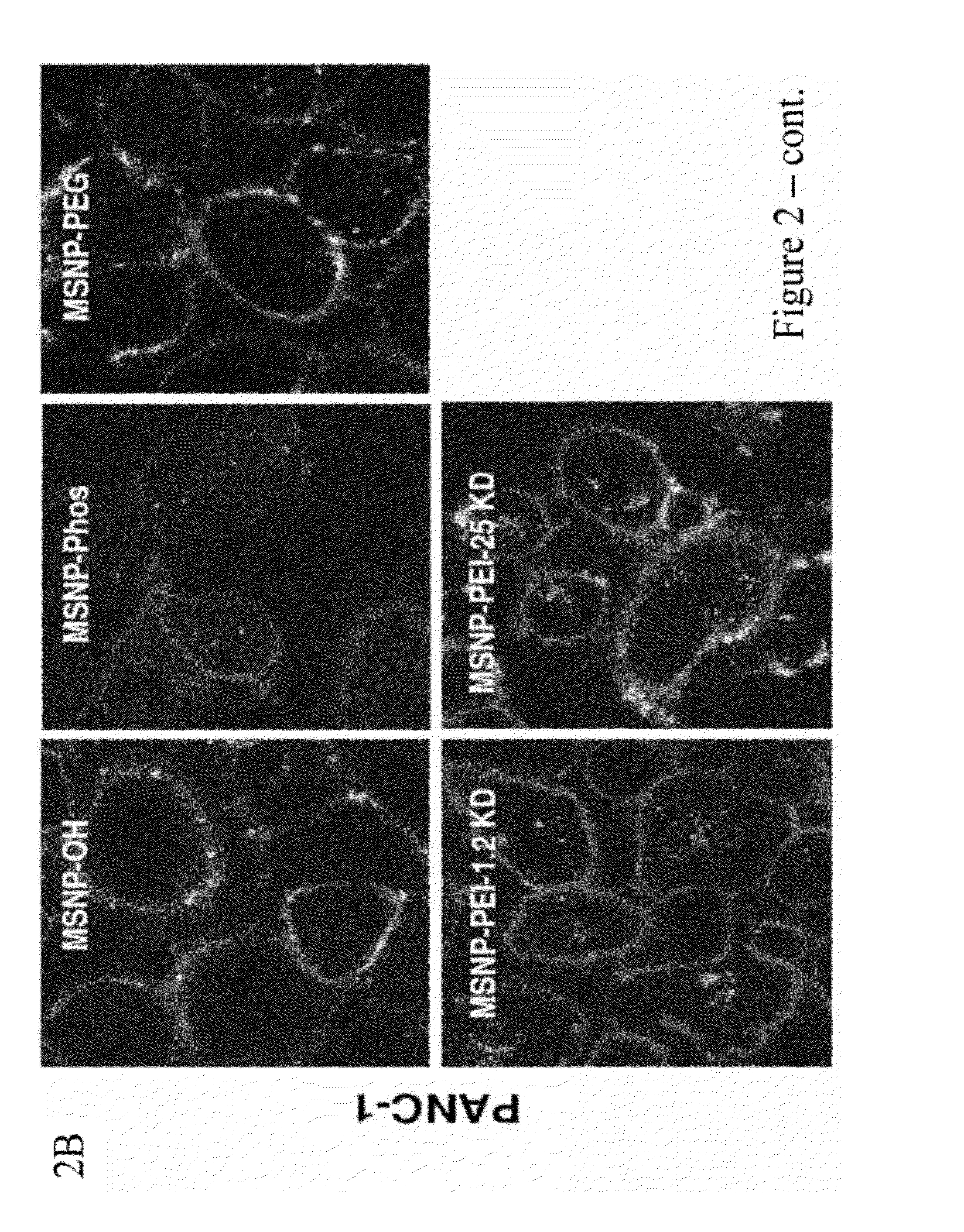
[0242]To screen for particle hazard, the MTS assay was used, which reflects dehydrogenase activity in healthy cells (Xia et al., ACS Nano, vol. 2, pp. 85-96, 2008). While most of the MSNP did not interfere in MTS activity in the PANC-1 and BxPC3 pancreatic cancer cell lines, particles coated with the 25 KD PEI polymer showed decreased cellular viability (FIG. 1). The particles coated with the 25 KD polymer also induced toxicity in macrophage (RAW 264.7) and bronchial epithelial (BEAS-2B) cell lines staining (FIG. 10). The toxicity was confirmed by propidium iodide (PI) staining, which showed that the rate of cell death was progressive over 15 hour period (FIG. 10). Similar to previous results with cationic polystyrene nanoparticles (Xia et al., ACS Nano, vol. 2, pp. 85-96, 2008), the toxicity of cationic MSNP involves an effect on mitochondria as determined by JC-1 fluorescence (FIG. 10). JC-1 measures mitochondrial m...


PUM

PropertyMeasurementUnit
ensemble average diameteraaaaaaaaaa
diameteraaaaaaaaaa

Abstract

A submicron structure having a silica body defining a plurality of pores is described. The submicron body may be spherical or non-spherical, and may include a cationic polymer or co-polymer on the surface of said silica body. The submicron structure may further include an oligonucleotide and be used to deliver the oligonucleotide to a cell. The submicron structure may further include a therapeutic agent and be used to deliver the therapeutic agent to a cell. An oligonucleotide and therapeutic agent may be used together. For example, when the oligonucleotide is an siRNA, the composition may be used to decrease cellular resistance to the therapeutic agent by decreasing translation of a resistance gene.

Description

CROSS-REFERENCE OF RELATED APPLICATION[0001]This application claims is a Continuation-in-Part of International Application Number PCT / US11 / 43874, filed Jul. 13, 2011, which claims priority to U.S. Provisional Application No. 61 / 363,945 filed Jul. 13, 2010. This application claims priority to U.S. Provisional Application No. 61 / 466,581 filed Mar. 23, 2011, U.S. Provisional Application No. 61 / 469,190 filed Mar. 30, 2011, and U.S. Provisional Application No. 61 / 479,751 filed Apr. 27, 2011. The entire contents of each are hereby incorporated by reference.[0002]This invention was made with Government support under Grant Nos. CA133697, ES016746, ES018766, and ES019528 awarded by the National Institutes of Health and Grant No. 0830117 awarded by the National Science Foundation. The Government has certain rights in this invention.BACKGROUND[0003]1. Field of Invention[0004]The current invention relates to submicron structures having a silica body defining a plurality of pores and an outer su...

Claims


Application Information

Patent Timeline
Patent Type & Authority Applications(United States)
IPC IPC(8): A61K31/713A61P35/00A61K9/00B32B3/26C12N5/071B82Y5/00
CPCA61K9/0019A61K9/5115A61K9/5146A61K9/5192A61K31/704Y10T428/2982B82Y5/00A61K31/713A61K2300/00A61P35/00
Inventor ZINK, JEFFREY I.NEL, ANDRE E.XIA, TIANJI, ZHAOXIAMENG, HUANLI, ZONGXILIONG, MONTYXUE, MINTARN, DERRICK Y.
Owner RGT UNIV OF CALIFORNIA
Learn More