Method for identifying senescent mesenchymal stem cells
a mesenchymal stem cell and stem cell technology, applied in the field of identifying senescent mesenchymal stem cells, can solve the problems of limiting the biomedical utility of the cell, and affecting the survival of the cell
- Summary
- Abstract
- Description
- Claims
- Application Information
AI Technical Summary
Benefits of technology
Problems solved by technology
Method used
Image

Examples
example 1
Cells and Culture Conditions
[0094]Human mesenchymal stem cells were obtained from the Inbiobank Stem Cell Bank (www.inbiobank.org) and came from adipose tissue. These cells exhibited the phenotype CD29+, CD73+ (SH3 and SH4), CD105+ (SH2), CD166+, CD45− and CD31−. All the cell donors were tested for HIV-1, HIV-2, hepatitis B and C and mycoplasma. The cell extraction process was carried out using liposuction and the cells were later treated with collagenase.
[0095]MSCs (1-2×10⁴ cells / ml) were cultured in a glucose-rich medium, Dulbecco's Modified Eagle's Medium (DMEM, Sigma, USA), supplemented with 10% foetal bovine serum (FBS, Sigma, USA), glutamine and penicillin / streptomycin. The cells were cultured in the presence of two different O2 concentrations, 20% and 3%, to obtain a senescent cell population and a non-senescent population, respectively, that could be compared. The cells capable of adhering to the plate were considered the first hMSC extract derived from adipose tissue. The m...
example 2
Measurement of Telomere Shortening in Human Mesenchymal Stem Cells Growing In Vitro
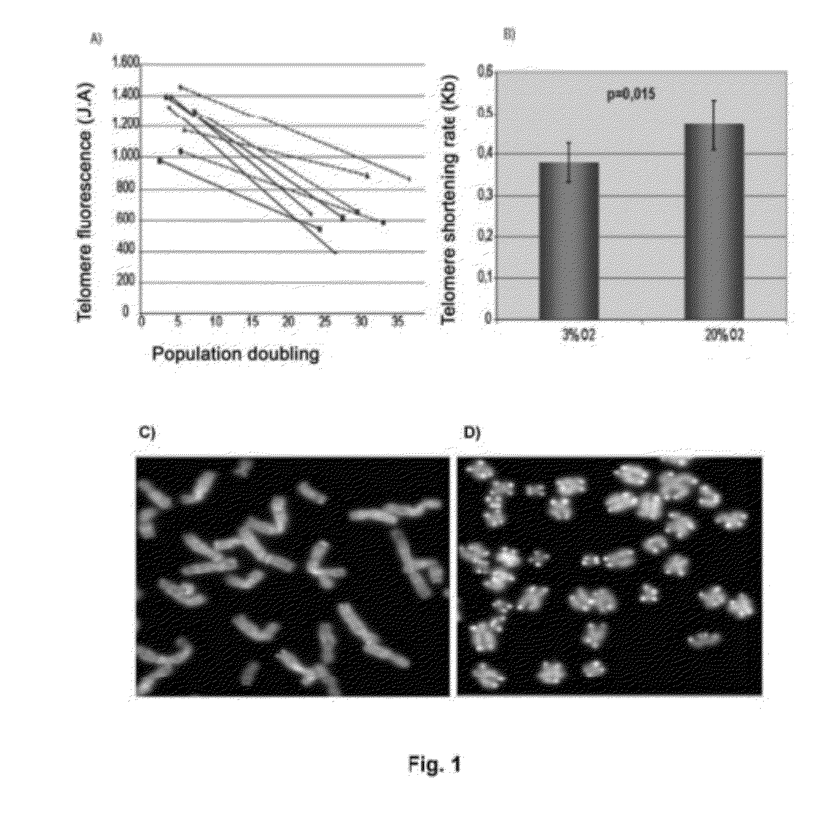
[0096]To determine if senescence in human mesenchymal stem cells is due to telomere shortening, a quantitative fluorescence in situ hybridization (Q-FISH) was carried out using a telomere PNA (Peptide Nucleic Acid) LL(CCCTAA)3 probe labelled with Cy3 (Eurogentec) in the nucleus of MSCs in four different lines in interphase, growing in 20% O2 and in 3% O2, in passage 2 and in passage 15 (see FIG. 1A). After hybridization, the cells were washed three times in 0.1% PBS Tween for 10 minutes at 60° C. and were dehydrated in ethanol for 5 minutes. Finally, they were contrast stained in Vectashield mountain medium with DAPI (Vector Laboratories Inc.). Images were taken with CytoVision. The telomere signals were captured at the same exposure time in all samples. Telomere length in Kb was calculated as a function of the fluorescence of 82-6 fibroblasts immortalized with hTert express...
example 3
Analysis of Ploidy Level of Human Mesenchymal Stem Cells
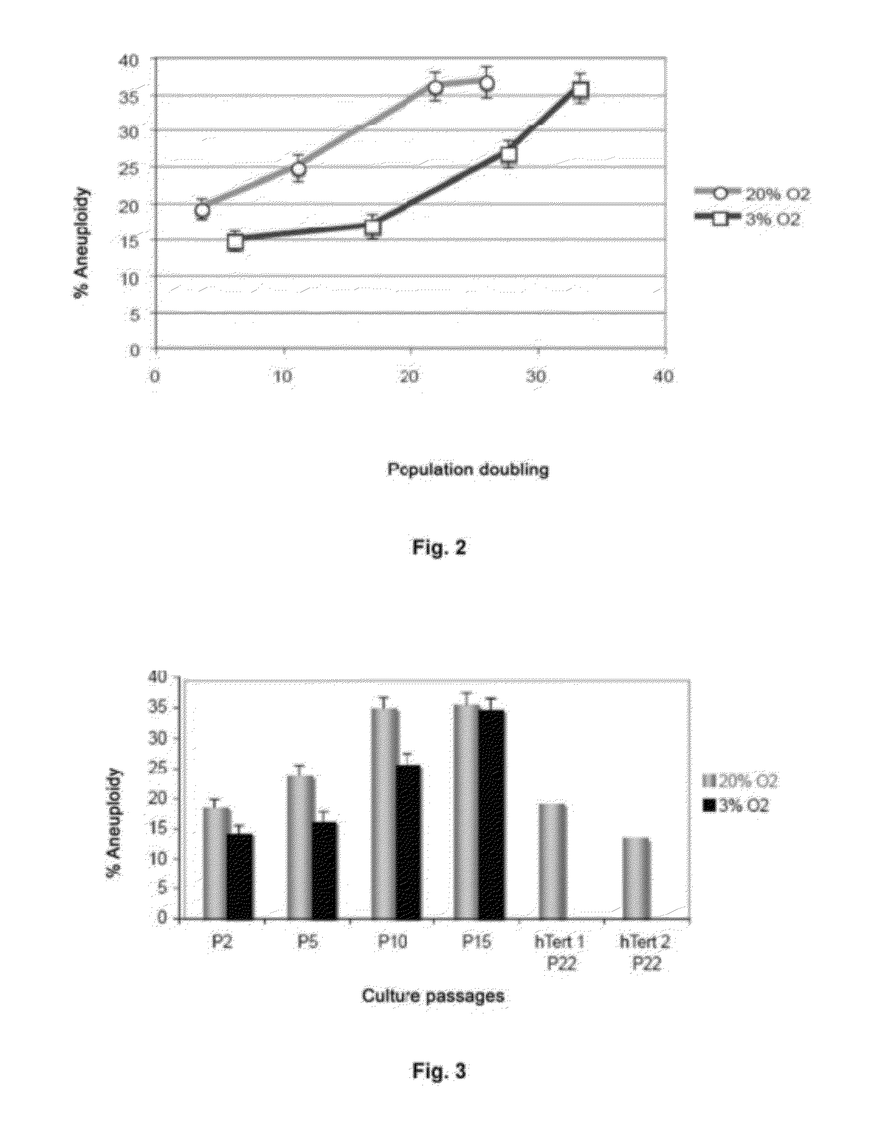
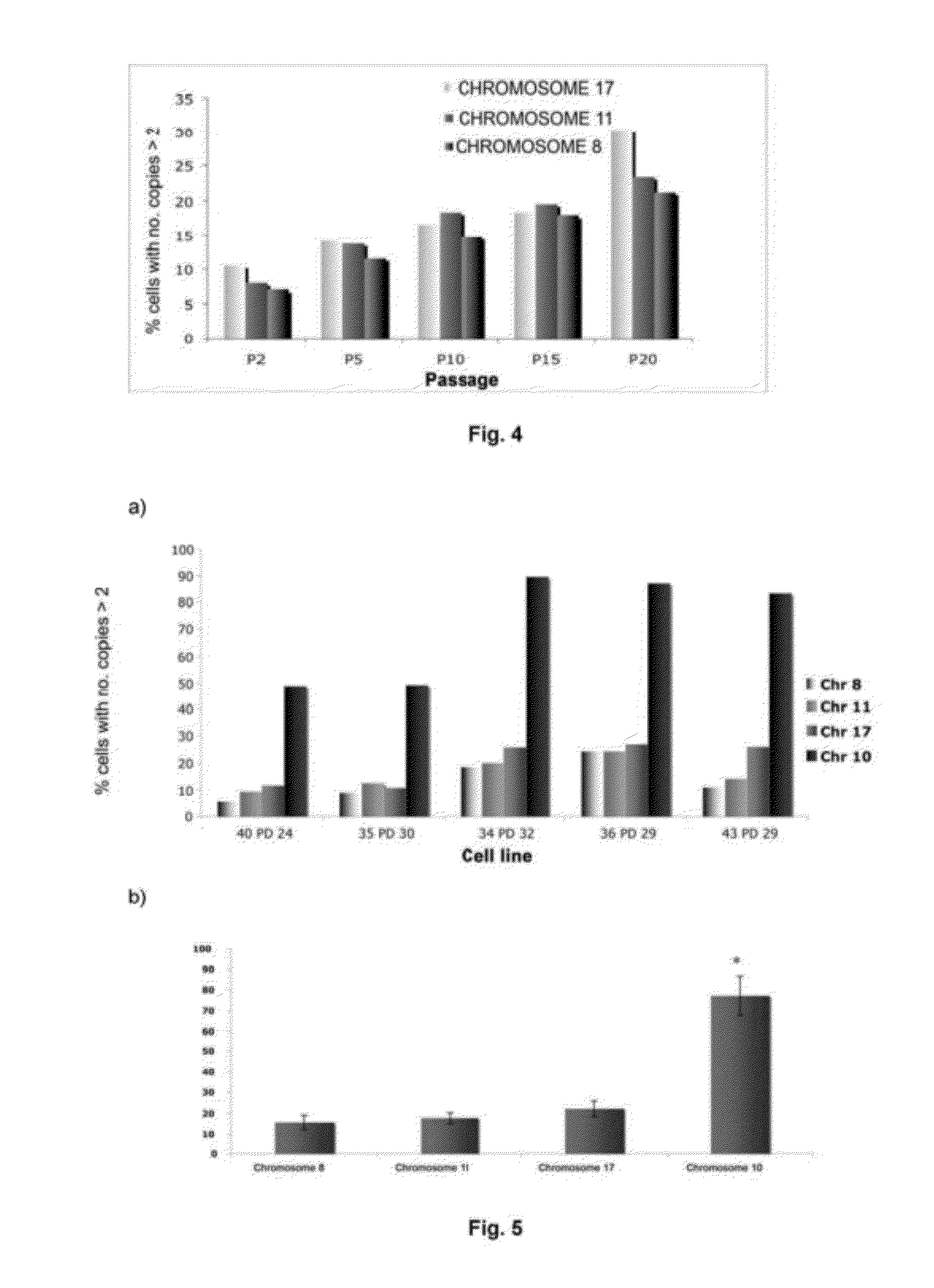
[0103]The ploidy levels in MSCs in interphase under both culture conditions with different oxygen concentrations were determined in passages 2 and 15 by FISH with centromere probes for chromosomes 8, 11 and 17 (see FIGS. 2 and 3). Cells were incubated with 10 μg / ml Colcemid for 4 hours at 37° C., then treated with 0.56% KCl for 15 minutes at 37° C. and fixed in methanol:acetic acid (3:1) three times. The cell suspensions were deposited on slides and dried in air for 24 hours. Before hybridization, the slides were treated with protease solution (0.1% pepsin and HCl) and fixed in 4% formaldehyde for 5 minutes at room temperature; next, they were dehydrated by serial incubations in increasing ethanol concentrations (70, 90 and 100%) for 5 minutes each. The cells were denatured in 70% deionized formamide and 2×SSC for 5 minutes at 73° C. Next, they were dehydrated as described above. After dehydration, the cells were incubated for ...
PUM
| Property | Measurement | Unit |
|---|---|---|
| temperature | aaaaa | aaaaa |
| length | aaaaa | aaaaa |
| fluorescent | aaaaa | aaaaa |
Abstract
Description
Claims
Application Information
 - R&D
- Intellectual Property
- Life Sciences
- Materials
- Tech Scout
- Unparalleled Data Quality
- Higher Quality Content
- 60% Fewer Hallucinations
Browse by: Latest US Patents, China's latest patents, Technical Efficacy Thesaurus, Application Domain, Technology Topic, Popular Technical Reports.
© 2025 PatSnap. All rights reserved.Legal|Privacy policy|Modern Slavery Act Transparency Statement|Sitemap|About US| Contact US: help@patsnap.com