
Lateral Flow Immunoassay for Complement Activation and Methods of Use for Point-of-Care Assessment of Complement-Associated Disorders

Active Publication Date: 2012-06-07
KYPHA
View PDF5 Cites 9 Cited by
  • Summary
  • Abstract
  • Description
  • Claims
  • Application Information

AI Technical Summary

Benefits of technology

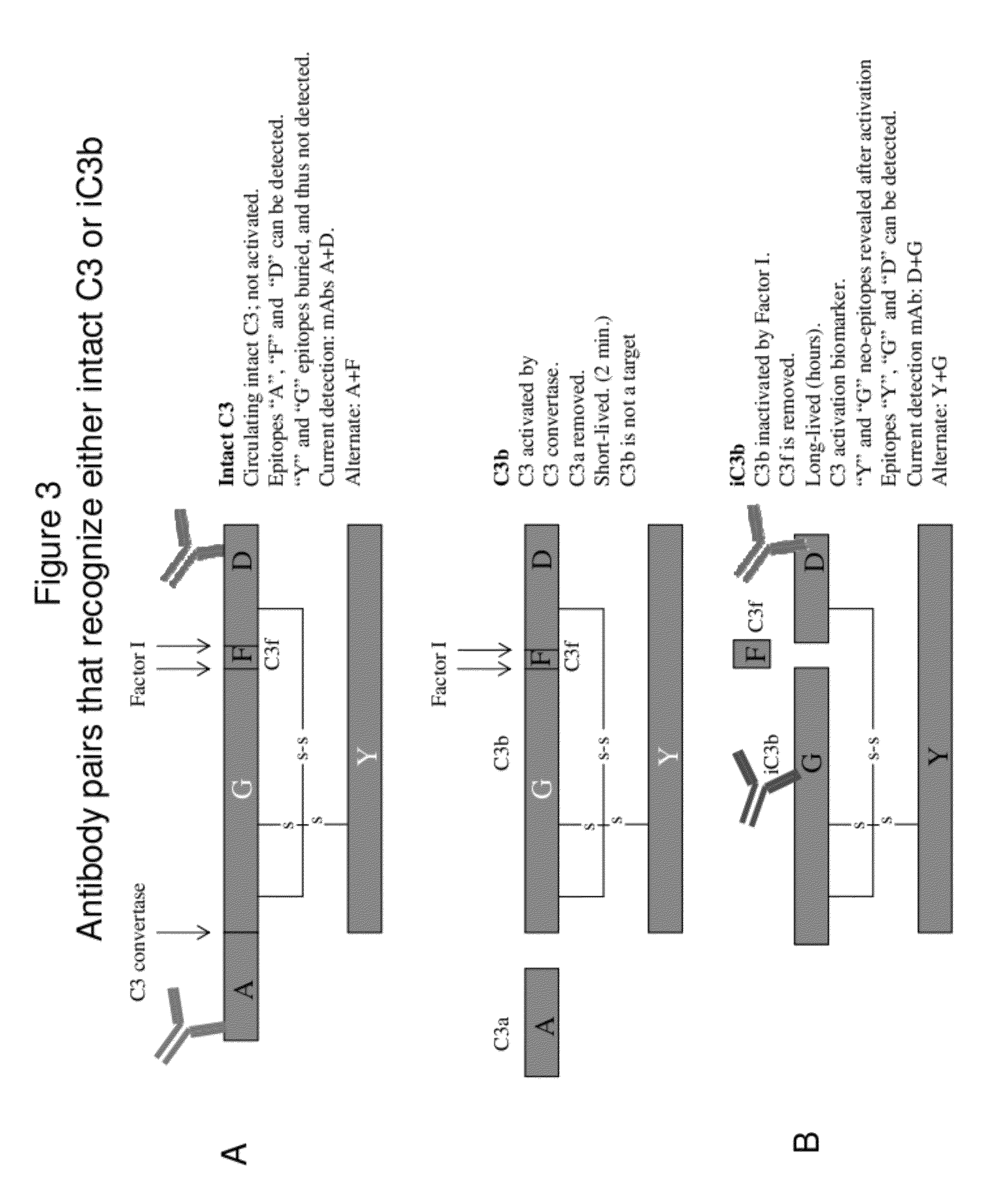
[0018]The present inventors have now developed a point-of-care method and assay for the qualitative and quantitative measurement of intact C3 and iC3b suitable for use in directing patient care at the earliest time points immediately following traumatic injury or other physiological crises. By carefully selecting capturing and detecting antibody pairs that avoid interfering cross-talk and applying the technology to a lateral flow immunoassay platform, the inventors have surprisingly found that it is possible to detect and quantify the biomarkers intact C3 and iC3b, while avoiding the false positive results that have plagued more conventional testing methods for these analytes.

Problems solved by technology

However, inflammation can also damage self-tissue.
Severe trauma patients present unique clinical challenges.
Those patients admitted to the ICU, even after initial stabilization, remain at high risk for secondary life-threatening complications involving organ dysfunction, respiratory distress and sepsis, among others.
Many of these conditions involve hyper-inflammatory events that can escalate rapidly and cause significant damage before clinical symptoms are detected.
Even in those instances, current assay methods have limitations.
However, other factors such as diet or exercise can cause lower steady state levels of C3.
Furthermore, a test that measures total C3 cannot monitor the real-time changes in C3 activation signature that would be useful in directing patient care.
However, the physician has difficulty in discerning whether the underlying disease process has been halted or just retarded sufficiently for homeostatic mechanisms to return C3 to physiologically normal levels.
A second limitation in current C3 testing is the time required to perform most assays.
This assay platform is therefore not useful for indications of inflammatory dysfunction, in which biomarkers change on the order of minutes and clinical intervention is required on a similar timescale.
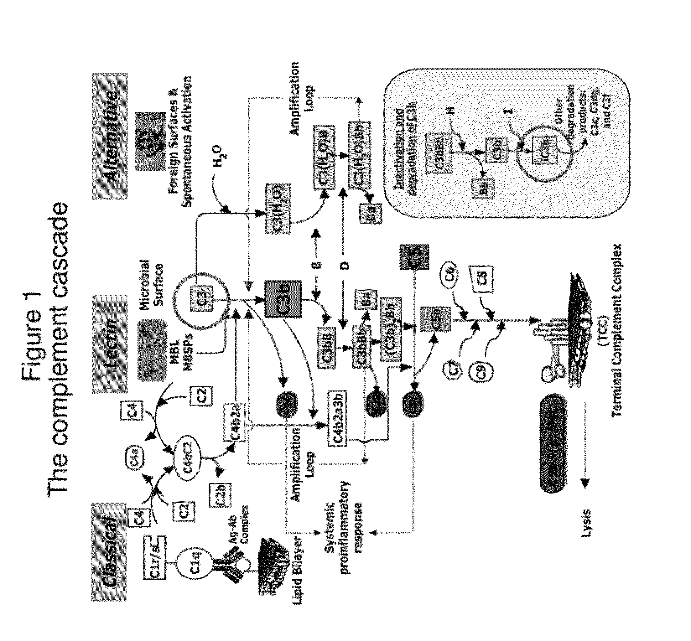
A third limitation in current C3 testing lies in the nature of the protein cascade itself.
Complement is notoriously fastidious and can become activated by virtue of standard analysis procedures (handling, storage, and exposure to foreign materials that contact C3 during analysis).
While this property is useful in directing an immune response to new foreign pathogens, this same property presents formidable challenges to experimental and diagnostic study.
Materials such as plastics used in sample handling, manipulation of the sample itself, and improper storage conditions can also trigger complement activation.
These false positives complicate traditional testing and render current testing methods unsuitable for use in directing patient care in near real-time.
Although technically easier to perform, total C3 assays cannot detect C3 depletion as accurately as intact C3 assays.
Intact C3 assays have historically proven difficult to administer or depend upon, in part because intact C3 is very labile and can denature or self-activate if not handled properly.
Hence, while a desirable marker of inflammation, heretofore iC3b has posed significant challenges in diagnostic testing.
However, Zhang et al. is limited to traditional sandwich-type immunoassays such as ELISA, requiring laboratory processing and the expertise of skilled technicians.
Further, the assays and methods of Zhang et al. require sample preparation, storage, and handling steps that are known to activate the labile intact C3 produce false positive test results, impeding the ability to accurately measure intact C3.
Moreover, the assays and methods of Zhang et al. require hours to process and are thus incapable of providing the near real-time data that can impact patient care in the earliest time points after physiological crisis.

Method used


Image

  • Lateral Flow Immunoassay for Complement Activation and Methods of Use for Point-of-Care Assessment of Complement-Associated Disorders
  • Lateral Flow Immunoassay for Complement Activation and Methods of Use for Point-of-Care Assessment of Complement-Associated Disorders
  • Lateral Flow Immunoassay for Complement Activation and Methods of Use for Point-of-Care Assessment of Complement-Associated Disorders

Examples


example 1

Patient Triage

[0122]Before the first test sample is assayed, a standard curve is performed using 10 ng / ml 30 ng / ml, 100 ng / ml, 300 ng / ml, and 1000 ng / ml of intact C3 and iC3b standards. Lateral flow immunoassay cassettes are read with an electronic reader after 20 minutes.

[0123]The test is used to gauge injury severity within 15, 30, or 60 minutes of injury. It is most useful for patients who may have suffered injuries not obvious by visual inspection. A drop of blood is collected either from an arterial line (A-line) or finger stick. A 10 ul sample is drawn up using a fixed volume pipet. The sample then mixed with 990 ul of sample buffer. The blood and sample buffer are mixed. Using a fixed volume pipet bulb, 100 ul is drawn up and pipetted onto the lateral flow immunoassay cassette containing integrated intact C3 and iC3b test strips. Alternatively, 100 ul can be applied to separate intact C3 and iC3b lateral flow assay cassettes. After 10 minutes but before 40 minutes, the casset...

example 2

Trajectory Monitoring of a Trauma Patient

[0124]At the beginning of the shift, ICU staff performs a standard curve using 10 ng / ml 30 ng / ml, 100 ng / ml, 300 ng / ml, and 1000 ng / ml of intact C3 and iC3b standards. Lateral flow immunoassay cassettes are read with an electronic reader after 20 minutes.

[0125]The objective of trajectory monitoring is to detect changes in inflammatory and immune status of patients that have been stabilized after severe trauma. In this example, respiratory distress caused by either pneumonia or inflammatory dysfunction is to be detected. The expected patient profile is one who has an injury severity score (ISS) equal or greater to 16 and who requires ventilator assistance for breathing.

[0126]The patient receives a complement test at frequent intervals, which aligns with the time points for testing glucose levels in blood. This interval between testing is usually about two hours. Blood is collected using the same method as for glucose testing, either by A-line ...

example 3

Determining Disease Severity and Effectiveness of Treatment in a Patient with Systemic Lupus Erythematosus (SLE)

[0128]Before the first test sample is assayed, a standard curve is performed using 10 ng / ml 30 ng / ml, 100 ng / ml, 300 ng / ml, and 1000 ng / ml of intact C3 and iC3b standards. Lateral flow immunoassay cassettes are read with an electronic reader after 20 minutes.

[0129]The test is used to gauge the initial severity of disease as well the effectiveness of therapy. One of the standard diagnostics performed on SLE patients is measurement of total C3 levels. C3 levels are normally depressed in SLE patients and return to normal (>1 mg / ml) following successful treatment. However, it not known generally whether the C3 activation has been abrogated or only slowed enough to allow normal replenishment mechanisms to restore C3 levels to normal.

[0130]At each doctor visit, a patient's blood is collected for total C3, intact C3, and iC3b tests. Only one drop is required for the combine 3 tes...


PUM

PropertyMeasurementUnit
Fractionaaaaaaaaaa
Fractionaaaaaaaaaa
Timeaaaaaaaaaa

Abstract

A method for treating an individual at risk for a complement-associated disorder is provided, including: (a) obtaining a sample of a body fluid from the individual; (b) measuring a complement activation level in the sample via a point-of-care lateral flow immunoassay; (c) correlating the complement activation level in the sample to a risk of a complement-associated disorder by comparing the complement activation level in the sample to a reference level in a control, wherein a deviation in complement activation level in the sample compared to the reference level in the control indicates the individual is at risk for a complement-associated disorder; (d) selecting a treatment for the individual, based on the correlating of step (c); and (e) treating the individual with the treatment selected in accordance with step (d). Lateral flow immunoassays and a method of monitoring an individual suffering from a complement-associated disorder are also provided herein.

Description

RELATED APPLICATION[0001]This application claims priority under 35 U.S.C. §119(e) to U.S. Provisional Application Ser. No. 61 / 409,297, filed Nov. 2, 2010, the contents of which are incorporated by reference herein in their entirety.FIELD OF THE INVENTION[0002]The presently disclosed subject matter relates to lateral flow immunoassays for the measurement of complement activation. Specifically, the presently disclosed subject matter relates to a lateral flow immunoassay for the quantitative measurement of intact C3 and iC3b in a sample and methods of using the same in the evaluation and treatment of individuals at risk for complement-associated disorders such as inflammatory distress.BACKGROUND[0003]Inflammation is the physiological response of vascularized tissue to injury, infection, and certain diseases. The inflammatory process is a biological requirement for wound healing after traumatic injury and for the clearance of infection. However, inflammation can also damage self-tissue....

Claims


Application Information

Patent Timeline
IPC IPC(8): A61K39/395G01N33/566A61P31/00A61P29/00A61P11/00A61P9/00A61P27/02A61P25/00A61P15/00A61P13/12A61K38/12A61P43/00
CPCG01N33/564G01N2333/4716G01N33/86G01N2800/26G01N33/558G01N2800/125A61P11/00A61P13/02A61P13/12A61P15/00A61P17/02A61P25/00A61P27/02A61P29/00A61P31/00A61P31/04A61P37/02A61P37/06A61P43/00A61P7/04A61P9/00A61P9/10G01N33/68G01N2800/7095G01N33/54388
Inventor OLSON, PAULMOSS, DON W.
Owner KYPHA
Learn More