
Bone Sonometer

Inactive Publication Date: 2012-02-02
BEAMMED
View PDF6 Cites 6 Cited by
  • Summary
  • Abstract
  • Description
  • Claims
  • Application Information

AI Technical Summary

Benefits of technology

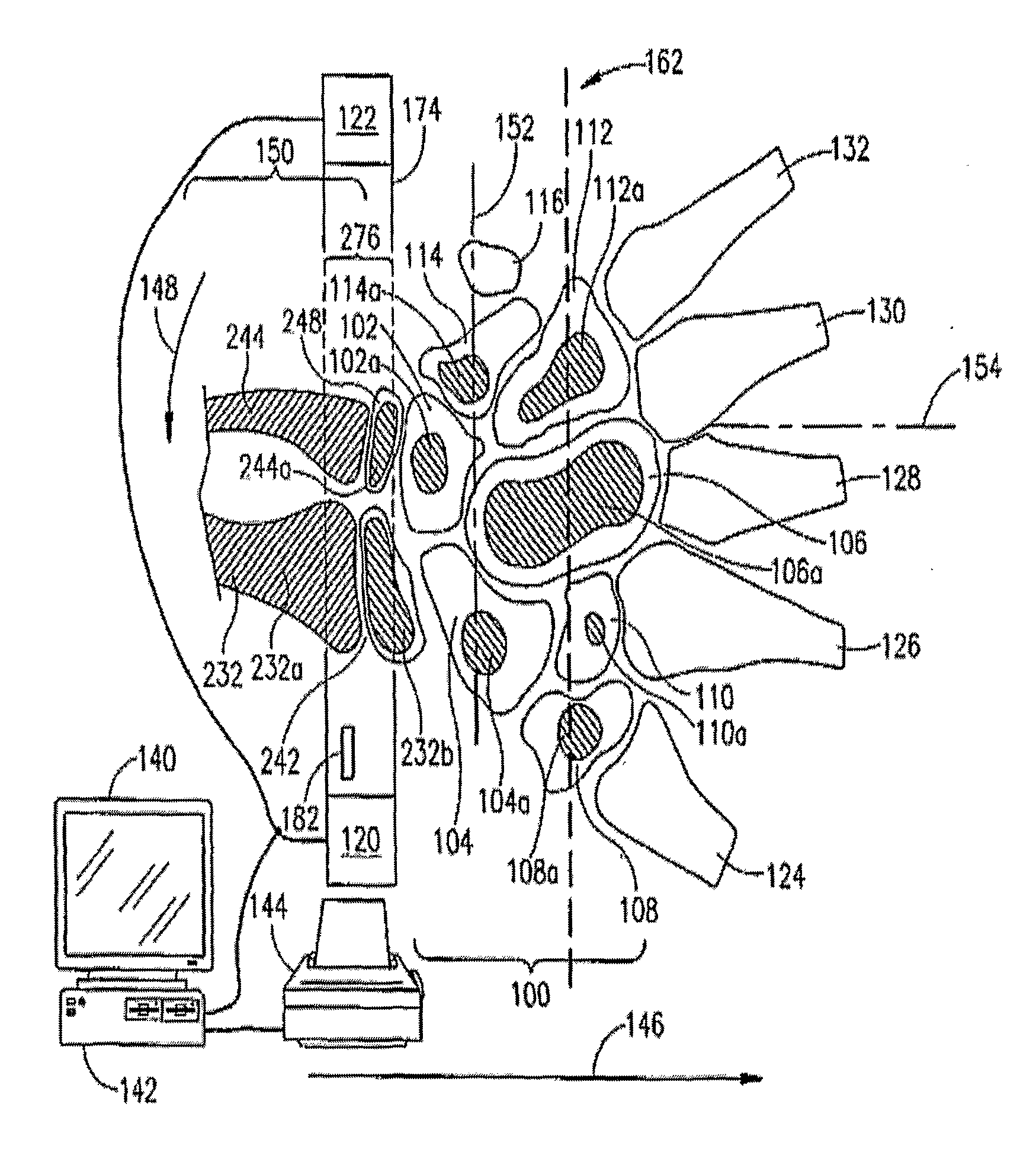
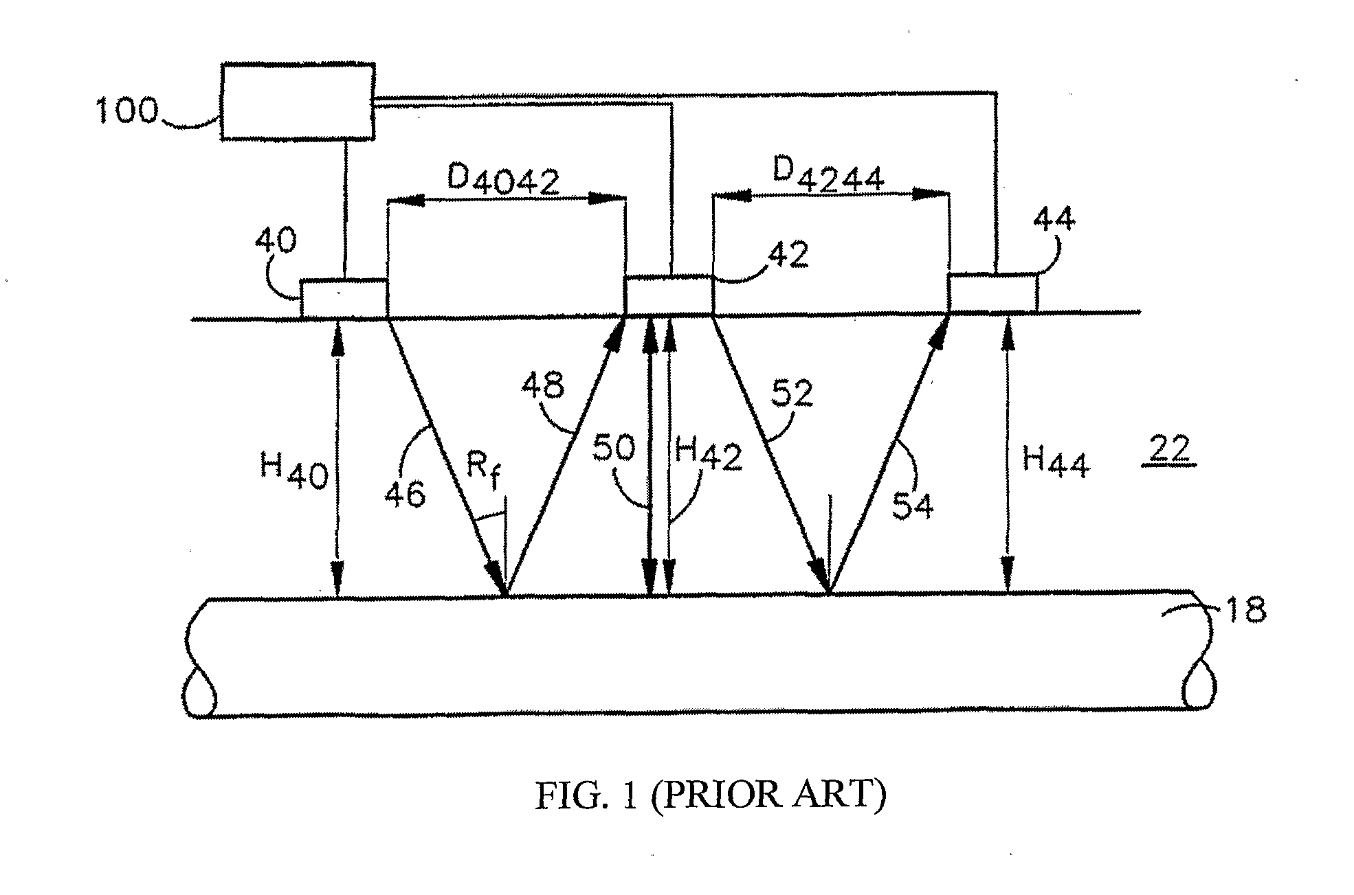
[0027]The present invention fulfills this long-felt need. It comprises a system and method for bone density measurement based on sound velocity differences in media of different density in which the means for measuring differences in arrival times of ultrasonic pulses are located within a dedicated detector located either within the measuring head or within a special probe. While the bone sonometry measurements are being made, the measuring head or probe is located near the body of the patient. Thus, the invention obviates the need for a special drop-in card, and enables performance of bone sonometry measurements without any need for a dedicated computer system.
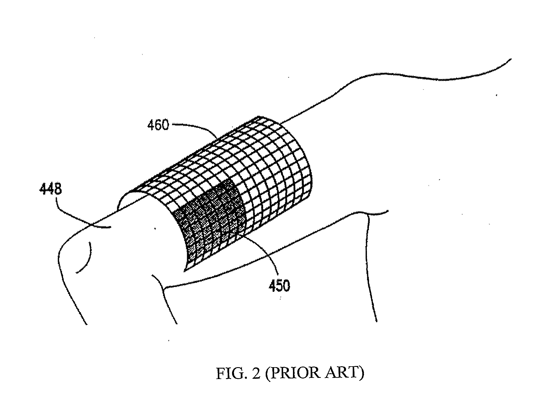
[0062]It is a further object of this invention to disclose a method for performing multi-site bone density measurements of a region of subject's body, said method comprising steps of: (a) obtaining an ultrasound bone sonometer, said sonometer comprising (1) a cuff-like measurement head adapted to enclose at least part of the circumference of said region of said subject's body, said measurement head comprising: (i) an array of ultrasound sources adapted for providing ultrasonic pulses; (ii) at least one measurement transducer adapted to measure the difference in arrival times of ultrasonic pulses; and (iii) at least one dedicated data processing element adapted for determining differences in arrival times of ultrasonic pulses; (2) means for transferring data from said at least one measurement transducer to said at least one dedicated data processing element; and (3) means for transmitting data from said dedicated data processing element to a non-dedicated computing means; (b) encircling said region of said subject's body by said flexible measurement head; (c) transmitting ultrasonic pulses from said ultrasound sources to said multiple sites encircled within said region; (d) detecting said ultrasonic pulses from said multiples sites encircled within said region; and (e) measuring the acoustic velocity of said ultrasonic pulses, thereby simultaneously performing said multi-site bone density measurements. It is within the essence of the invention wherein said step of performing said multi-site bone density measurements is provided by said step of encircling said region of said subject's body by said flexible measurement head without having to relocate said measurement head from one site to another.
[0085]It is a further object of this invention to disclose a method for measuring bone age comprising steps of: (a) providing a compact measurement head adapted to transmit an acoustic energy into the body of a subject; (b) receiving an acoustic signal from one or more structures including an ossification-actuated skeletal structure or a cranial structure that changes with age, responsive to said transmitted acoustic energy; (c) providing computing means adapted for analyzing the acoustic signal to determine at least one effect of said structure on said signal; (d) providing communications means adapted to transmit acoustic signal information from said measurement head to said computing means; and (e) estimating the age of the structure from said determined effect with said computing means. It is within the essence of the invention wherein bone age measurements are obtained in a system with separate measurement and computation means, allowing increased portability of said measurement head.
[0088]It is a further object of this invention to disclose a method for increasing the number of patients on whom bone status measurements are made, comprising steps of: (a) providing a bone sonometer adapted for performing at least one measurement chosen from the group consisting of bone fragility measurements, bone density measurements, bone strength measurements, and bone age measurements; (b) providing said bone sonometer with multifunctional multipurpose screening means; (c) locating said bone sonometer at accessible locations; (d) analyzing at least one rigid or semi-rigid site within a measured region of the body of a patient with said bone sonometer; and (e) screening a preset matter with said screening means, wherein said accessible location is selected from the group consisting of pharmacies, clinics, medical vehicles, ambulances, hospitals, medical departments, homes, points of care, pre-hospital sites, sports clubs, exercise facilities, stadiums, government offices, post offices, relief agencies, workout gyms, malls, and cosmetic counters; and further wherein said number of patients at high risk for bone fragility on whom bone status measurements are made is increased by the accessibility of said bone sonometer.

Problems solved by technology

The gradual depletion of a person's bone mass can make the bone prone to fracture and / or deformation and cause numerous accompanying adverse effects, including pain and discomfort.
The condition of osteoporosis manifests itself as a decrease in bone tissue mass and often leads to fractures of the vertebrae, hip, femur, and distal end of the wrist bone.
Once major deterioration has occurred, it is difficult to restore the lost bone.
Current treatment and / or prevention protocols fail to adequately account for or incorporate such information.
All of these methods expose the patient and operator to x-ray radiation.
Due to the ionizing nature of X-radiation the frequency with which such measurements may be carried out is limited, to limit dosage of ionizing radiation.
As a further example, x-ray based equipment requires a licensed technician to operate the equipment due to the ionizing radiation hazards involved.
In addition, this equipment is structurally large, requiring an x-ray source, detector, power supply, cooling equipment, as well as an examination area usually consisting of a bed.
Amongst known problems with the DXA method are its relatively low accuracy, the radiation dosage involved in the measurement, and the size of the apparatus.
This is especially so given that repeated measurements (which in principle could be used to find a reliable average value despite the scatter of individual measurements) cannot be used in the case of X-ray based measurement.
Moreover, the microstructure of the medium, as well as macro-structures on the order of a wavelength of the ultrasound, affects the speed.
The use of a foot well limits the portability of the device and the possible applications thereof, however.
Since the device requires proprietary processing equipment on this drop-in card, the portability of the unit is limited by the requirement to provide a computer containing the proper card along with the measurement head.
Furthermore, current methods are based on single point methods that indicate only a gross overall measure of bone density and strength, and cannot discover small points of compromised bone density.

Method used


Image

  • Bone Sonometer
  • Bone Sonometer
  • Bone Sonometer

Examples


example 1

[0216]In order to assess the properties of the device herein disclosed, a series of tests were performed to determine its precision, accuracy, compliance with international standards, and the effects of cleaning and disinfection on the device.

[0217]Two different in vivo precision tests for the device of the current invention were conducted. Included in these precision tests were: (1) a reproducibility study which involved the assessment of in vivo precision between different instruments, connecting slot configurations and probes; and (2) a reproducibility study which measured the in vivo precision between different operators and probes. The objectives of both studies were to estimate the variability, between device components and between operators, of SOS measurements of the distal one-third of the radius. The in vivo precision (reproducibility), expressed by the coefficient of variation (CV), ranged from 0.60% to 0.73%.

[0218]In addition, accuracy tests were performed as part of the...

example 2

[0220]Two clinical studies were undertaken in order to create normative reference databases for SOS in a Caucasian female population. The first of these was a multisector study performed in North America. This study was conducted on a group of Caucasian females between the ages of 20 and 90 years old by five investigators at five geographically diverse investigational sites in North America (4 in the U.S. and 1 in Canada). Potential subjects were identified by placing advertisements in the newspaper, contacting potential subjects from drivers' license listings, recruiting at college and university campuses, and recruiting at nursing homes. Eligible women had a negative history of osteoporosis fracture or chronic conditions affecting bone metabolism, and were not taking medications that affect bone metabolism. Of the 573 subjects recruited, 545 subjects were found eligible according to the inclusion / exclusion criteria of the study; SOS measurements at the distal third of the radius w...

example 3

[0228]The second study designed to create normative reference databases for SOS in a Caucasian female population was conducted in Caucasian females between the ages of 20 and 90 years old by a single investigator at Asaf-Harophe Medical Center, Israel. The eligibility criteria were met by 1,132 subjects who had their SOS measurements of the distal third of the radius taken. The mean age of the study subjects was 49.3±17.6 years with a range of 20 to 89 years. Each decade was roughly comparable in size except for the decade 40-49, in which there were 266 subjects. Sixty percent of the subjects in this study were pre-menopausal.

[0229]The mean SOS was 4082±151 m s−1 with a range of 3510 to 4602. Ninety percent of the SOS measurements were between 3800 and 4300 m s−1. Over half of the measurements (52.5%) were between 4000 and 4200 m / sec. Table 4 presents mean SOS result by age decade.

[0230]The moving average SOS increases to a peak of 4173 m s−1 at the age of 39, with population standa...


PUM


Abstract

A probe device is for performing a bone density measurement is disclosed. The device comprises at least one ultrasound source for providing ultrasonic pulses; a plurality of ultrasound detectors for measuring the differences in arrival times of said ultrasonic pulses; at least one dedicated data processing element adapted for determining differences in arrival times of ultrasonic pulses; means for transferring data from said at least one measurement transducer to said at least one dedicated data processing element; and communication means adapted to transmit data from said dedicated data processing element to a non-dedicated computing means. Unlike systems known in the art, the measurements of the times of arrival of the ultrasonic pulses are performed within the probe itself, obviating the need for a dedicated computer system or measurement card.

Description

FIELD OF THE INVENTION[0001]The present invention relates to systems for bone sonometery e.g., densitometry and methods for the use thereof.BACKGROUND OF THE INVENTION[0002]Bone mass loss is a widespread medical condition, appearing with particular frequency in the elderly and in women. The gradual depletion of a person's bone mass can make the bone prone to fracture and / or deformation and cause numerous accompanying adverse effects, including pain and discomfort. The condition of osteoporosis manifests itself as a decrease in bone tissue mass and often leads to fractures of the vertebrae, hip, femur, and distal end of the wrist bone.[0003]The World Health Organization defines four diagnostic categories: normal, osteopenia, osteoporosis, and established osteoporosis, and further defines those categories using diagnostic value ranges. Currently, within the United States, osteoporosis affects about 20-25 million people. By age 80, the percentage of women with normal bone density decre...

Claims


Application Information

Patent Timeline
IPC IPC(8): A61B8/00
CPCA61B8/4472A61B8/0875A61B8/4427A61B8/4477A61B8/4488
Inventor MAROM, TALZAMIR, GILAD
Owner BEAMMED
Learn More