
Bioluminescence imaging of myeloperoxidase activity in vivo, methods, compositions and apparatuses therefor

a technology of bioluminescence imaging and myeloperoxidase, which is applied in the directions of diagnostic recording/measure, ultrasonic/sonic/infrasonic diagnostics, organic chemistry, etc., can solve the problems of limited studies and achieve the effect of improving tissue penetration and in vivo optical properties

Inactive Publication Date: 2011-10-13
WASHINGTON UNIV IN SAINT LOUIS
View PDF1 Cites 10 Cited by
  • Summary
  • Abstract
  • Description
  • Claims
  • Application Information

AI Technical Summary

Benefits of technology

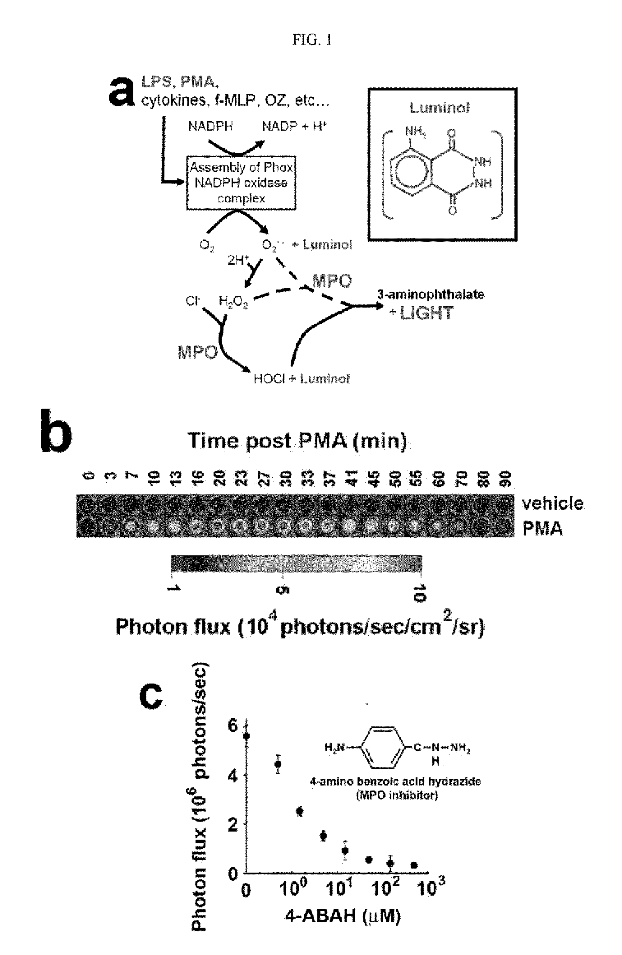
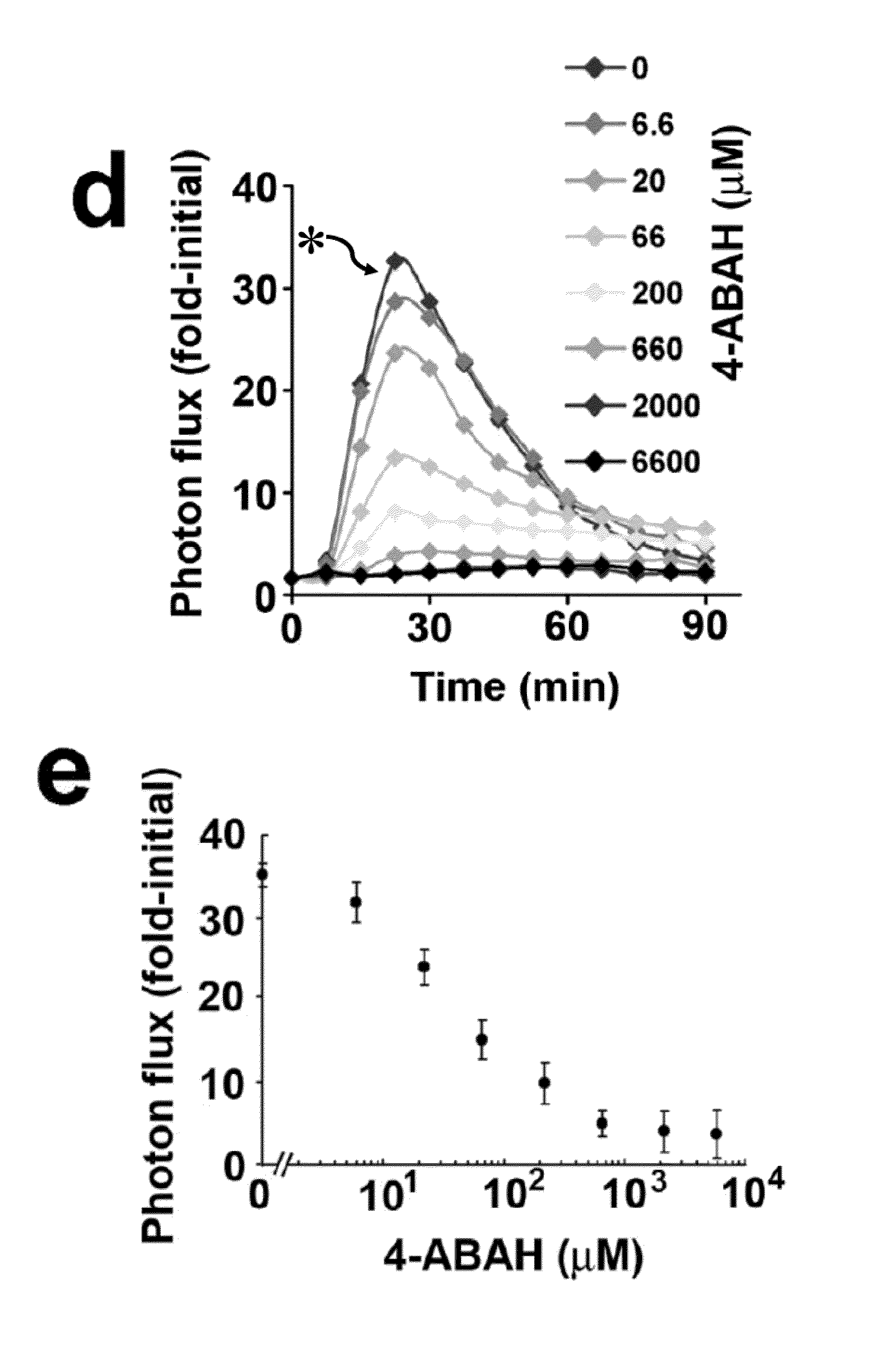
The present patent is about methods for imaging biological activity in a subject by using a compound that emits light upon contact with an oxidizing agent. The methods can be used to diagnose and assess the risk of developing diseases associated with high levels of MPO activity, such as cardiovascular disease and inflammation. The imaging can be done non-invasively and can provide information about the distribution and levels of MPO activity in a subject. The methods can also be used to evaluate the effectiveness of therapy and to establish a prognosis in patients with a pathological condition. The patent also describes the use of a composition comprising a compound that emits light upon contact with an oxidizing agent for imaging inflammation sites in a subject.

Problems solved by technology

While showing some promise, these studies were limited to phantoms in vitro, MPO+GOX implants or LPS muscle injections in vivo, and the relevance of these approaches to pathophysiology remains to be established.

Method used


Image

  • Bioluminescence imaging of myeloperoxidase activity in vivo, methods, compositions and apparatuses therefor
  • Bioluminescence imaging of myeloperoxidase activity in vivo, methods, compositions and apparatuses therefor
  • Bioluminescence imaging of myeloperoxidase activity in vivo, methods, compositions and apparatuses therefor

Examples


example 1

[0135]This Example illustrate the synthesis of Luminogenic-Optical Probe ILOG.

[0136]6-(N-(4-aminobutyl)-N-ethylamino)-2,3-dihydrophthalazine-1,4-dione (IL) dissolved in dimethylsulfoxide (0.5 ml) was treated with the succinimidylester of Oregon Green (1.1 equivalents) in the presence of trimethylamine (100 equivalents) at room temperature. Contents were stirred in an amber colored vial overnight. The product was purified on a Water's HPLC equipped with a dual wavelength detector, using an RP-C18 (Vydac) column, employing a mobile phase gradient of 2-90% acetonitrile with 0.1% TFA in water over 30 minutes. The fraction eluting at Rt=20.8 min was collected, lyophilized to obtain orange-red solid and identified via ESMS; Calcd for C35H28F2N4O8, 670.2; Found; 670.9 (FIG. 6, FIG. 7).

example 2

[0137]This Example illustrate the synthesis of Luminogenic-Optical Probe ILCY.

[0138]6-(N-(4-aminobutyl)-N-ethylamino)-2,3-dihydrophthalazine-1,4-dione (IL) dissolved in dimethylsulfoxide (0.5 ml) was treated with the succinimidylester of Cascade Yellow (1.1 equivalents) in the presence of trimethylamine (100 equivalents) at room temperature. Contents were stirred in an amber colored vial overnight. The product was purified on a Waters HPLC equipped with a dual wavelength detector, using an RP-C18 (Vydac) column, employing a mobile phase gradient of 10-90% acetonitrile with 0.1% TFA in water over 30 minutes. The fraction eluting at Rt=16 min was collected, lyophilized to obtain an orange-red solid and identified via ESMS; Calcd for C37H37N6O8S, 725.2; Found; 724.8. (FIG. 8, FIG. 9).

example 3

[0139]This Example illustrate the synthesis of Luminogenic-Optical Probe ILAF680.

[0140]6-(N-(4-aminobutyl)-N-ethylamino)-2,3-dihydrophthalazine-1,4-dione (IL) dissolved in dimethylsulfoxide (0.5 ml) was treated with succinimidylester of AlexaFluor 680 (1.1 equivalents) in the presence of trimethylamine (100 equivalents) at room temperature. Contents were stirred in an amber colored vial overnight. The product was purified on a Water's HPLC equipped with a dual wavelength detector, using an RP-C18 (Vydac) column, employing a mobile phase gradient of 10-70% acetonitrile with 0.1% TFA in water over 30 minutes. The fraction eluting at Rt=16.7 min was collected, lyophilized to obtain dark blue-purple solid and identified via ESMS; Calcd for C35H28F2N4O8, 670.2; Found; 670.9. (FIG. 10, FIG. 11)


PUM

PropertyMeasurementUnit
emission intensity maximum at a wavelengthaaaaaaaaaa
wavelengthsaaaaaaaaaa

Abstract

Methods of imaging distribution of myeloperoxidase activity in a subject are disclosed. These methods include the use of bioluminescent substrates, including luminol and wavelength-shifted analogues of luminol. Bioluminescent myeloperoxidase substrates that emit light at longer wavelengths compared to luminol are shown to be useful for imaging myeloperoxidase activity in vivo. The disclosed methods can be used for imaging sites of inflammation and other pathological conditions associated with abnormal levels of MPO activity in vivo. Methods of synthesis of luminol analogues are also disclosed.

Description

CROSS-REFERENCE TO RELATED APPLICATIONS[0001]This application claims priority from U.S. Provisional Application Ser. No. 61 / 110,727 filed on Nov. 3, 2008, which is incorporated herein by reference in its entirety.STATEMENT OF GOVERNMENT SUPPORT[0002]This invention was made with the support of government grants P50 CA94056, CA105218, CA10073, CA63417 and PO1 HL030086 from the National Institutes of Health. The government of the United States of America has certain rights in this invention.INTRODUCTION[0003]A need exists for diagnostic tests to aid in the characterization of subjects at risk for developing diseases characterized at least in part by high levels of MPO-mediated oxidation products and damage, in particular, cardiovascular disease. Furthermore, there is a need to establish whether a specific therapy is having the appropriate effect in individuals suffering from such conditions. Thus, prognostic indicators or biomarkers to monitor the effects of such therapy are also neede...

Claims


Application Information

Patent Timeline
Patent Type & Authority Applications(United States)
IPC IPC(8): A61K49/00C07D413/14C07D405/12C07D403/14C07D237/32C07D491/22
CPCC12Q1/28A61K49/0013
Inventor SHARMA, VIJAYGAMMON, SETHPIWNICA-WORMS, DAVIDGROSS, SHIMON
Owner WASHINGTON UNIV IN SAINT LOUIS
Learn More