
ASSAY FOR CARDIAC TROPONIN-T (cTnT)

a troponin-i and autoantibodies technology, applied in the field of cardiac troponin-i detection kits, can solve the problems of cardiac troponin-i reactive autoantibodies, the detection of human and the assay for ctnt fails to address the problem of circulating endogenous antibodies, or “autoantibodies”, and the measurement of ctni is reportedly limited

Inactive Publication Date: 2011-06-02
ABBOTT LAB INC
View PDF35 Cites 7 Cited by
  • Summary
  • Abstract
  • Description
  • Claims
  • Application Information

AI Technical Summary

Benefits of technology

The present patent provides an immunoassay for quantifying cardiac troponin-T (cTnT) in a test sample. The assay involves contacting the sample with capture antibodies that bind to cTnT and detection antibodies that bind to cTnT, resulting in a measurable assembly. The detection antibodies can be labeled with a detectable label, such as an enzyme, oligonucleotide, nanoparticle chemiluminophore, fluorescine, or biotin. The assay can be performed using optical, electrical, or change-of-state signals. The capture and detection antibodies can be humanized. The immunoassay provides a reliable and sensitive method for measuring cTnT in biological samples.

Problems solved by technology

The assay is reportedly limited to detecting human cTnT from human heart tissue to only 7%.
Additionally, like many others, assays for cTnT are subject to interference heterophilic endogeneous antibodies, which produce false positive results and thus misdiagnosis (G. H. White et al., Clin Chem 2002; 48:201-3; N. Shayanfar et al., Swiss Med Wkly 2008; 138:470).
Additionally, the known assay for cTnT fails to address the problem of circulating endogenous antibodies, or “autoantibodies”.
For example, cardiac troponin-I reactive autoantibodies may interfere with the measurement of cTnI using conventional midfragment-specific immunoassays.
Thus, interference from autoantibodies can also produce erroneous results, particularly near the cut-off values established for clinical diagnoses, thereby increasing the risk of false negative diagnostic results and the risk that individuals will not obtain a timely diagnosis.
However, this effort has been largely unsuccessful.

Method used


Image

  • ASSAY FOR CARDIAC TROPONIN-T (cTnT)
  • ASSAY FOR CARDIAC TROPONIN-T (cTnT)
  • ASSAY FOR CARDIAC TROPONIN-T (cTnT)

Examples


example 1

Detection Antibody Conjugates

[0180]General Conjugation Procedure: Detection antibody was dissolved in a conjugation buffer (100 mM sodium phosphate, 150 mM NaCl, pH 8.0) to give a concentration of 1-10 mg / mL (6.25-62.5 μM). Acridinium, 9-[[[4-[(2,5-dioxo-1-pyrrolidinyl)oxy]-4-oxobutyl][(4-methylphenyl)sulfonyl]amino]carbonyl]-10-(3-sulfopropyl)-, inner salt, 2 (Adamczyk, M.; Chen, Y.-Y.; Mattingly, P. G.; Pan, Y. J. Org. Chem. 1998, 63, 5636-5639) labeling reagent was prepared in N,N-dimethylformamide (DMF) at a concentration of 1-50 mM, as shown in formula 2 below:

[0181]The selected antibody was treated with the acridinium-labeling reagent in a molar excess of 1-35 fold for 3-14 h at ambient temperature in the dark. Afterwards, the acridinium-9-carboxamide-antibody conjugate solution was dialyzed at ambient temperature over 20 hours using a 10 kilodalton molecular weight cutoff membrane against three volumes (1000× conjugate solution volume) of a dialysis buffer consisting of 10 mM...

example 2

Capture Antibodies on Magnetic Microparticles

[0192]Carboxy paramagnetic microparticles (5% solids, nominally 5 micron diameter, Polymer Labs, Varian, Inc. Amherst, Mass.) were diluted to a concentration of 1% solids in 2-(N-morpholino)ethanesulfonic acid buffer (MES, 2 mL, pH 6.2, 50 mM) then washed with MES buffer (3×, 2 mL), and finally, resuspended in MES (2 mL). The particles were activated by mixing with 1-ethyl-3-[3-dimethylaminopropyl]carbodiimide hydrochloride (20 μL of 11 mg / 1.129 mL in water) for 20 min, then washed (MES, 2 mL) and re-suspended in MES (2 mL). Murine anti-cardiac troponin-T 1C11 (Fitzgerald Ind Intnl, Concord, Mass., cat no. 10R-T127D) was added to the suspension at 60 μg / mL. After mixing for 60 min the antigen coated particles were magnetically sequestered, and the antigen solution was replaced with a blocking solution consisting of 1% BSA in PBS (2 mL). After mixing for 30 min, the particles were washed with 1% BSA in PBS (3×, 2 mL) and finally, resuspend...

example 3

Chemiluminescent Immunoassay Dose-Response for Combinations of Capture and Detection Antibodies

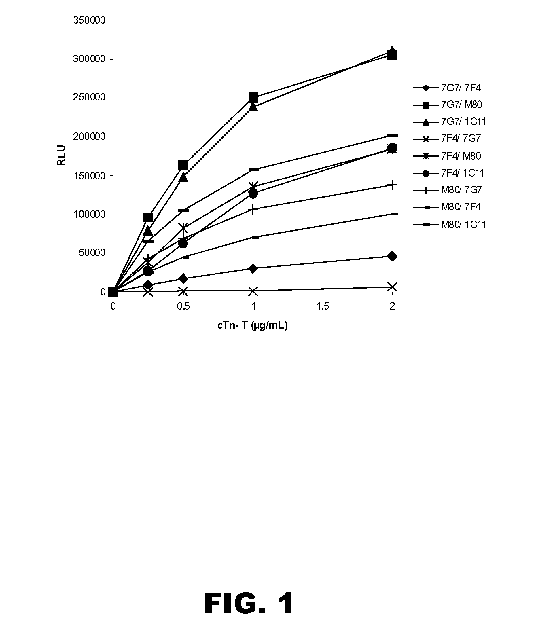
[0194]A working suspension of each capture antibody microparticles prepared in Example 2 was prepared by dilution of the stock suspension to 0.05% solids in MES buffer (20 mM, pH 6.6) containing sucrose (13.6%) and antimicrobial agents. A working solution of each detection antibody conjugate was prepared by dilution of the stock solution to 10 ng / mL. Cardiac troponin-T (Biospacific, cat no. J34510359) standard solutions were prepared at 0, 0.25, 0.5, 1.0 and 2.0 μg / mL.

[0195]The assays were carried out on an ARCHITECT® i2000 instrument (Abbott Laboratories, Abbott Park, Ill.). Briefly, the cardiac troponin-T standard solution (10 μL) was diluted with ARCHITECT® PreIncubation Diluent (50 μL) and the capture antibody microparticles (50 μL) and incubated in the instrument reaction vessel. Following incubation, the microparticles were magnetically sequestered and washed with ARCHITECT® wash buf...


PUM

PropertyMeasurementUnit
molecular weightaaaaaaaaaa

Abstract

The present disclosure describes immunoassays for detecting cardiac troponin-T (cTnT) in a test sample, and in particular immunoassays and kits for detecting cTnT in a test sample suspected of containing substances that may interfere with the determination of cTnT, such as heterophilic endogenous antibodies and autoantibodies to cTnT. The methods use more than one capture phase antibody and more than one detection antibody to improve specificity, and provide for the use of humanized immunoreagents to overcome heterophilic antibody interferences.

Description

RELATED APPLICATION INFORMATION[0001]None.TECHNICAL FIELD[0002]The present disclosure relates generally to assays and kits for detecting cardiac troponin-T (cTnT) in a test sample, and in particular to methods and kits for detecting cTnT in a test sample suspected of containing substances that may interfere with the determination of cTnT.BACKGROUND OF THE INVENTION[0003]Immunoassay techniques have been known for the last few decades and are now commonly used in medicine for a wide variety of diagnostic purposes to detect target analytes in a biological sample. Immunoassays exploit the highly specific binding of an antibody to its corresponding antigen, wherein the antigen is the target analyte. Typically, quantification of either the antibody or antigen is achieved through some form of labeling such as radio- or fluorescence-labeling. Sandwich immunoassays involve binding the target analyte in the sample to the antibody site (which is frequently bound to a solid support), binding la...

Claims


Application Information

Patent Timeline
Patent Type & Authority Applications(United States)
IPC IPC(8): C12Q1/68G01N33/53
CPCG01N33/6893G01N2800/324G01N2800/32G01N2333/4712
Inventor ADAMCZYK, MACIEJBRASHEAR, JEFFREY R.MATTINGLY, PHILLIP G.
Owner ABBOTT LAB INC
Learn More