
Single domain antibodies directed against epidermal growth factor receptor and uses therefor

a technology of epidermal growth factor and single-domain antibodies, which is applied in the field of single-domain antibodies, can solve the problems of inability to completely treat cancer with these antibodies, inability to fully develop, and inability to meet the needs of patients, so as to improve the permeability of the intestinal mucosa, and improve the effect of permeability

Inactive Publication Date: 2011-05-26
ABLYNX NV
View PDF63 Cites 45 Cited by
  • Summary
  • Abstract
  • Description
  • Claims
  • Application Information

AI Technical Summary

Benefits of technology

The present invention provides polypeptides that can inhibit the biological activity of Epidermal Growth Factor Receptor (EGFR) by binding to it and preventing its natural ligand from binding or homodimerization. These polypeptides can be single domain antibodies or other types of single domain antibodies. The invention also provides methods for administering these polypeptides to humans and animals for the treatment of allergies and other disorders. The polypeptides can be delivered through various routes such as injection, orally, nasally, and via internalizing receptors. The invention also provides a therapeutic agent for the treatment of allergies.

Problems solved by technology

Yet none of these antibodies nor the presently available drugs are completely effective for the treatment of cancer, and most are limited by severe toxicity.
In addition, it is extremely difficult and a lengthy process to develop a new chemical entity (NCE) with sufficient potency and selectivity to such target sequence.
However, conventional antibodies are difficult to raise against multimeric proteins where the receptor-binding domain of the ligand is embedded in a groove or at the interphase between the two subunits, as is the case with Epidermal Growth Factor Receptor.
The use of antibodies derived from sources such as mouse, sheep, goat, rabbit etc., and humanized derivatives thereof as a treatment for conditions which require a cytostatic or cytotoxic effect on tumor cells is problematic for several reasons.
Traditional antibodies are not stable at room temperature, and have to be refrigerated for preparation and storage, requiring necessary refrigerated laboratory equipment, storage and transport, which contribute towards time consumption and expense.
Furthermore, the manufacture or small-scale production of said antibodies is expensive because the mammalian cellular systems necessary for the expression of intact and active antibodies require high levels of support in terms of time and equipment, and yields are very low.
Furthermore the large size of conventional antibodies, would restrict tissue penetration, for example, at the site of a solid tumor.
Furthermore, traditional antibodies have a binding activity which depends upon pH, and hence are unsuitable for use in environments outside the usual physiological pH range such as, for example, in treating colorectal cancer.
Furthermore, traditional antibodies are unstable at low or high pH and hence are not suitable for oral administration.
Furthermore, traditional antibodies have a binding activity, which depends upon temperature, and hence are unsuitable for use in assays or kits performed at temperatures outside biologically active-temperature ranges (e.g. 37±20° C.).
Another important drawback of conventional antibodies is that they are complex, large molecules and therefore relatively unstable, and they are sensitive to breakdown by proteases.
This means that conventional antibody drugs cannot be administered orally, sublingually, topically, nasally, vaginally, rectally or by inhalation because they are not resistant to the low pH at these sites, the action of proteases at these sites and in the blood and / or because of their large size.
Furthermore, subjects commonly experience physical and psychological stress prior to and upon receiving an injection.
However, they have one important drawback: these are complex, large molecules and therefore relatively unstable, and they are sensitive to breakdown by proteases.
Because the degradation they undergo during passage through, for instance, the gastrointestinal tract, administration of conventional antibodies and their derived fragments or single-chain formats (e.g. scFv's) is not very effective.
This means that conventional antibody drugs cannot be administered orally, sublingually, topically, nasally, vaginally, rectally or by inhalation because they are not resistant to the low pH at these sites, the action of proteases at these sites and in the blood and / or because of their large size.
Administration by injection is therefore the most frequently used method of administration although the method has many disadvantages, for example: (a) poor tolerance by patients, especially when treating chronic disorder; (b) a consequent risk of poor compliance with the dosage when the drug is not a ‘life saver’; (c) difficulty of carrying out self-administration by the patient; (d) possible non-availability of suitable surroundings for carrying out the procedure in an aseptic manner; (e) requires specialist training in order to use a hypodermic syringe or needle correctly and safely.
However, they have important drawbacks: these antibodies are complex, large molecules and therefore relatively unstable, and they are sensitive to breakdown by proteases.
Moreover, the domains of such antibodies are held together by disulphide bonds that dissociate in the reducing environment of the cytoplasm leading to a substantial loss of binding activity.
Therefore, they cannot be used to target intracellular proteins.

Method used


Image

  • Single domain antibodies directed against epidermal growth factor receptor and uses therefor
  • Single domain antibodies directed against epidermal growth factor receptor and uses therefor
  • Single domain antibodies directed against epidermal growth factor receptor and uses therefor

Examples


example 1

Immunization

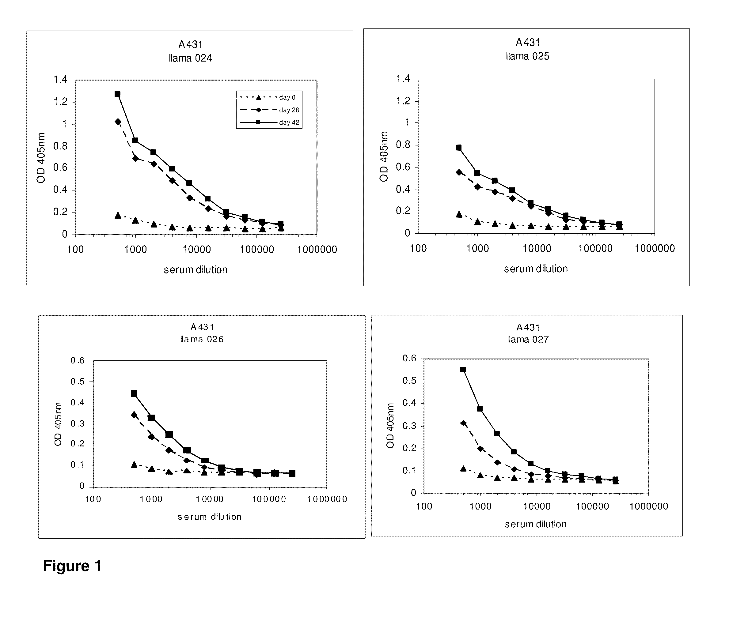
[0610]After approval of the Ethical Committee of the Faculty of Veterinary Medicine (University Ghent, Belgium), 4 llamas (024, 025, 026 and 027) were immunized with the tumor antigen epidermal growth factor receptor (EGFR) according to all current animal welfare regulations. To generate an antibody dependent immune response (table 1), two animals were injected with intact human vulvar squamous carcinoma cells (A431, ATCC CRL 1555), expressing EGFR on its cell surface, while A431 derived membrane extracts were administered to two other llamas (026 and 027). Each animal received seven doses of subcutaneously administered antigens at weekly intervals (table 1). When immunizing with intact cells, each dose consisted of 108 freshly harvested A431 cells. The dose for immunization with membrane extracts consisted of vesicles prepared from 108 A431 cells. Vesicles were prepared according to Cohen and colleagues (Cohen S, Ushiro H, Stoscheck C, Chinkers M, 1982. A native 170,000...

example 2

Evaluation of Immune Response

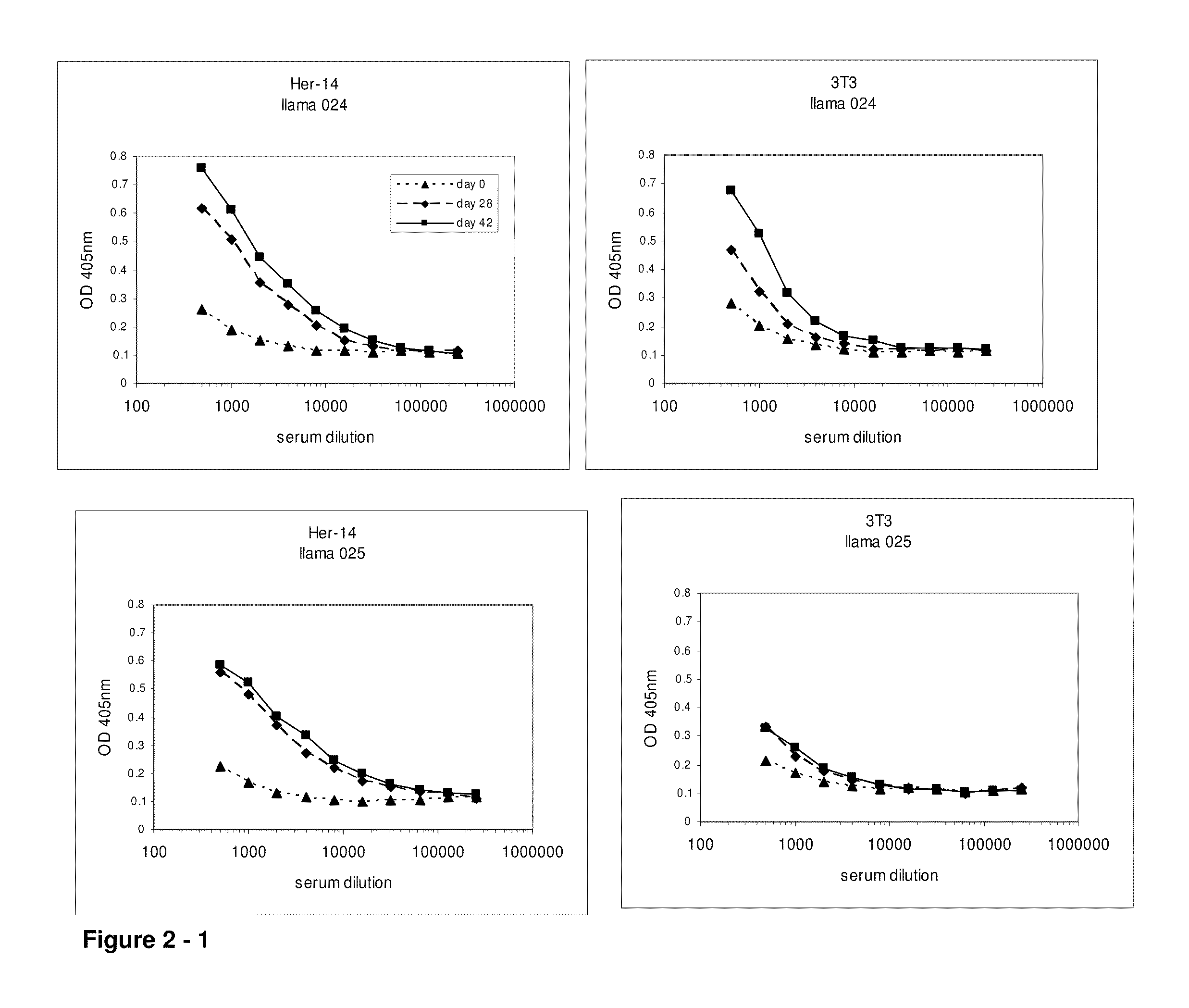
[0611]At day 0, 28 and 42, 10 ml of (pre-)immune blood was collected and serum was used to evaluate the induction of the immune responses in the 4 animals. A first ELISA was performed to verify whether the animals generated antibodies that recognized A431 epitopes. After coating a tissue-culture treated 96-well plate with gelatin (0.5% in PBS for 10 minutes), the excess of gelatin was removed and A431 cells were grown overnight in the microwells to confluency. Cells were fixed with 4% paraformaldehyde in PBS for 30 minutes at room temperature. Subsequently, the fixative was blocked with 100 mM glycine in PBS for 10 minutes, followed by blocking of the wells with a 4% skim milk-PBS solution, again for 10 minutes. Serum dilutions of immunized animals were applied and A431 specific antibodies were detected with a polyclonal anti-llama antiserum developed in rabbit, followed by a secondary goat anti-rabbit horse radish peroxidase (HRP) conjugate (Dako, Denma...

example 3

Cloning of the Heavy-Chain Antibody Fragment (VHH) Repertoire

[0613]Since little is known on the immunoglobulin ontogeny of camelids, B-cell containing tissues of distinct origin and of different time points were collected for each animal (table 1). After tissue collection, total RNA was isolated according to the procedure described by Chomczynski and Sacchi. (Chomczynski P and Sacchi N. 1987. Single-step method of RNA isolation by acid guanidinium thiocyanate-phenol-chloroform extraction. Anal Biochem 162:156-159). The procedure to clone the VHH repertoire is based on a method described in patent application WO 03 / 054016. cDNA was prepared on total RNA with MMLV Reverse Transcriptase (Invitrogen) using oligo d(T) oligonucleotides (de Haard H J, van Neer N, Reurs A, Hufton S E, Roovers R C, Henderikx P, de Bruine A P, Arends J W, Hoogenboom H R. 1999. A large non-immunized human Fab fragment phage library that permits rapid isolation and kinetic analysis of high affinity antibodies. ...


PUM

PropertyMeasurementUnit
temperaturesaaaaaaaaaa
particle sizeaaaaaaaaaa

Abstract

The present invention relates to polypeptides derived from single domain heavy chain antibodies directed to Epidermal Growth Factor Receptor. It further relates to single domain antibodies that are Camelidae VHHs. It further relates to methods of administering said polypeptides orally, sublingually, topically, intravenously, subcutaneously, nasally, vaginally, rectally or by inhalation. It further relates to protocols for screening for agents that modulate the Epidermal Growth Factor Receptor, and the agents resulting from said screening. The invention further a method for delivering therapeutic molecules to the interior of cells.

Description

RELATED APPLICATIONS[0001]This application is a continuation of U.S. patent application Ser. No. 12 / 431,403, filed Apr. 28, 2009, which is a continuation-in-part application of U.S. patent application Ser. No. 10 / 553,105, filed Oct. 12, 2005, which is a national stage filing under 35 U.S.C. §371 of international application PCT / BE2003 / 000189, filed Nov. 7, 2003, which was published under PCT Article 21(2) in English; which is also a continuation-in-part application of U.S. patent application Ser. No. 10 / 534,292, filed May 9, 2005, which is a national stage filing under 35 U.S.C. §371 of international application PCT / BE03 / 00190, filed Nov. 7, 2003, which was published under PCT Article 21(2) in English, which claims priority to international application PCT / EP03 / 06581, filed Jun. 23, 2003, and international application PCT / EP03 / 07313, filed Jul. 8, 2003. This application claims the benefit under 35 U.S.C. 119(e) of U.S. provisional application Ser. No. 60 / 425,073, filed Nov. 8, 2002,...

Claims


Application Information

Patent Timeline
Patent Type & Authority Applications(United States)
IPC IPC(8): A61K39/395C07K16/00C07H21/00C07K2/00C12N15/63C12N5/10C12P21/00C12N5/071
CPCA61K2039/505C07K16/18C07K16/241C07K16/249C07K16/2863C07K16/40C07K2317/80C07K2316/96C07K2317/22C07K2317/31C07K2317/569C07K2317/77C07K16/4291C07K2317/76
Inventor LAEREMANS, TOONVAN BERGEN EN HENEGOUWEN, PAUL M. P.SILENCE, KARENVAECK, MARK
Owner ABLYNX NV
Learn More