
Methods for drug delivery comprising unfolding and folding proteins and peptide nanoparticles

a technology of unfolding and folding proteins and nanoparticles, which is applied in the field of in vivo delivery of pharmacologically active agents, can solve the problems of high pressure and relatively high temperature required for obtaining nanoparticles and removing organic solvents, and the use of organic solvents is undesirable, and the drug loading capacity is limited

Active Publication Date: 2011-03-10
NANJING UNIV +1
View PDF17 Cites 21 Cited by
  • Summary
  • Abstract
  • Description
  • Claims
  • Application Information

AI Technical Summary

Benefits of technology

[0026]The current invention represents an improvement over the prior art in several aspects. First, nanoparticles formed by the methods of the current invention may be able to bind up to 90% of the added pharmacologically active agent, an amount that exceeds prior art methods and creates a more efficient process and cost-effective method. Additionally, the current invention allows for a drug loading capacity of up to about 40%, such that about 40% of the weight of the nanoparticle may comprise the pharmacologically active agent. Due to the higher drug loading capacity, the skilled artisan will be able to achieve smaller drug volumes for administration to the patient, shorter administration times, and thus better patient compliance. The higher drug loading capacity decreases the amount of protein used to deliver the pharmacologically active agent, which also improves the cost-efficiency of the product. Further, prior art drug delivery systems with smaller drug loading capacities were not capable of adequately delivering pharmacologically active agents requiring high doses, due to the large volume of delivery particles that would be required to deliver an effective dose. However, with the high drug loading capacity of the nanoparticles created by the process of the current invention, this limitation is decreased significantly.
[0027]Another benefit of the current invention is that it can selectively deliver pharmacologically active agents for clinical use. Particles prepared from the methods described herein allow some targeting to certain organs and body systems. For instance, when the protein carrier is albumin, targeting to the liver, lungs, spleen or lymphatic circulation is feasible by varying the size of particles. When the protein carriers are transferrin or insulin, whose receptors are highly expressed on the tumor cells surface, pharmacologically active agents can be selectively delivered to tumor tissue because of the affinity between ligands and receptors expressed on the surface of tumor cells. When the protein carrier is endostatin, whose receptor is mainly located on the new blood vessel, the nanoparticle will accumulate in the tumor tissue because there are a huge amount of new blood vessels in tumor tissue. In some way, targeting to different tissues would be allowed by varying different protein carriers. Accordingly, the current invention provides a more efficient, cost effective method for delivering pharmacologically active agents to targeted areas of the body.

Problems solved by technology

Besides the complexity of these methods, high pressure and relatively high temperature are required for obtaining nanoparticles and removing organic solvent.
Use of organic solvents is undesirable due to its toxicity and the need to control residual.
In addition, drug loading capabilities are limited likely due to the limitation of relying on physical forces to open disulfide bridges.
Meanwhile, due to the high pressure and shearing force during the preparation, proteins or peptides may denature permanently, and therefore may lose their bioactivity.
However, the drug delivery systems cited in the literature are limited by their drug loading capacity as well as limitations as to the therapeutic agents that may be incorporated into the drug delivery systems.
The reported methods for preparation of protein nanoparticles provide difficulty in encapsulating water-insoluble agents.
This limitation is caused by the method itself that relies on crosslinking of proteins in the aqueous phase of an emulsion.
Any aqueous-soluble agent dissolved in the protein-containing aqueous phase may be entrapped within crosslinked protein matrix, but a poorly water-soluble or oil-soluble agent cannot be incorporated into a protein matrix formed by these techniques.
Furthermore, the protein used for encapsulation is often already denatured and their bioactivity is easily lost during the preparation.

Method used


Image

  • Methods for drug delivery comprising unfolding and folding proteins and peptide nanoparticles
  • Methods for drug delivery comprising unfolding and folding proteins and peptide nanoparticles
  • Methods for drug delivery comprising unfolding and folding proteins and peptide nanoparticles

Examples


example 1

Preparation of Paclitaxel-Albumin Nanoparticles

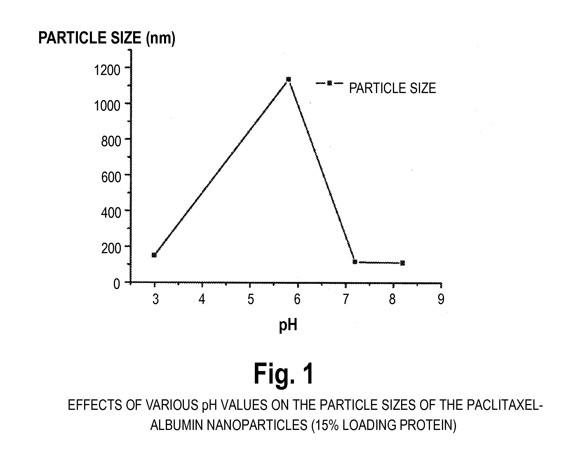
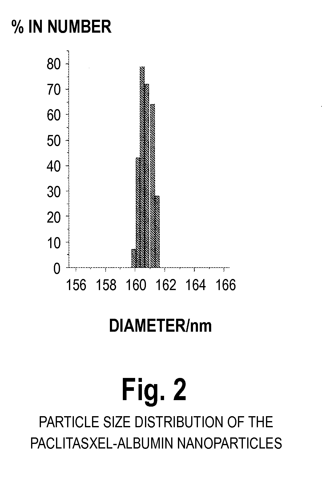
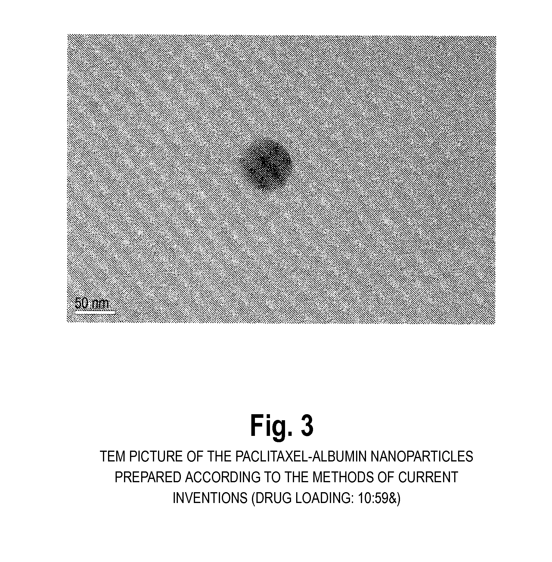
[0047]100 mg of albumin was dissolved in 10 ml 0.1M phosphate buffer pH 6.0 containing 0.5 mg / mL ethylenediaminetetraacetate and 0.05M mercaptoethanol. The reaction was carried out for two hours. At the end of reaction, the protein was precipitated and washed with 5% trichloroacetic acid. Next, 16 mg paclitaxel was dissolved in 1.6 mL ethanol and mixed with the precipitated protein. After 10 minutes mixing, 50 mL 0.1M phosphate buffer pH 8.0 was added to dissolve the mixture. The resulting dispersion is translucent, and the typical diameter of the resulting paclitaxel particles was 80 nm-200 nm (BIC 90plus particle size analyzer). HPLC analysis of the paclitaxel content revealed that more than 90% of the paclitaxel was encapsulated into the albumin.

example 2

Preparation of Paclitaxel-Albumin Nanoparticles without Protein Precipitation

[0048]100 mg of albumin was dissolved in 50 mL tris-buffer (pH 7.4) with stifling at 37° C. and 350 μL mercaptoethanol was added. The unfolding reaction was carried out for 10 minutes. Two mL of ethanol solution containing 20 mg paclitaxel was added. After 30 minutes, the sample was dialyzed in tris-buffer (pH 7.4) for 24 h. The dispersion was further lyophilized for 48 hours. The resulting cake could be easily reconstituted to the original dispersion by addition of sterile water or saline. The particle size after reconstitution was the same as before lyophillization. The typical diameter of the resulting paclitaxel particles was 80 nm-200 nm (BIC 90plus particle size analyzer). HPLC analysis of the paclitaxel content revealed that more than 90% of the paclitaxel was encapsulated into the nanoparticles.

example 3

Preparation of Paclitaxel-Albumin Nanoparticles

[0049]100 mg human albumin was dissolved in 10 ml buffer, pH 4.8, and kept in an ice bath for 30 minutes. 7.5 mL cold acetone (0° C.) was added (to induce precipitation) and kept in the ice bath for an additional 60 minutes. The solution was then centrifuged and precipitates were collected. A 10 mg / ml acetone solution of paclitaxel was added and mixed with the precipitates by applying sonication. A physiological buffer was added and mixed using a magnetic stirrer to form a solution. The mean particle size of the paclitaxel-albumin nanoparticles thus obtained was analyzed using BIC 90plus particle size analyzer, and found to be 150-220 nm. The nanoparticle solution was then dried by lyophillization. The freeze-dried cake was reconstituted. The paclitaxel loading of the nanoparticles was 8.3% using a HPLC assay.

[0050]Additional experiments showed that glycine, mannitol, trehalose or lactose can be used as a bulking agent for lyophillizati...


PUM

PropertyMeasurementUnit
Temperatureaaaaaaaaaa

Abstract

The present invention provides preparation methods of protein nanoparticles for in vivo delivery of pharmacologically active agents, wherein said methods are to encase pharmaceutically active agents into proteins or peptides to form nanoparticles by unfolding the protein, and subsequently refolding or assembling the protein to produce a pharmacologically active agent encased within a protein nanoparticle.

Description

FIELD OF THE INVENTION[0001]The present invention relates to in vivo delivery of pharmacologically active agents and clinical use thereof. Specifically, the invention provides methods for encasing pharmaceutically active agents into proteins or peptides to form nanoparticles by unfolding the protein, and subsequently refolding or assembling the protein to create a nanoparticle. Produced particles comprise about 1 to about 40% by weight of pharmacologically active agents and have the diameter in the range of about 5 to about 500 nm or more.BACKGROUND OF THE INVENTION[0002]Intravenous administration of pharmacologically active agents offers rapid and direct equilibration with the blood stream which carries the medication to the rest of the body. To decrease the side effects of the intravenous administrated agents, encapsulation of agents to micro- or nano-particles is a useful method. In one aspect, intravenous injection of the therapeutic particles would allow gradual release of the ...

Claims


Application Information

Patent Timeline
IPC IPC(8): A61K9/14A61K31/337A61K31/352A61P35/00B05D7/00B82Y5/00B82Y40/00
CPCA61K9/19A61K9/5192A61K9/5138A61K31/337A61P35/00
Inventor HU, YIQIAOWU, JINHUIDING, DAWEIGONG, GUANGMINGTANG, XIAOLEITONG, CHUNHUIZHU, YANLI, SHAOLING
Owner NANJING UNIV
Learn More