
Hydrophilic, High Protein Binding, Low Fluorescence, Western Blotting Membrane

a technology of western blotting membrane and hydrophilic protein, which is applied in the direction of liquid/fluent solid measurement, fluid pressure measurement, peptides, etc., can solve the problems of increasing sorption and membrane fouling, reducing membrane permeability and productivity, and reducing the permeability and productivity of membranes, etc., to achieve excellent retention of protein sample spot morphology, low background fluorescence, and high protein binding

Active Publication Date: 2011-02-24
MILLIPORE CORP
View PDF29 Cites 2 Cited by
  • Summary
  • Abstract
  • Description
  • Claims
  • Application Information

AI Technical Summary

Benefits of technology

The patent text describes a new method for creating a hydrophilic membrane that can be used for blotting applications. The membrane is made by contacting a substrate with a monomer solution and then subjecting it to polymerizing conditions. The resulting membrane is permanently hydrophilic and has low background fluorescence, high protein binding, and excellent retention of protein sample spot morphology. It is also stable to various chemical environments and is resistant to biomolecule adsorption. The membrane is water-wettable and can be used without an alcohol pre-wet step. Overall, this method provides a solution for creating a high-quality hydrophilic membrane that is suitable for blotting applications.

Problems solved by technology

However, it is known that polymers from such monomers are not resistant to strong alkaline solutions.
Such acrylic acid containing polymers are ionically charged under certain pH conditions, and will attract and bind oppositely charged proteins or biomolecules, thus increasing sorption and membrane fouling.
In addition, acrylic acid containing polymers swell in water to an extent that they constrict pore passages, thus reducing membrane permeability and productivity.
However, initial starting levels of each monomer had to be severely decreased before observing satisfactory dot blot morphology and blotting transfer performance.

Method used


Image

  • Hydrophilic, High Protein Binding, Low Fluorescence, Western Blotting Membrane
  • Hydrophilic, High Protein Binding, Low Fluorescence, Western Blotting Membrane
  • Hydrophilic, High Protein Binding, Low Fluorescence, Western Blotting Membrane

Examples


example 1

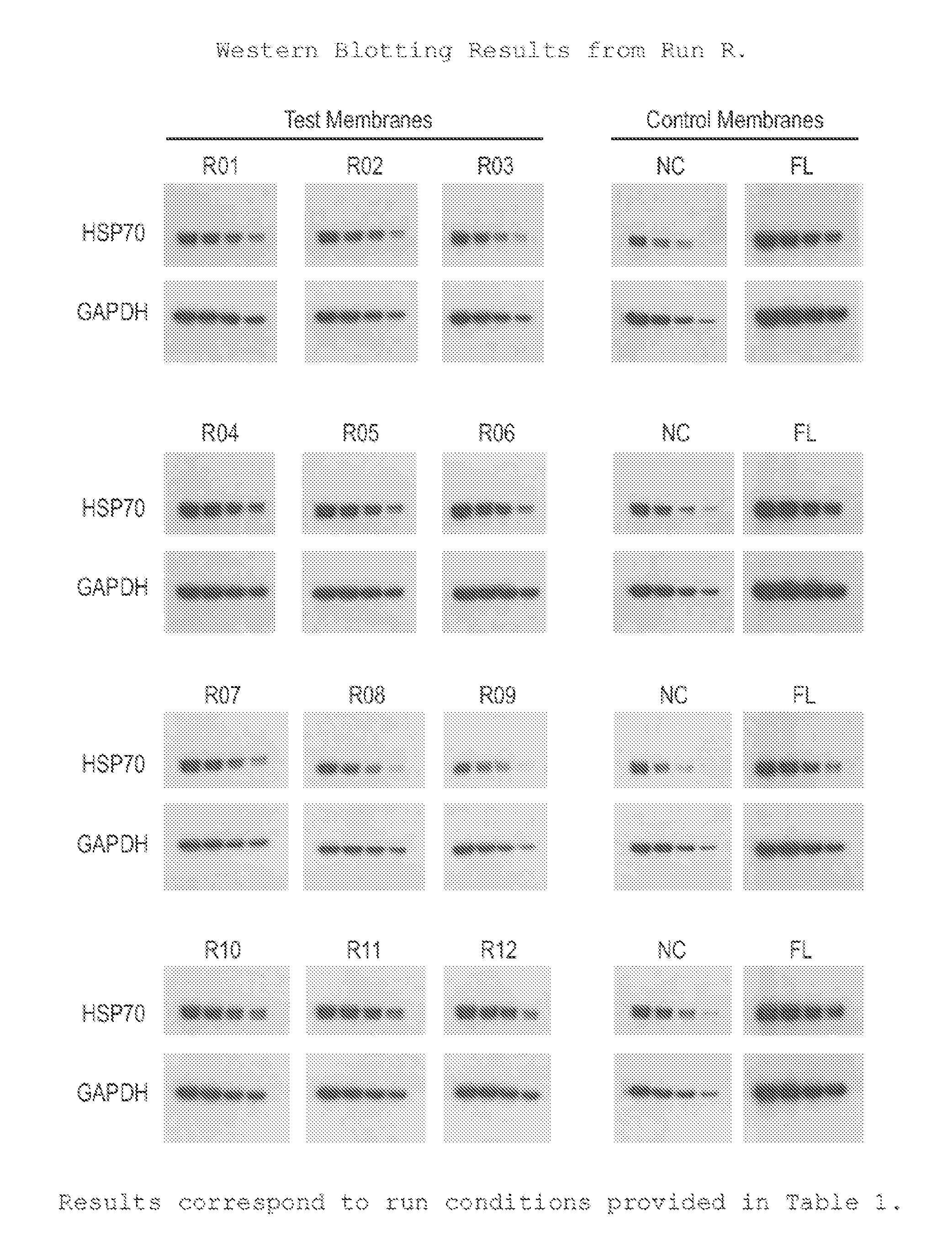
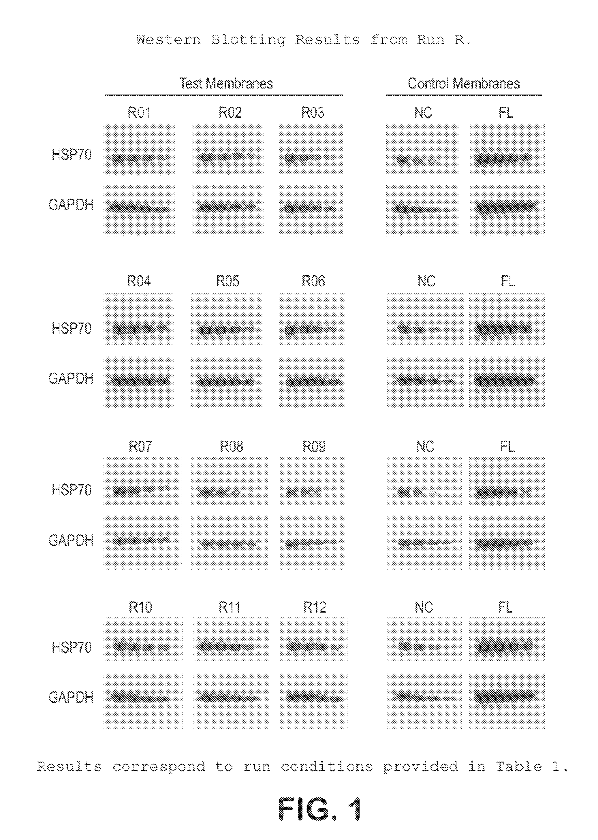
[0029]Commercially available hydrophobic PVDF membrane from Millipore Corporation (IPFL00000) was immersed in methyl alcohol. The membrane was withdrawn and immersed in water with agitation to extract methanol for 1 minute. The membrane was withdrawn and immersed in fresh water for an additional 2-minute interval and then placed in fresh water before immersion in reactant monomer solution. Excess water was drained from the membrane and the membrane was then immersed in monomer reactant solution with gentle agitation for 2 minutes. Membrane was then exposed to UV radiation from both sides in a UV curing process at a line speed of 15 to 25 fpm. Membrane was recovered and placed into a water bath to remove unreacted monomer and non-adhering oligomers and polymer. Samples were dried either in air at room temperature overnight, or in a static forced-air oven between 60° C. and 80° C. for 10 minutes, or on an impingement dryer at 90-110° C. at a line speed of 15 to 25 fpm. Average membran...

example 2

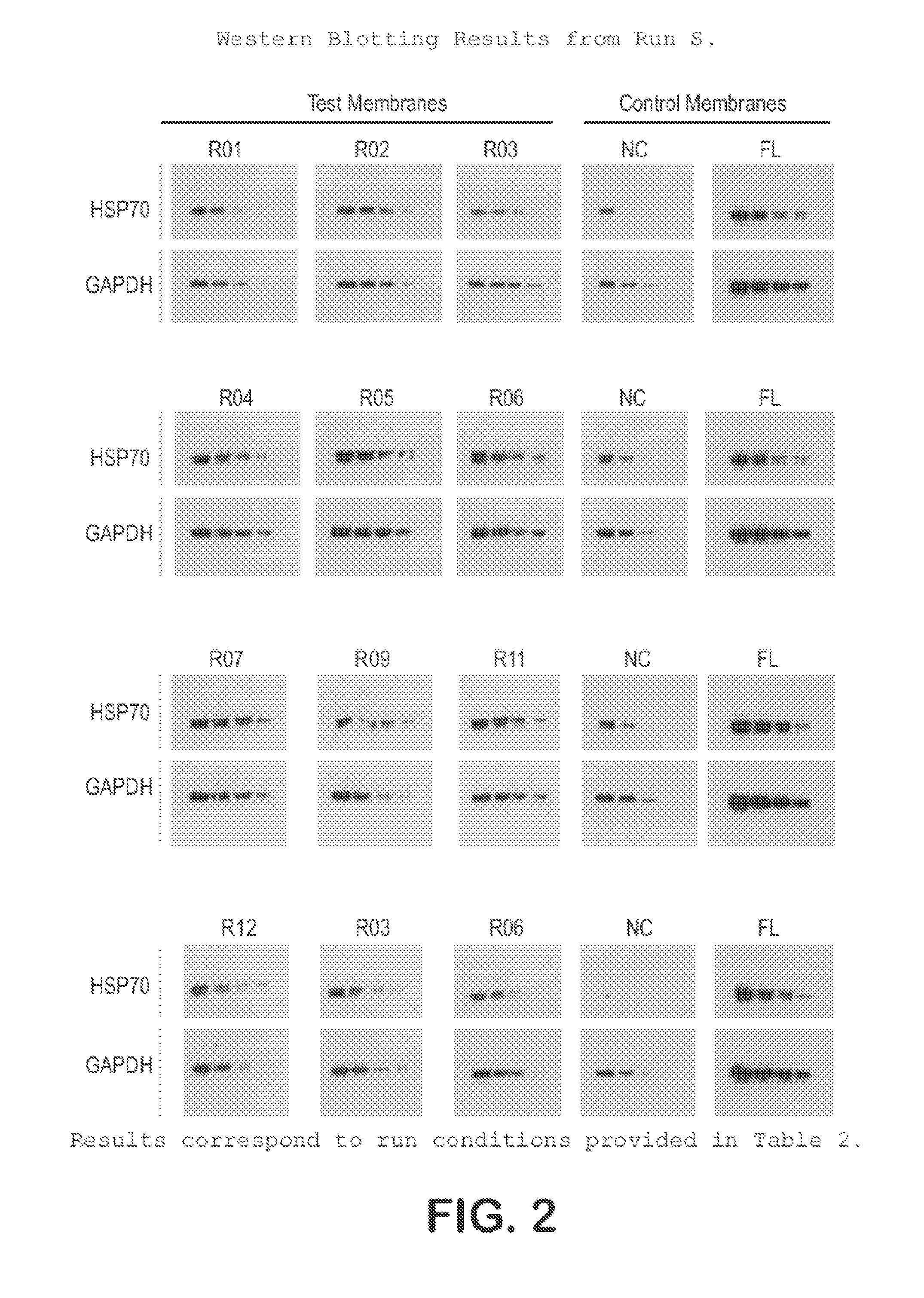
[0031]The procedure of Example 1 was used to treat PVDF membranes having the specifications, treatment conditions and reactant solutions shown in Tables 3A-D, 4A-D, and 5A-D.

TABLE 3AMODIFICATION DATA for Hydrophilic PVDF Western Blotting MembraneMembraneMonomerStartingCastingStarting Membrane PropertiesRollMixMembraneDryerThickPorosityBbl PtFlow TimeNo.Lot#Lot DataTemperature (F.)(um)(%)(psi)(seconds)RUN R - No Mix Ajustment - Footage VS Monomer Mix ConcentrationNANANANANANANANAR01R Mix 1IPFL 071007 T21430011272.89.456.0R02R Mix 1IPFL 071007 T20530011373.09.463.0R03R Mix 1IPFL 071007 T20530011373.09.463.0R04R Mix 1IPFL 071007 T20530011373.09.463.0R05R Mix 1IPFL 071007 T20530011373.09.463.0R06R Mix 1IPFL 071007 T20630011172.49.565.0R07R Mix 1IPFL 071007 T20630011172.49.565.0R08R Mix 1IPFL 071007 T20630011172.49.565.0R09R Mix 1IPFL 071007 T20630011172.49.565.0R10R Mix 1IPFL 071007 T21430011272.89.456.0R11R Mix 1IPFL 071007 T21430011272.89.456.0R12R Mix 1IPFL 071007 T21430011272.89.456...

example 3

[0032]The protocol used for Western blotting and chemiluminescent detection is as follows:[0033]Protein samples are electrophoretically separated using Bis:Tris (4˜12%) midi gradient gel (Invitrogen, WG1402BOX).[0034]Samples are electro-blotted at 45V for 1 hr 15 min using BioRad tank transfer apparatus (Criterion Blotter #165-6024) onto a hydrophilic membrane prepared as in Example 1.[0035]Blots are washed 2× (3 min each) in TBS-T (0.1% Tween)[0036]Blots are blocked for 1 hr at RT in TBS-T with 3% NFM (non-fat milk, Carnation).[0037]Blots are washed 2× (3 min each) in TBS-T (0.1% Tween)[0038]Blots are incubated with primary antibody in TBS-T for 1 hr.[0039]Blots are washed 3× (5 min each) in TBS-T (0.1% Tween)[0040]Blots are incubated with secondary antibody in TBS-T for 1 hr[0041]Blots are washed 4× (5 min each) in TBS-T (0.1% Tween)[0042]Protein bands are visualized using ECL (Millipore Immobilon-HRP) and x-ray film.

[0043]This protocol was used on the samples prepared in Example ...


PUM

PropertyMeasurementUnit
timeaaaaaaaaaa
pore sizesaaaaaaaaaa

Abstract

Hydrophilic membrane particularly suited for blotting applications, preferably Western blotting. A pre-wet hydrophobic membrane substrate, preferably made of PVDF, is contacted with a monomer solution and subjected to a UV-initiated free radical polymerization step to render the substrate permanently hydrophilic. The resulting membrane exhibits low background fluorescence, high protein binding, excellent retention of protein sample spot morphology, and extended dynamic range (high signal-to-noise ratio, enhanced sample detectability). The membrane demonstrates comparable or higher performance in Western blotting applications than conventional nitrocellulose Western blotting membranes, particularly for protein detection at low sample concentrations, and is directly water-wettable, eliminating the need for an alcohol pre-wet step prior to use.

Description

[0001]This application is a continuation of Ser. No. 12 / 462,635 filed Aug. 6, 2009, which claims priority of Provisional Application Ser. No. 61 / 189,302 filed Aug. 18, 2008, the disclosures of which are hereby incorporated by reference.BACKGROUND OF THE INVENTION[0002]“Blotting” or “electro-blotting” refers to the process used to transfer biological samples from a gel to a membrane under the influence of an electric field. The process requires a membrane that can immobilize biomolecular samples for subsequent detection. This places specific requirements on the membranes related to surface area, porosity, and protein binding capacity.[0003]Western blotting is one modification of this technique that involves the immobilization of proteins on membranes before detection using monoclonal or polyclonal antibodies. Prior to protein immobilization on the membrane, sample proteins are separated using SDS polyacrylamide gel electrophoresis (SDS-PAGE) to separate native or denatured proteins. ...

Claims


Application Information

Patent Timeline
Patent Type & Authority Applications(United States)
IPC IPC(8): G01N33/53
CPCB01D67/0093B01D2323/02Y10T436/24B01D2325/36G01N33/54393B01D2323/30B01D67/00933
Inventor PETERS, ANTONIGODDARD, PHILIPCHARKOUDIAN, JOHNSOICE, NEILBREWSTER, DAVEDEDEO, ANJA
Owner MILLIPORE CORP
Learn More