
DHA and PEDF, a Therapeutic Composition for Nerve and Retinal Pigment Epithelial Cells

Inactive Publication Date: 2010-12-02
BOARD OF SUPERVISORS OF LOUISIANA STATE UNIV & AGRI & MECHANICAL COLLEGE
View PDF16 Cites 4 Cited by
  • Summary
  • Abstract
  • Description
  • Claims
  • Application Information

AI Technical Summary

Benefits of technology

[0018]I have discovered that the unique combination of PEDF and DHA acts synergistically to enhance cell survival and decrease apoptosis. Blinding retinal degeneration (e.g., retinitis pigmentosa) is due to the progressive degeneration of photoreceptors and retinal pigment epithelial cells (RPE cells). I have discovered that PEDF (pigment epithelium-derived factor) plus DHA (docosahexaenoic acid) act synergistically in protecting the survival of the RPE cells. PEDF and DHA (a member of the omega-3 essential fatty acid family), protected human retinal pigment epithelial cells (RPE) confronted with oxidative stress. This synergistic cytoprotection is reflected in the ability of this combination to block apoptotic cell death. In addition, the combination of PEDF and DHA acted synergistically in modifying the molecular mechanisms that lead to cell death. In particular, the combined use of PEDF and DHA increased the synthesis of the important mediator neuroprotectin D1.
[0019]NPD1 was shown to be involved in the interaction between these two cell types in the retina, RPE cells and photoreceptors, when under oxidative stress. Therefore, PEDF plus DHA may be useful as a therapeutic approach in several eye diseases. In the dry form of age-related macular degeneration, apoptosis of macular photoreceptors occurs. Therefore, administering a composition comprising PEDF and DHA will elicit cytoprotection and thus halt or slow down the initiation and progression of death of the macular photoreceptors and thus decrease the amount of blindness of the patient. In the wet-form of age-related macular degeneration (AMD), one of the major problems in this form of AMD is the pathological growth of blood vessels in the retina. The combination of PEDF and DHA acts synergistically to increase the amount of NPD1 produced in the retina. This administration of this combination to the retina would increase the NPD1 concentration and thus cause a decrease in the amount of pathological angiogenesis.
[0020]The diseases retinitis pigmentosa and retinal degeneration that are caused by gene mutations can also be helped using this new combination. Although the ultimate cure will be to correct or replace the defective gene, the administration of PEDF plus DHA will slow down the initiation and progression of photoreceptor death by inhibiting the apoptotic cell death that occurs in most of those conditions. Even in glaucoma, cell death of retinal ganglion cells occurs and is a cause of some of the clinical symptoms. Again, the administration of the combination of PEDF and DHA can be used to slow down or decrease the amount of retinal cells dying. Thus, in general, symptoms of any retinal disease that involves cell death, or cell injury leading to death caused by other forms of damage, can be alleviated by administering the combination of PEDF and DHA.
[0021]The administration of the combination of PEDF plus DHA may also be protective in neurological conditions where cell damage and death takes place, such as stroke, epilepsy, Alzheimer' disease, Huntington's, Parkinson's, amyotrophic lateral sclerosis, multiple sclerosis, head injury, and spinal cord injury. Several diseases commonly described in psychiatry—depression, post-traumatic stress disorder, and schizophrenia—also involve neuronal cell death and may be alleviated by PEDF plus DHA. It is believed that the combination of PEDF and DHA administered to neural cells will increase the production of NPD1, and will thus work synergistically to promote brain cell survival, and be an effective treatment for stroke, neurotrauma, spinal cord injury, and neurodegenerative diseases, such as Alzheimer's disease, as well as in certain psychiatric illnesses.
[0022]Refractive surgery, dry eye, herpetic keratitis and several other diseases result in damage to the cornea nerves. I have also shown that the topical application of the combination of PEDF and DHA promotes cornea nerve regeneration, and thus this combination could be used to prevent the complications of refractive surgery and certain diseases, e.g., neurotrophic keratitis due to Herpes virus.

Problems solved by technology

In the wet-form of age-related macular degeneration (AMD), one of the major problems in this form of AMD is the pathological growth of blood vessels in the retina.

Method used


Image

  • DHA and PEDF, a Therapeutic Composition for Nerve and Retinal Pigment Epithelial Cells
  • DHA and PEDF, a Therapeutic Composition for Nerve and Retinal Pigment Epithelial Cells
  • DHA and PEDF, a Therapeutic Composition for Nerve and Retinal Pigment Epithelial Cells

Examples


example 1

Promotion of Cornea Nerve Regeneration

[0042]The effect of treatment with PEDF and DHA after either LASIK or PRK surgery was evaluated for its potential to increase the corneal nerve surface area, and thus decrease the complications seen in patients of corneal hypoesthesia and dry eye. Using a recognized experimental model, a lamellar dissection was performed on six rabbits with the use of a crescent blade and scissors creating a pocket in the cornea using techniques similar to S. Esquenazi et al., “Topical combination of NGF and DHA increases rabbit corneal nerve regeneration after photorefractive keratectomy,” Investigative Opthalmology & Visual Science, vol. 46, pp. 3121-3127 (2005). The six rabbits were divided into two groups: (1) one group treated with PEDF and DHA (100 μg), and (2) one group treated with a vehicle (saline plus albumin). In both groups, the treatment solution was delivered in collagen shields applied to the eye that would dissolve in 72 hr, and were changed twi...

example 2

Materials and Methods

[0043]Human RPE cells. Cultures of retinal pigment epithelial (RPE) cells were prepared. RPE cells were seeded onto Millicell-HA culture plate inserts (Millipore, Bradford, Mass.), placed in 24-well plates, and allowed to reach confluence. Consent for use of tissue for research was obtained in compliance with Federal and State law and institutional regulations. Cultures were maintained in Chee's essential replacement medium until the experiments were performed as described in J. Hu and D. Bok (2001) Mol Vis 7:14-19. The medium includes Eagle's minimal essential medium with calcium (MEM, Irvine Scientific, Irvine, Calif.), 1% heat-inactivated calf serum (JRH Bioscience, Lennexo, Kans.), amino acid supplements, and 1% bovine retinal extract. The Millicell-HA filter inserts allow separate manipulation of the culture media bathing the apical and basal surfaces of the RPE monolayer and measurement of the transepithelial resistance (TER), which provides a measure of c...

example 3

A2E-mediated ARPE-19 Cell Damage is Attenuated by Neuroprotectin D1

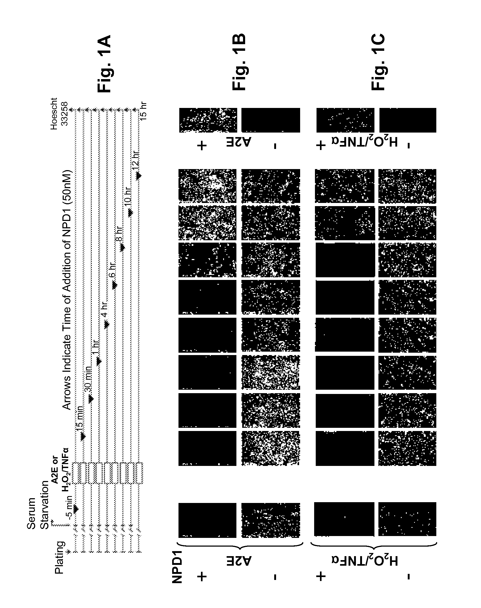
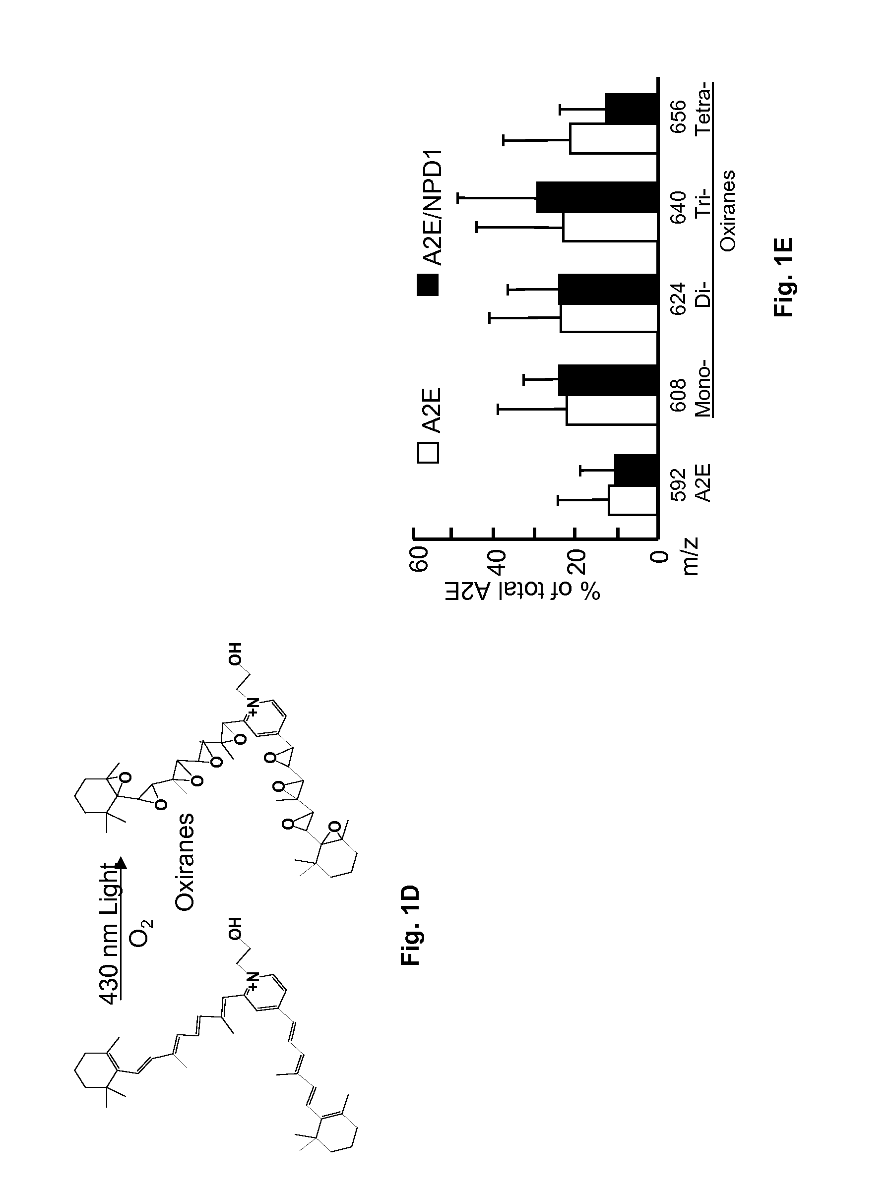
[0050]A2E, a lipofusin component, accumulates in the RPE during aging, and, in an exaggerated manner, in age-related macular degeneration (AMD) and Stargardt macular dystrophy (an early onset form of AMD). As a consequence, RPE apoptosis precedes the demise of photoreceptors. A2E-induced oxidative stress in ARPE-19 cells was shown to be inhibited by NPD1. Seventy-two (72) hours after plating, ARPE-19 cells were serum starved for 4 hours. A2E (20 μM) was added in the presence of 430 nM light and O2 (see Materials and Methods). Other cells were exposed instead to H2O2 / TNFα. NPD1 was added prior to, or at different times after, A2E or H2O2 / TNFα, up to 12 hours. Time of NPD1 addition is indicated by black arrows on FIG. 1A. Hoechst 33258 was analyzed at 15 hours. FIG. 1B illustrates the appearance of Hoechst 33258 positive cells upon exposure to A2E and the effect of NPD1. FIG. 1C is similar to FIG. 1B, except that H2O2 / ...


PUM

PropertyMeasurementUnit
Compositionaaaaaaaaaa
Therapeuticaaaaaaaaaa
Transparencyaaaaaaaaaa

Abstract

The combination of pigment epithelium-derived factor (PEDF) and docosahexaenoic acid (DHA) has been discovered to act synergistically to enhance cell survival and decrease apoptosis in retinal pigment epithelial (RPE) cells. PEDF and DHA synergistically protected RPE cells by confronted with oxidative stress by blocking apoptotic cell death and increasing the synthesis of the important mediator neuroprotectin D 1. Administering a composition comprising PEDF and DHA will halt or slow down the initiation and progression of macular degeneration, retinitis pigmentosa and retinal degeneration. In addition, the topical application of the combination of PEDF and DHA was found to promote cornea nerve regeneration after refractive surgery, and thus this combination could be used to prevent the complications of refractive surgery and certain diseases, e.g., neurotrophic keratitis due to Herpes virus.

Description

[0001](In countries other than the United States:) The benefit of the 30 Jul. 2007 filing date of U.S. patent application Ser. No. 60 / 952,725 is claimed under applicable treaties and conventions. (In the United States:) The benefit of the 30 Jul. 2007 filing date of provisional patent application No. 60 / 952,725 is claimed under 35 U.S.C. §119(e).[0002]The development of this invention was partially funded by the Government under grant number EY05121 from the National Institutes of Health National Eye Institute, and grant number P20 RR016816 from the National Institutes of Health National Center for Research Resources. The Government has certain rights in this invention.TECHNICAL FIELD[0003]This invention pertains to the use of the combination of pigment epithelium-derived factor (PEDF) and docosahexaenoic acid (DHA) to protect the survival of nerve cells and retinal pigment epithelial (RPE) cells. This unique combination acts synergistically in nerve cells and RPE cells to increase ...

Claims


Application Information

Patent Timeline
IPC IPC(8): A61K9/00A61K38/22A61P27/02A61P25/00
CPCA61K31/202A61K38/482A61K2300/00A61P25/00A61P25/28A61P27/02
Inventor BAZAN, NICOLAS G.
Owner BOARD OF SUPERVISORS OF LOUISIANA STATE UNIV & AGRI & MECHANICAL COLLEGE
Learn More