
System and method for site specific therapy

Inactive Publication Date: 2010-11-11
TWIN STAR MEDICAL
View PDF19 Cites 18 Cited by
  • Summary
  • Abstract
  • Description
  • Claims
  • Application Information

AI Technical Summary

Benefits of technology

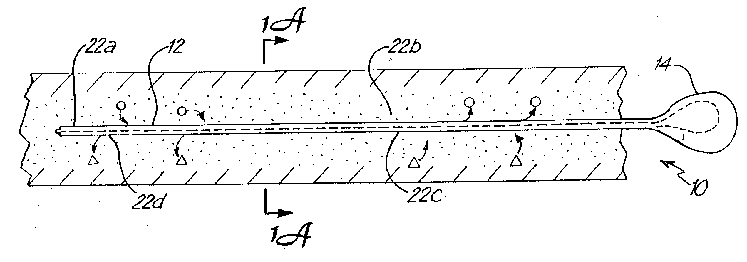
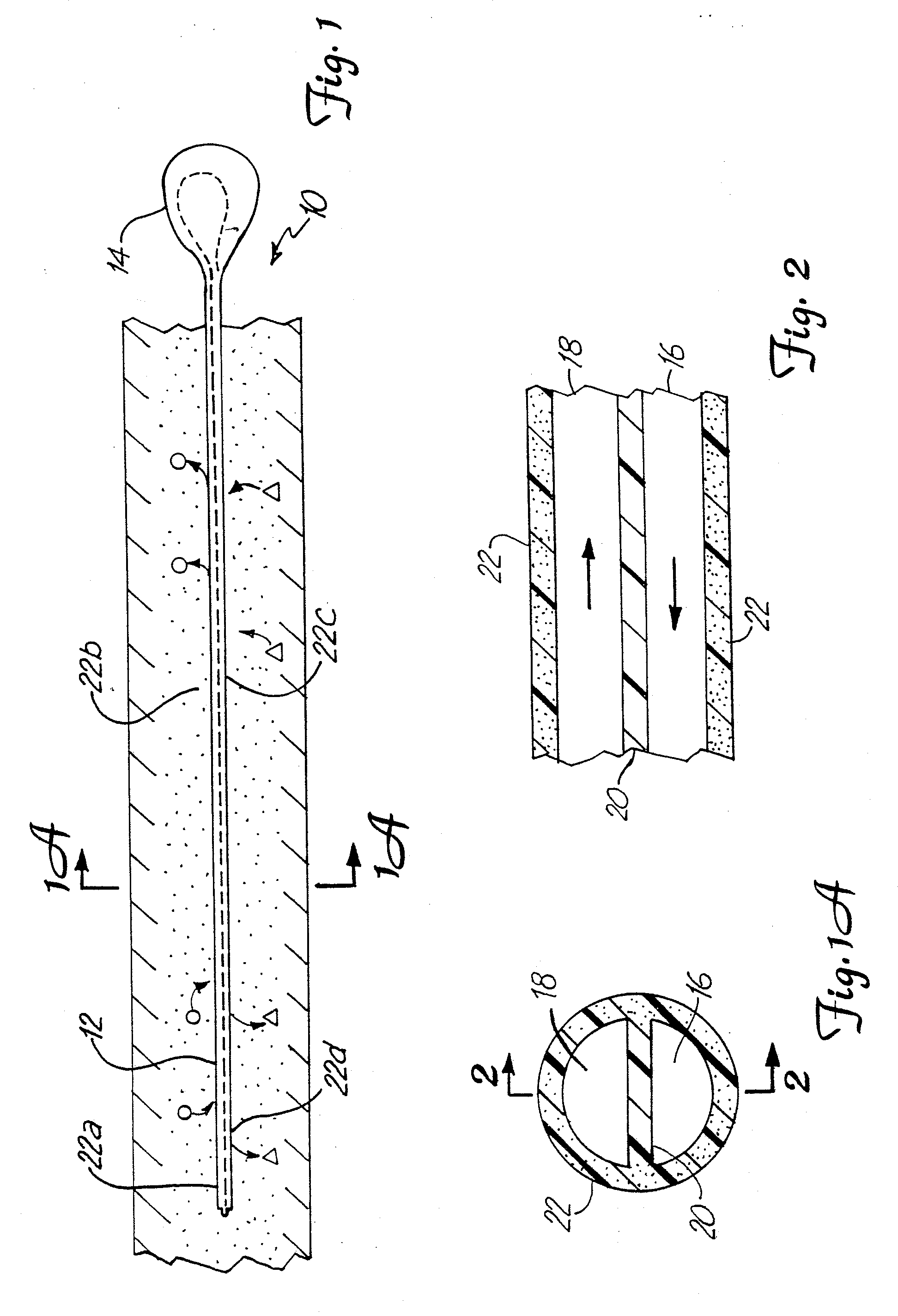
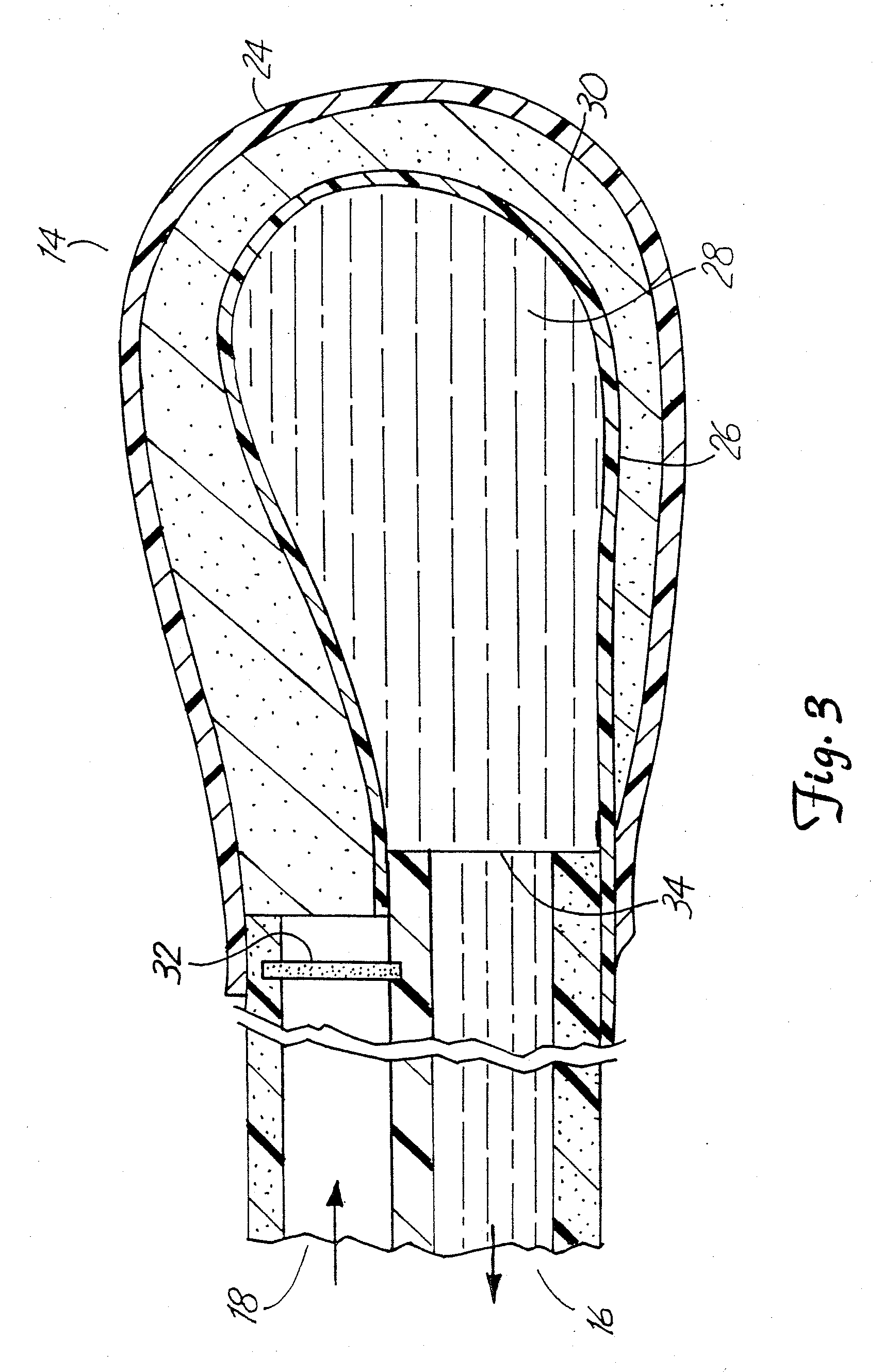
"The present invention provides a method and system for using catheters to control the movement of fluids and active fluid components within or between tissue portions in a tissue site-specific manner for therapeutic purposes. The catheters can be semipermeable microcatheters that can be positioned within the tissue site to deliver and remove fluids and active fluid components. The system can be used to affect the osmolar nature of a remote fluid or to move biologically active molecules between healthy and diseased portions of the same tissue. The apparatus can be used for various purposes, such as treating reperfusion injury, delivering toxic agents directly to a tissue site, and preparing bone and soft tissue grafts. The invention provides a commercially viable in vitro tissue engineering technique based on the principle of microdialysis."

Problems solved by technology

A number of conditions involve poor blood supply to the bone, leading to bone necrosis.
There is no treatment presently available that can predictably alter the natural history of the disorder.
Several technical barriers to the treatment of AVN of the femoral head and neck include the limited blood supply of the site, difficult surgical access, and the accelerated progression of the disease due to biomechanical demands of walking on the hip joint.
Acute compartment syndrome generally involves impaired circulation within an enclosed fascial space, leading to increased tissue pressure and necrosis of muscle and nerves.
Direct trauma, ischemia, or excessive, unaccustomed exercise can result in hemorrhage and swelling inside the anterior compartment.
In addition, the prolonged compression of nerves can destroy their ability to function.
These symptoms must be identified quickly, since misdiagnosis can lead to permanent neuromuscular damage and physical disability.
The characteristics of acute tissue edema are well known, and the condition continues to be a clinical problem, particularly since edema can be detrimental to the tissue as a result of disruption of the microcirculation.
Tissue swelling results in increased diffusion distances, which in turn decreases interstitial nutrient delivery.
Irreversible disruption of the microcirculatory system can occur as a result of unresolved acute injury.
Resolution of tissue edema is problematic since natural mechanisms by which edema resolves are also affected by the edema.
Pharmacologic treatment is often not effective since blood borne agents have difficulty reaching their target tissue.
Opening the skull generally cannot be done to relieve pressure, because the brain tissue would herniate out the opening causing significant tissue damage.
Giving intravenous treatments is also not effective because the brain microcirculation is disrupted so deilivery to the brain is impaired.
Brain swelling leading to dangerously elevated ICP develops in 40-50% of TBI patients with a Glascow Coma Scale (GCS) of 8 or less, and higher ICP levels have been repeatedly shown to lead to poor prognosis or outcome.
While the placement of an ICP monitor is invasive, the benefits of ICP monitoring are felt to offset this factor, carry a relatively small risk of complications (e.g., infection, hemorrhage, malfunction, obstruction or malposition), and rarely result in increased patient morbidity.
As fluid is removed, however, brain swelling often progresses to the point where the ventricular system is compressed and the ability to drain CSF can be compromised.
This may be exacerbated by overdrainage, leading to the ventricular walls or the choroid plexus actually collapsing in a manner that occludes the orifices of the catheter.
The therapeutic efficacy of convective CSF drainage by conventional ventriculostomy catheters, therefore, is limited.
Also, ventriculostomy catheters can become occluded with tissue debris and clots during convective fluid removal.
As briefly outlined below, it can be seen that each of these therapeutic strategies is a “double-edged sword” since each treatment is typically associated with potential adverse consequences and each has limited efficacy.Hyperventilation: Prophylactic hyperventilation of TBI patients is currently questioned since it has been reported to worsen outcomes, does not consistently reduce ICP, and may cause loss of autoregulation and potentiate secondary ischemia due to its actions on reducing cerebral blood flow.Mannitol: This osmotic diuretic is currently the most widely used, and probably the safest, treatment for short-term control of elevated ICP in patients with TBI.
Although it has become the cornerstone for control of elevated ICP after severe TBI, mannitol administration is not without risks.
Although the use of mannitol affects osmolarity within the site, this approach is not site-specific, rather, it is systemically administered.
Since this approach is also chemically based, rather than device based, it does not employ a device that is itself provides an osmotic barrier.Barbiturates: Prophylactic barbiturate therapy is currently discouraged, due to variable and unpredictable positive effects on ICP.
While edema can be controlled in many patients by the use of drug treatments, there are many patients for whom such treatment is not effective.
To date, however, Applicant is unaware of any description of the use of such dialysis techniques or apparatuses in the treatment of cerebral edema or ICP.
In the course of inserting microdialysis probes into brain parenchyma, in order to monitor neurochemical alterations in patients, it has been found that there is minimal trauma to brain tissue and that complications are extremely rare.
However, most, if not all, current microdialysis procedures rely on the slow, pump-driven infusion of dialysis fluid which travels through inlet lines past the dialysis fiber and then through outlet lines to enable collection of the dialysate.
These techniques, however, continue to be plagued by problems having to do with survival of the skin flaps, which in turn, is believed to rely, at least in part, on efficient revascularization of the site.
To Applicant's knowledge, however, there is no present teaching, let alone clinically acceptable approach for the application of tissue microdialysis in site specific therapy, or in particular, a microdialysis apparatus useful for prolonged periods, difficult sites, and in clinical settings.
In turn, current therapies for treating elevated ICP and cerebral edema, in humans with severe traumatic brain injury, have limited efficacy and continue to be associated with serious risks (particularly with prolonged use).
In some patients, cerebral edema simply remains untreatable or nonresponsive to treatment.

Method used


Image

  • System and method for site specific therapy
  • System and method for site specific therapy
  • System and method for site specific therapy

Examples


example 1

Rat Model

[0215]Cerebral edema is induced in rats, and thereafter reduced by the use of microdialysis fibers implanted directly into injured brain tissue. As an initial experimental phase, a hyperosmolar solution of albumin is infused into the ventricles of the rat after experimental brain injury has been induced. Conventional methods (e.g., Onal et al.) are used to induce the injury, with the following exceptions: a contusion injury is used and infusion is performed over a longer period to see if the benefits of infusion can be prolonged. Twelve hours after experimental TBI is induced, an infusion catheter is placed into the lateral ventricle. Thereafter a 20% (w / v) solution of albumin in saline is infused at 0.5 ul / hour for a period of six hours. Brain water content is determined at 24 hours.

[0216]In a subsequent phase, the effect of intraventricular microdialysis on brain water content is determined. Some impermeant solutes are already present in the ventricles (and the concentrat...

example 2

Osmolarity of Human Traumatic CSF

[0220]A study was performed using banked human CSF. Osmolarity was determined in control patients (no head trauma), and three patients who suffered closed head injury. Normal osmolarity of CSF in the controls was 305 mosmols / L. As seen in Table I (see FIG. 49), the TBI patients had an increase in CSF osmolarity in the first 3 days, with an even greater increase in the next three days.

[0221]These findings of a delayed increase in CSF osmolarity are consistent the osmotic fluid shift premise described herein. Although CSF from trauma patients has apparently not been examined for changes in osmolarity before, and it is difficult to make conclusions based on this small number, it nevertheless appears that CSF osmolarity does increase after head trauma. This finding, together with findings in the literature that CSF osmolarity increases after a cryogenic injury in rats, and that brain tissue osmolarity increases within hours after ischemia provides suppor...

example 3

Spinal Microdialysis

[0272]The method and system of the present invention are used to perform spinal microdialysis using the following materials:[0273]1. A 3 inch needle, 18-20 gauge, similar to contemporary spinal tap needles. This is placed between the third and fourth lumbar vertebral space. The distal 2 mm of the needle is unique in that it has a memory to bend 50-70 degrees when the obturator of the needle is removed.[0274]2. The obturator of the needle is straight, rigid, and has a lumen.[0275]3. The patient assumes a lateral decubitus position with knees drawn up. The L3-4 interspace is palpated and marked after sterile scrub of the skin.[0276]4. The spinal needle with obturator in place is advanced until a slight decrease in resistance is felt as the needle tip pierces the posterior spinal ligament and enters the spinal canal CSF space.[0277]5. Once CSF is confirmed to drip from the lumen of the obturator, the obturator is held stationary and the needle is advanced another 2-...


PUM


Abstract

A system, including catheter apparatus, and related method for performing site specific therapy. The catheter apparatus can include one or more semipermeable microcatheters for use in performing site specific microdialysis. The system and method are particularly suited for use in addressing cerebral edema by affecting the osmolar relationship between fluids making up the brain tissue.Also disclosed is an apparatus having a delivery / recovery mechanism in the form of a pump reservoir and one or more catheters in the form of semipermeable microcatheters, for use in delivering and / or recovering fluid to and / or from a tissue site or for performing tissue engineering outside of the body. The apparatus can be used in a method to perform site specific microtherapy, including for the treatment of avascular necrosis, compartment syndrome, cerebral edema, and to improve skin flap survival in the course of reconstructive surgery.

Description

CROSS REFERENCE TO RELATED APPLICATIONS[0001]The present application is a continuation of U.S. patent application filed 24 Mar. 2003 and assigned Ser. No. 10 / 395,573, which is a continuation of U.S. patent application filed 17 May 1999 and assigned Ser. No. 09 / 313,341, which is a continuation of an international patent application filed 7 Aug. 1998 and assigned Serial No. PCT / US98 / 16416 which is a continuation-in-part of U.S. patent application filed 8 Aug. 1997 and assigned Ser. No. 08 / 908,555, the entire disclosure of which is incorporated herein by reference.TECHNICAL FIELD[0002]In one aspect, the present invention relates to methods and apparatuses for treating microcirculatory problems, including transient and reversible conditions that do no involve structural injury, as well as permanent or chronic conditions that do involve structural injury to the microcirculation. In another aspect, the invention relates to methods and apparatuses for augmenting normal microcirculation. In...

Claims


Application Information

Patent Timeline
Patent Type & Authority Applications(United States)
IPC IPC(8): A61M25/00A61M27/00A61M1/16A61M1/20A61M25/14
CPCA61M1/1678A61M2210/0693A61M2025/0057A61M2025/0042A61M1/26
Inventor ODLAND, RICK MATHEW
Owner TWIN STAR MEDICAL
Learn More