Methods of detection of factor xia and tissue factor
a tissue factor and factor xia technology, applied in the field of methods of detecting tissue factor, can solve the problems of complex detection of fxia in plasma, and achieve the effect of “propensity to develop, greater likelihood of developing and/or suffering
- Summary
- Abstract
- Description
- Claims
- Application Information
AI Technical Summary
Benefits of technology
Problems solved by technology
Method used
Image
Examples
example 1
Selection of Subjects
ACS
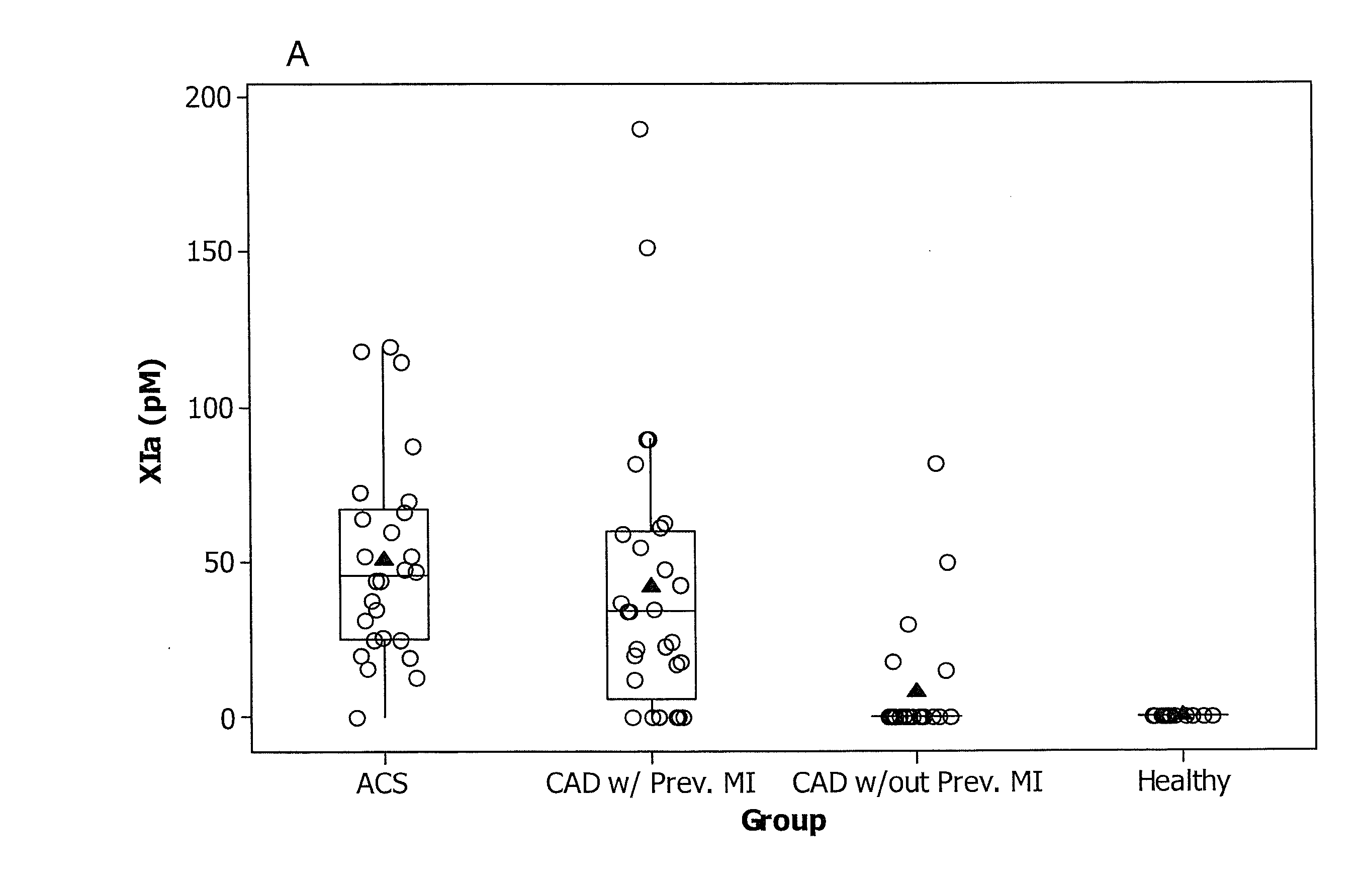
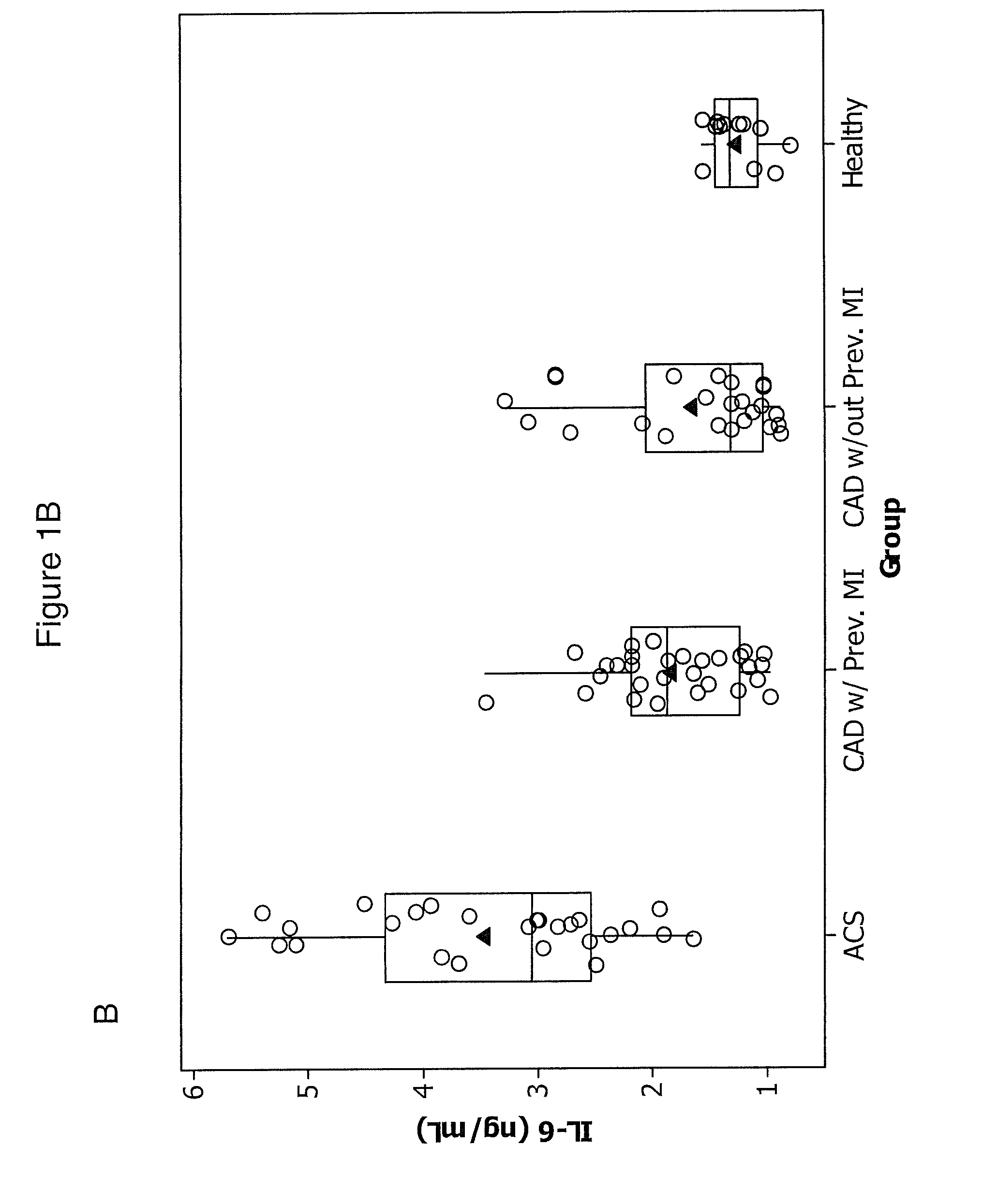
[0092]We studied 30 patients with ACS were admitted to the Department of Hemodynamics of the Jagiellonian University and 53 patients with angiographically confirmed stable CAD (>50% stenosis in at least one major coronary artery) recruited from an outpatient clinic. Twenty-nine of the stable CAD patients (55%) had a history of previous myocardial infarction (MI) 0.5-3 years before enrollment. ACS patients had experienced chest pain for up to 12 hours prior to seeking treatment.
[0093]Upon arrival at the hospital, patients were enrolled and blood was taken before treatment was initiated. All patients took 300 mg aspirin before the study. None of the subjects received thienopyridines or other anticoagulants prior to blood collection. Four ACS patients received 5000 U of unfractionated heparin prior to the blood draw and were excluded.
[0094]Inclusion criteria for ACS patients were typical chest pain and either ST-segment elevation ˜0.1 mV or ST-segment depression...
example 2
Sample Collection and Characterization
[0102]Blood was drawn from an antecubital vein with minimal stasis within 15 minutes upon admission in the case of ACS patients, and as soon as reasonably possible for all other acute event subjects, preferably within 15 minutes of admission, and after an overnight fast between 7 to 9 a.m. in the case of stable CAD patients and healthy volunteers. Serum and citrate plasma samples (9:1 of 3.2% sodium citrate a calcium chelator that inhibits coagulation) were centrifuged at 2540 g for 15 minutes at 24° C. within 20 minutes of collection, immediately frozen, and stored in aliquots at −80° C. until further use. Lipid profiles, blood morphology, glucose, creatinine, albumin, aminotransferases, and creatine kinase were assayed by routine laboratory techniques. Fibrinogen was determined using the Clauss method. High-sensitivity CRP was measured by latex nephelometry (Dade Behring, Marburg, Germany). Commercially available immunoenzymatic assays were us...
example 3
Factor XIa Plasma Clotting Assay
[0103]Citrate plasma (200 ul) was thawed at 37° C. in the presence of 4 ul corn trypsin inhibitor (5 mg / ml) (CTI; prevents contact pathway initiation of coagulation; prepared as previously described in Cawthern et al. (Blood 1998; 91:4581-4592, incorporated herein by reference). Plasma was divided into 2-100 ul aliquots, one test and one control.
[0104]An aliquot of the CTI-containing plasma was placed in a cuvette in an ST8 instrument (Diagnostica Stago, Parsippany, N.J.) and maintained at 37° C. CaCl2 was added to a final concentration of 15 mM (1.5 ul of 1M stock) and the plasma incubated for 1 min; clotting was initiated by the addition of 2 μM phospholipid vesicles (PCPS) (4 ul of 5 uM stock) composed of 25% dioleoyl-sn-glycero-3-phospho-L-serine and 75% of 1,2-dioleoyl-sn-glycero-3-phosphocholine (both from Avanti Polar Lipids, Inc; Alabaster, Ala.) and prepared as described in Higgins and Mann (J Biol Chem 1983; 258(10):6503-6508, incorporated h...
PUM
| Property | Measurement | Unit |
|---|---|---|
| clotting time | aaaaa | aaaaa |
| clotting time | aaaaa | aaaaa |
| clotting time | aaaaa | aaaaa |
Abstract
Description
Claims
Application Information
 - R&D
- Intellectual Property
- Life Sciences
- Materials
- Tech Scout
- Unparalleled Data Quality
- Higher Quality Content
- 60% Fewer Hallucinations
Browse by: Latest US Patents, China's latest patents, Technical Efficacy Thesaurus, Application Domain, Technology Topic, Popular Technical Reports.
© 2025 PatSnap. All rights reserved.Legal|Privacy policy|Modern Slavery Act Transparency Statement|Sitemap|About US| Contact US: help@patsnap.com


