Method of reducing the effects of cytostatic drugs on bone marrow derived cells, and methods of screening
a cytostatic drug and bone marrow technology, applied in the field of reducing the effects of cytostatic drugs on can solve the problems of unfavorable endothelial cell effects of both agents, affecting the survival of bone marrow derived cells, and affecting the survival of endoluminal procedures. , to achieve the effect of reducing the number of patients, preventing restenosis, and simplifying the treatment method
- Summary
- Abstract
- Description
- Claims
- Application Information
AI Technical Summary
Benefits of technology
Problems solved by technology
Method used
Image
Examples
example 1
Material and Methods
Cell Isolation and Culture
[0073]Six-week old C57BL / 6 female mice (Jackson Laboratoiries, Bar Harbor, Mass.) were used as bone marrow donors. The mice were euthanized by a ketamine hydrochloride (Bioniche, Belleville, ON) and xylazine (Rompum, Bayer's Inc, Toronto, ON) injections. Total bone marrow cells were collected from femurs and tibia, pooled, and washed twice with phosphate-buffered saline (PBS, Invitrogen corp., Carlsbad, Calif.) contained 2% fetal bovine serum (FBS, Hyclone Laboratories, Logan, Utah). All the bone marrow derived cells (BMDCs) were plated in 6-wells culture plates (Corning, Corning, N.Y.) precoated with rat fibronectin (Calbiochem, San Diego, Calif.), in Hematopoietic Growth Medium (HPGM Lonza, Walkersvelle, Md.) contained 2% fetal bovine serum (FBS, Hyclone Laboratories) and antibiotics (1% penicillin-streptomycin, Invitrogen corp.). Cells were grown in presence of 10 ng / mL platelet derived growth factor (PDGF, Peprotech. Inc, Rocky Hill,...
example 2
Effect of Estradiol on the Number of Living and Dead Bone Marrow Derived Cells
[0084]BMDCs were incubated with a dose range of E2 and the total number of living cells and percentage of dead cells were evaluated after a one-week treatment as described in Example 1 above in the section Proliferation and Toxicity assays. The number of living cells (FIG. 1A) and dead cells as evaluated by trypan blue staining and manual count (FIG. 1B) were not affected by the E2 treatments when compared to cells maintained in HPGM.
example 3
Effect of Paclitaxel, or Rapamycin on the Proliferation Rate of Bone Marrow Derived Cells
[0085]In order to later evaluate the combined effect of paclitaxel or rapamycin and estradiol the 50% inhibition concentration (IC50) for cell growth for each drug was first determined.
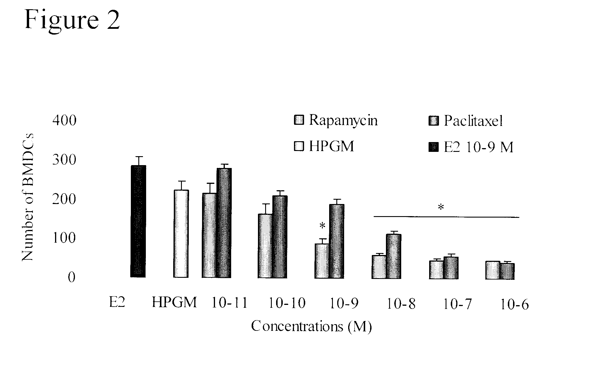
[0086]BMDCs (cultured and isolated as described in Example 1 above) were treated during one week with log scale concentration of each drug (FIG. 2). Cultures without drug were used as negative control and cultures with 10−9 M of E2 were included as reference samples. The IC50 for rapamycin was found to be 10−10 M and with paclitaxel, an IC50 of 5×10⁻⁹ M was found. This value is a balance between the proliferative effect of paclitaxel observed at very low doses and its anti-proliferative effects at higher doses.
PUM
| Property | Measurement | Unit |
|---|---|---|
| Concentration | aaaaa | aaaaa |
| Biological properties | aaaaa | aaaaa |
| Toxicity | aaaaa | aaaaa |
Abstract
Description
Claims
Application Information
 - R&D
- Intellectual Property
- Life Sciences
- Materials
- Tech Scout
- Unparalleled Data Quality
- Higher Quality Content
- 60% Fewer Hallucinations
Browse by: Latest US Patents, China's latest patents, Technical Efficacy Thesaurus, Application Domain, Technology Topic, Popular Technical Reports.
© 2025 PatSnap. All rights reserved.Legal|Privacy policy|Modern Slavery Act Transparency Statement|Sitemap|About US| Contact US: help@patsnap.com


