Microparticle-Based Transfection and Activation of Dendritic Cells
a technology of dendritic cells and microparticles, applied in the direction of genetically modified cells, immunological disorders, antibody medical ingredients, etc., can solve the problems of limited progress toward effective dendritic-cell-based immunotherapy, poor efficiency of vitro transfection of dendritic cells by non-viral methods, and low efficiency of transfection, so as to avoid obvious safety concerns with the use of live viral vectors, rapid internalization and expression
- Summary
- Abstract
- Description
- Claims
- Application Information
AI Technical Summary
Benefits of technology
Problems solved by technology
Method used
Image
Examples
example 1
[0098]Plasmids and DNA formulations. pCMVgag plasmid encoding HIV p55 gag protein under the control of the cytomegalovirus early promoter was purified by ion-exchange chromatography using an Qiagen Endo Free Giga Kit and determined to be endotoxin free (<2.5 EU / ml). For uptake and reporter gene expression experiments, a rhodamine PNA-clamp plasmid encoding B-galactosidase was purchased from Gene Therapy Systems (San Diego, Calif.).
[0099]Cationic microparticles were prepared using a modified solvent evaporation process. The microparticles were prepared by emulsifying 10 ml of a 5% w / v polymer (RG 504 PLG (Boehringer Ingelheim)) solution in methylene chloride with 1 ml of PBS (Phosphate-Buffered Saline) at high speed using an IKA homogenizer. The primary emulsion was then added to 50 ml of distilled water containing cetyl trimethyl ammonium bromide (CTAB) (0.5% w / v). This resulted in the formation of a w / o / w emulsion, which was stirred at 6000 rpm for 12 hours at room temperature, all...
example 2
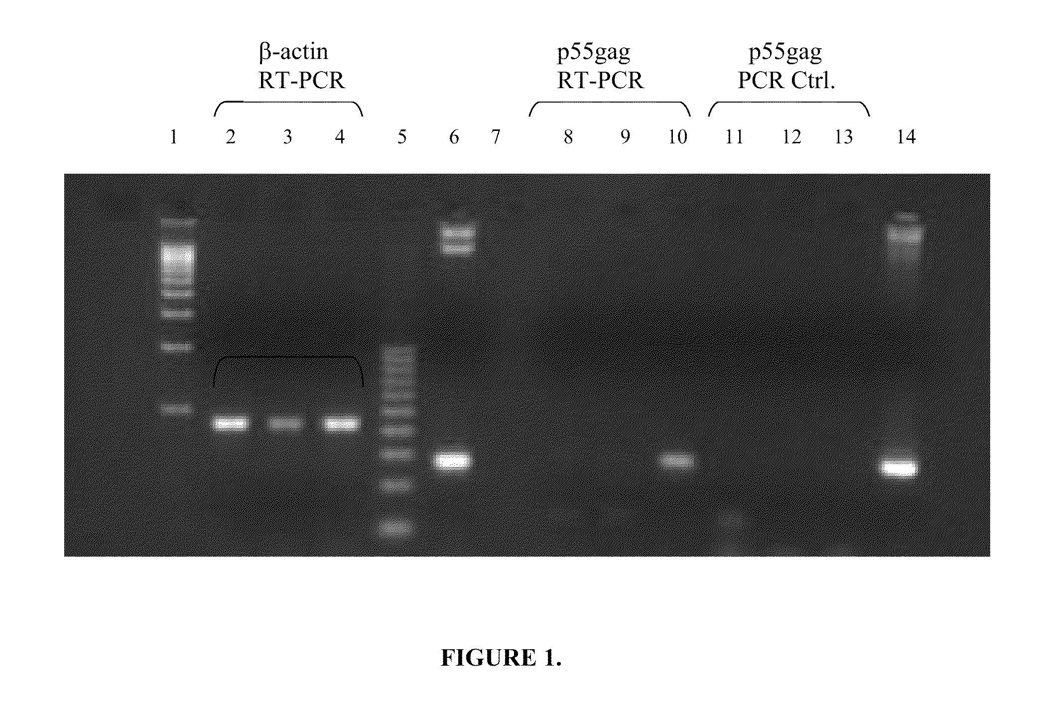
[0106]RNA isolation and RT-PCR. BMDCs were plated at a density of 0.5×10⁶ cells / ml in RPMI+GM-CSF on day 6 of culture. Cells were either left untreated as negative control, or incubated in the presence of 1 μg / ml pCMV-gag DNA either alone (naked) or formulated on PLG-CTAB microspheres. Following 24 h incubation, 2×10⁵ cells were removed, washed 2× in cold PBS (Life Technologies), then lysed per manufacturer's instructions for the mRNA Capture kit (Roche) and frozen at −80° C. Samples were thawed on ice with the addition of RNase-free DNase and RNase inhibitor (Roche). The mRNA isolation protocol was then followed for isolation of biotin-hybridized mRNA in streptavidin PCR tubes. The Promega Reverse Transcription System (Madison, Wis.) was utilized for cDNA synthesis according to manufacturer's instructions, and the reaction was run at 45° C. for 45 min, followed by heat inactivation at 99° C. for 5 min. PCR control tubes were treated as stated above but without the addition of AMV-r...
example 3
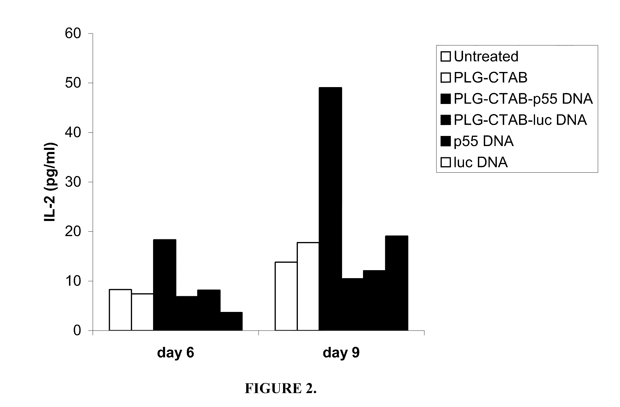
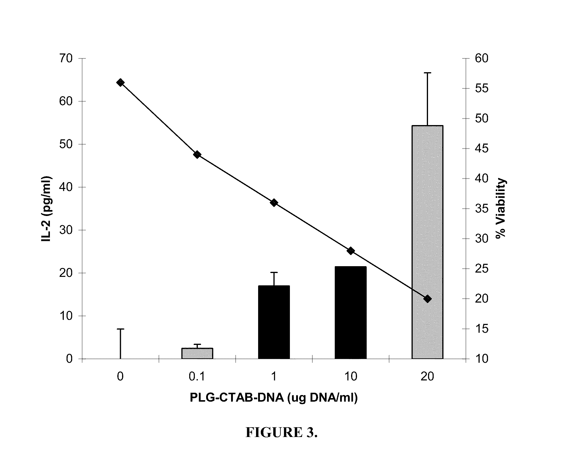
[0108]Stimulation of T cells. Bone marrow cells differentiated in the presence of GM-CSF for 6 days were classified as immature as determined by FACS analysis of cell surface phenotype CD11c+, CD11b+, H-dKd+, I-Ad(low), CD80(low), and CD86(low), and mature by day 9 (CD11c+, CD11b+, H-2Kd+, I-Ad(bright), CD80+, CD86+) (R. C. Fields, J. J. O., J. A. Fuller, E. K. Thomas, P. J. Geraghty, and J. J. Mule'. 1998. Comparative analysis of murine dendritic cells derived from spleen and bone marrow. J. Immunother. 21:323). Both immature and mature BMDCs were stimulated for 24 h with PLG-CTAB-pCMVgag DNA or naked pCMVgag DNA. Controls included untreated cells, microparticles alone or formulated with non-specific plasmid DNA (pCMV-luciferase) as well as non-specific naked DNA. T cell hybridoma 12.2 (a d-restricted T cell hybridoma specific for the p7g epitope (AMQMLKETI) of HIV p55 gag) was plated at 1×10⁵ cells per well of a 96 well, U-bottom microtiter plates. Varying numbers of BMDCs were pl...
PUM
| Property | Measurement | Unit |
|---|---|---|
| diameter | aaaaa | aaaaa |
| diameter | aaaaa | aaaaa |
| diameter | aaaaa | aaaaa |
Abstract
Description
Claims
Application Information
 - R&D
- Intellectual Property
- Life Sciences
- Materials
- Tech Scout
- Unparalleled Data Quality
- Higher Quality Content
- 60% Fewer Hallucinations
Browse by: Latest US Patents, China's latest patents, Technical Efficacy Thesaurus, Application Domain, Technology Topic, Popular Technical Reports.
© 2025 PatSnap. All rights reserved.Legal|Privacy policy|Modern Slavery Act Transparency Statement|Sitemap|About US| Contact US: help@patsnap.com


