Antibiotic dressing for the treatment of infected wounds

a technology for wounds and antibiotics, applied in the field of antimicrobial wound dressings, can solve the problems of inability to demonstrate clinical efficacy of wound dressings loaded with silver ions, the burden of health insurers on the treatment of chronic wounds, and the inability to choose alternative systemic antibiotics, etc., to prolong the retention time of wound dressings and improve the method of wound treatment.

Inactive Publication Date: 2009-12-03
SPINTECH ENG
View PDF2 Cites 25 Cited by
  • Summary
  • Abstract
  • Description
  • Claims
  • Application Information

AI Technical Summary

Benefits of technology

[0010]The object of the present invention is to improve the method for the treatment of wounds. This object is achieved in one aspect of the invention by using a silk protein membrane as the wound dressing for the treatment of infected wounds loaded with HDPs. The silk protein membrane can be kept in place on the infected wound bed for at least four days and for up to six days (or even longer) without the need of changing the wound dressing (silk protein membrane). This enables for the first time a single use wound dressing therapy with integrated antimicrobial therapeutic activity which eliminates the currently recommended daily change of the wound dressing. This is achieved by extending the retention time of a wound dressing from (currently) one day for at least four days and generally to up to six days (although longer times are not excluded). It will be noted that another advantageous aspect of the invention is that the silk protein membrane has pore sizes smaller than 200 nm for protection of the wounds against microbial and viral contamination.

Problems solved by technology

The treatment of the infected chronic wounds represents an enormous burden to health insurers.
Unfortunately, the choice of alternatives to the systemic antibiotics is limited.
In addition, wound dressings loaded with silver ions have failed to demonstrate clinical efficacy (Vermeulen et al., Cochrane Database of Systematic Reviews 2007).
However, despite their excellent activity and broad spectrum, rapid clearance of the HDPs and unfavourable pharmacokinetics, due to proteolytic degradation, has severely restricted their applicability as drugs.
However, the authors only demonstrated bacterial killing in a radial diffusion assay and do not teach or demonstrate a method of treatment of infected wounds.
A disadvantage when using non-native, regenerated silk fibroin as feedstock for casting silk protein membranes like those reported by Saido-Sakanaka et al. is the presence of a distinct granular or globular morphology (so called ultrastructure) when analysed by SEM (scanning electron microscopy).
Because of the granular ultrastructure of regenerated silk protein membranes, it has not been possible yet to manufacture mechanically strong, regenerated silk protein membrane.

Method used


Image

  • Antibiotic dressing for the treatment of infected wounds
  • Antibiotic dressing for the treatment of infected wounds
  • Antibiotic dressing for the treatment of infected wounds

Examples


example 1

Resistance to Proteolysis of Silk Protein Membranes in Wound Fluid

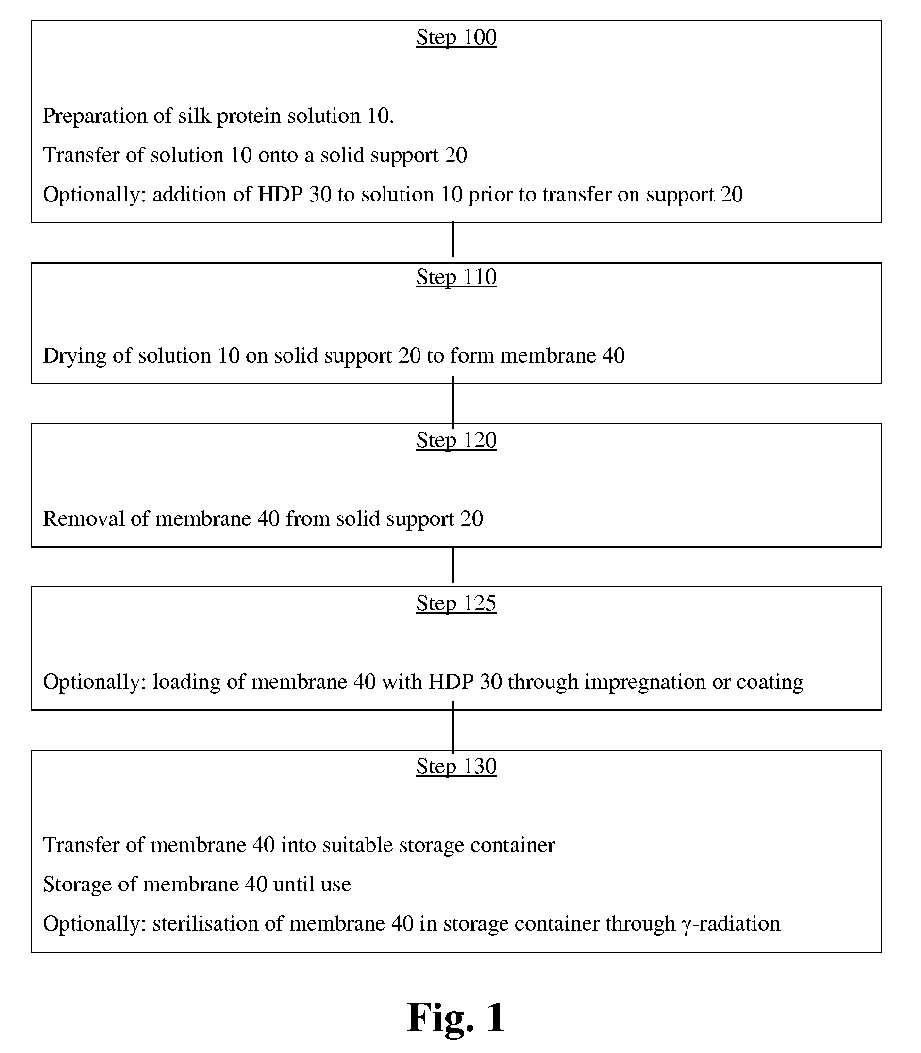
[0035]Silk protein membranes 40 as silk fibroin membranes were made by transferring the protein solution 10 into the solid support 20. The solid support 20 was a casting form made from polytetrafluoroethylene of size 250×110×0.7 mm. The protein solution 10 was produced with the apparatus described in the international application PCT / EP2007 / 001775. After filling with the protein solution 10, the casting form 20 was left to dry over night at room temperature to yield the silk protein membranes 40 of 80 μm thickness. Without further physical treatment (e.g. heat, mechanical stress) or chemical treatment (e.g. protein denaturing agents, alcohols, cross-linking agents), the silk protein membranes 40 were then cut into rectangular samples (of size 10×3 mm) and transferred individually into 1.5 ml sample tubes. 400 μl of freshly harvested undiluted wound exudates from pig and human wounds were added to each ones of the samp...

example 2

Ultrastructural Analysis of Silk Protein Membranes by SEM

[0036]Cross-sections of the silk protein membranes, which were prepared according to the method described in Example 1, were analysed by SEM at high resolution. FIG. 9 (scale bar 2 μm) demonstrates a homogenous SEM ultrastructure of the silk fibroin membrane without detectable pores and without a detectable granular or micellar-like morphology.

example 3

Controlled Release of Colistin Out of Silk Protein Membranes

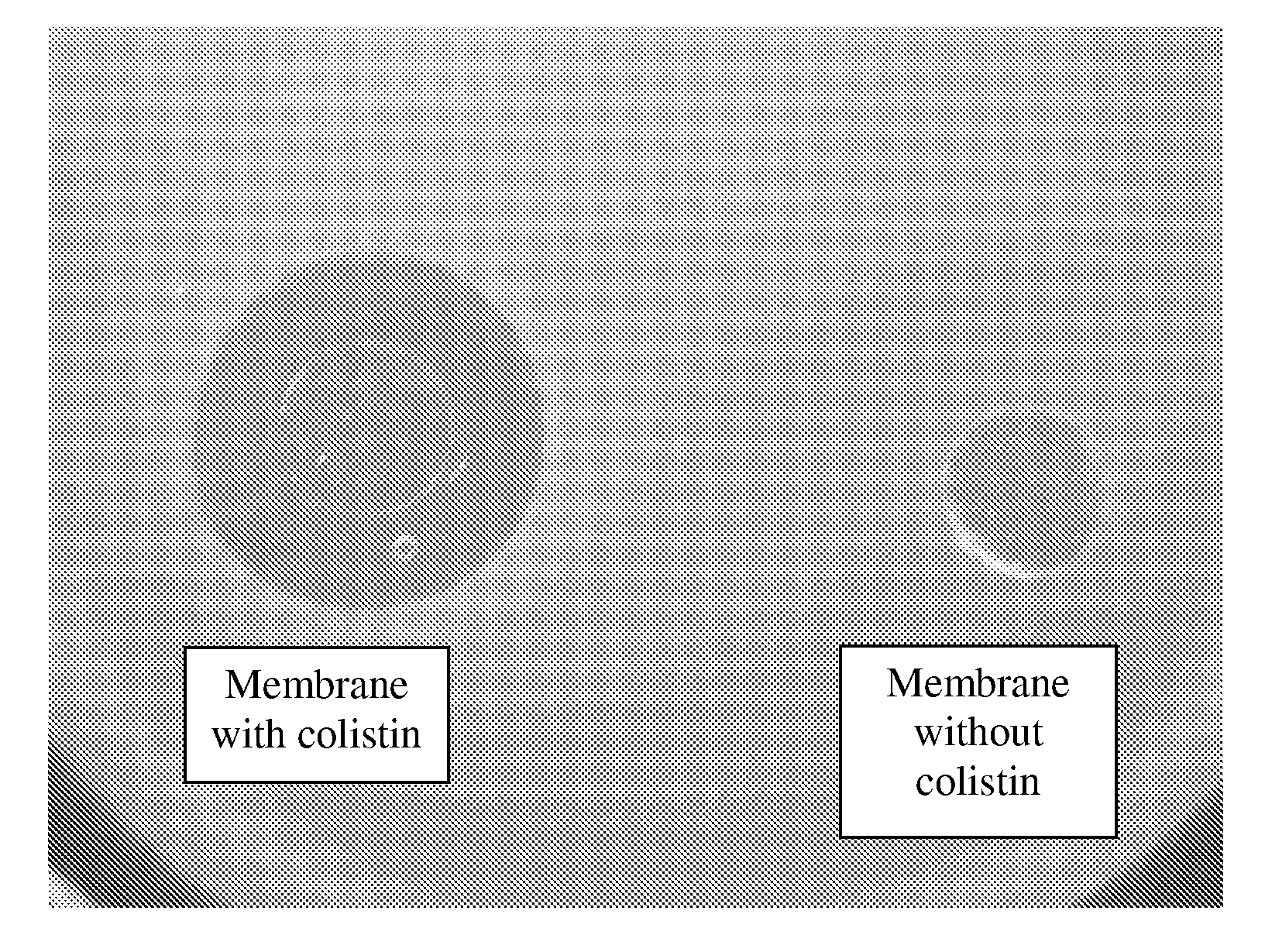
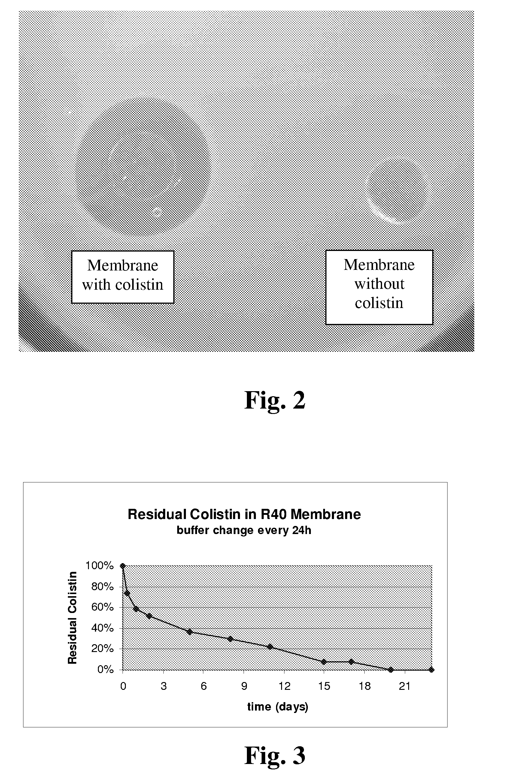
[0037]Round membrane samples 50 with a diameter of 6 mm were stamped out of the silk protein membranes 40 which had been prepared according to Example 1. The round membrane samples 50 were loaded with colistin sulphate (supplied by Carl Roth) through incubation in a colistin solution (10 mg / ml) for 18 hours at room temperature. The incubated round samples 50 were then kept individually in 2 ml solution at room temperature for up to 23 days. Each solution was refreshed every 24 hours in order to simulate wash-out. At defined time points (8 hours and 1, 2, 5, 8, 11, 15, 17, 20, 23 days), the round membrane samples 50 were retrieved, dried and analysed through radial diffusion assay.

[0038]This radial diffusion assay was performed by transferring each one of the round membrane samples 50 onto top-agar plates made by dissolving 32 g LB-Agar Lennox (from Carl Roth) in 400 ml water and adding log-phase E. coli BL21-T1 cells (from ...


PUM

PropertyMeasurementUnit
Lengthaaaaaaaaaa
Timeaaaaaaaaaa
Timeaaaaaaaaaa

Abstract

A silk protein membrane is described which is loaded with an antimicrobial compound and has a substantially non-granular ultrastructure which is (i) substantially devoid of micellar silk fibroin substructures and (ii) substantially devoid of pores when analysed by scanning electron microscopy at 0.2 μm resolution. The antimicrobial compound comprises, in one aspect of the invention, a host defense peptide. The silk protein membrane of the invention can be used in a method for the treatment of wounds and allows the wound dressing to be kept in place after removal of that wound dressing from a wound the wound has less than 105 colony forming units per gram. A method for manufacturing a wound dressing is also disclosed which comprises transferring a cast precursor material and optionally a host defense peptide, into a solid support and then drying the precursor material on the solid support to form a silk protein membrane for use as the wound dressing.

Description

FIELD OF THE INVENTION[0001]This invention relates to the use of an antimicrobial wound dressing for effective control of wound infections and to a method for the treatment of wound infectionsBACKGROUND OF THE INVENTION[0002]Understanding and managing microbial contamination and colonisation (bioburden) with appropriate treatments is required for the successful healing of chronic wounds (Attinger et al., Plast. Reconstr. Surg. 2006, 177 (Suppl.) 72S). Normally, a chronic wound is in balance with the natural microflora of the skin and mucosal surfaces. However, when the bioburden exceeds a level of greater than 105 bacteria per gram of tissue which can give rise to colony forming units (cfu's) (Robson M C, Surg Clin North Am 1997, 77: 637-650), the chronic wound is considered infected and requires antibiotic treatment. The treatment of the infected chronic wounds represents an enormous burden to health insurers. For example, about 6 million diabetic patients are estimated worldwide t...

Claims


Application Information

Patent Timeline
IPC IPC(8): A61L15/16A61K38/00A61P19/00
CPCA61L15/32A61L15/44A61L2300/602A61L2300/406A61L2300/25A61P19/00
Inventor RHEINNECKER, MICHAELZIMMAT, ROLFSTEINSTRAESSER, LARS
Owner SPINTECH ENG
Why Eureka
Try Eureka