
Inhibition of microtubule protrusion in cancer cells

a cancer cell and microtubule technology, applied in the field of cell biology, molecular biology, cancer, etc., can solve the problems of resistance to apoptosis cell death not necessarily preventing early stress response, specific role of microtubules in this process, and currently not well-characterized, so as to reduce microtentacles and increase crosslinking in the actin cortex

Active Publication Date: 2009-05-28
UNIV OF MARYLAND
View PDF5 Cites 19 Cited by
  • Summary
  • Abstract
  • Description
  • Claims
  • Application Information

AI Technical Summary

Benefits of technology

[0011]It is described herein that mammary epithelial cell lines generate long and dynamic microtubule-driven protrusions of the plasma membrane after detachment. We also determine that detachment produces rapid detyrosination of α-tubulin, and the concentration of detyrosinated tubulin in protrusions. Full length α-tubulin contains a tyrosine residue at its c-terminus, and is termed Tyr-tubulin. Cleavage of this c-terminal tyrosine by a tubulin carboxypeptidase exposes a glutamic acid residue, yielding a detyrosinated form (Glu-tubulin) (Argarana, Arce et al. 1977; Argarana, Barra et al. 1978). Although this α-tubulin modification has been appreciated for thirty years, the identity of the gene encoding tubulin carboxypeptidase remains unknown. Glu-tubulin is post-translationally converted back to Tyr-tubulin, by a tubulin tyrosine ligase (TTL), which has recently been cloned in mice and humans (Erck, MacLeod et al. 2003). While microtubules containing Tyr-tubulin have a relatively short half-life, measured in minutes, Glu-tubulin is enriched in a more stable subset of microtubules (Webster, Gundersen et al. 1987). Microtubules containing Glu-tubulin can persist for hours and have been observed to remain for as long as 16 hours in nondividing cells (Webster, Gundersen et al. 1987). In breast tumor samples, increased levels of Glu-tubulin are associated with poor patient prognosis and an increased risk of cancer-related complications, but the mechanism for this effect is still unknown (Mialhe, Lafanechere et al. 2001).
[0034]In an additional embodiment of the invention, there is a method of reducing the risk of a tumor cell from a first organ in an individual to establish a malignancy in a second organ in the individual, comprising delivering a therapeutically effective amount of an agent that targets one or more microtubule protrusions of the tumor cell. In a specific embodiment, the tumor cell releases from the tumor upon therapy for the tumor, such as upon surgery in the individual, for example. Releasing of the tumor cell from a primary tumor mass may be caused directly or indirectly upon utilizing the therapy. In further embodiments, the method may be further defined as the tumor cell releasing from the tumor upon excision of the tumor from the individual. In specific aspects, the tumor cell is released from a primary tumor mass during excision of at least part of the primary tumor mass from the individual. Therefore, in particular aspects this clarifies the difference between the primary tumor mass and the residual tumor cells left in the body (which would be at risk of spreading metastatically to distant tissues during wound healing). In particular cases, the method is further defined as the tumor cell releasing from the primary tumor mass during removal of the primary tumor mass from the individual and / or during therapy for the primary tumor mass for the individual. In other cases, the method is further defined as the tumor cell being one that remains in the body of the individual during removal of a primary tumor mass from the individual and / or during therapy for the primary tumor mass for the individual. In additional embodiments, the agent is delivered to the individual before, during, and / or after the surgery.

Problems solved by technology

However, any specific role for microtubules in this process remains unclear.
Bloodborne tumor cells therefore attach to capillary vessel walls via a cytoskeletal mechanism that is distinct from that of adherent cells, and is currently not well-characterized.
However, evidence is accumulating that resistance to apoptotic cell death does not necessarily prevent early stress responses from occurring in cells.

Method used


Image

  • Inhibition of microtubule protrusion in cancer cells
  • Inhibition of microtubule protrusion in cancer cells
  • Inhibition of microtubule protrusion in cancer cells

Examples


example 1

Exemplary Materials and Methods

[0223]This example provides exemplary materials and methods for use in the invention, although the skilled artisan is aware of other materials and methods that are sufficiently suitable.

Exemplary Cell Lines and Materials

[0224]MCF10A human mammary epithelial cells were kindly provided by Fred Miller and Robert Pauley of the Barbara Ann Karmanos Cancer Institute (Detroit. Mich.) and are a high-passage clone designated MCF10A1. MCF10A cells were grown in DMEM / F12 (Gibco) supplemented with 5% horse serum, insulin (5 μg / ml), EGF (20 ng / ml), hydrocortisone (500 ng / ml), penicillin-streptomycin (100 μg / ml each), and L-glutamine (2 mmol / L). EpH4 mouse mammary epithelial cells and those stably expressing pcDNA3.1-Bcl2 (B19) were previously described (Pinkas et al., 2004; Lopez-Barahona et al., 1995), and maintained in DMEM (Gibco) supplemented with 10% bovine calf serum, penicillin-streptomycin (100 μg / ml each), and L-glutamine (2 mmol / L). For all inhibitor and ...

example 2

Characterization of Microtubule Protrusions and Inhibition Thereof

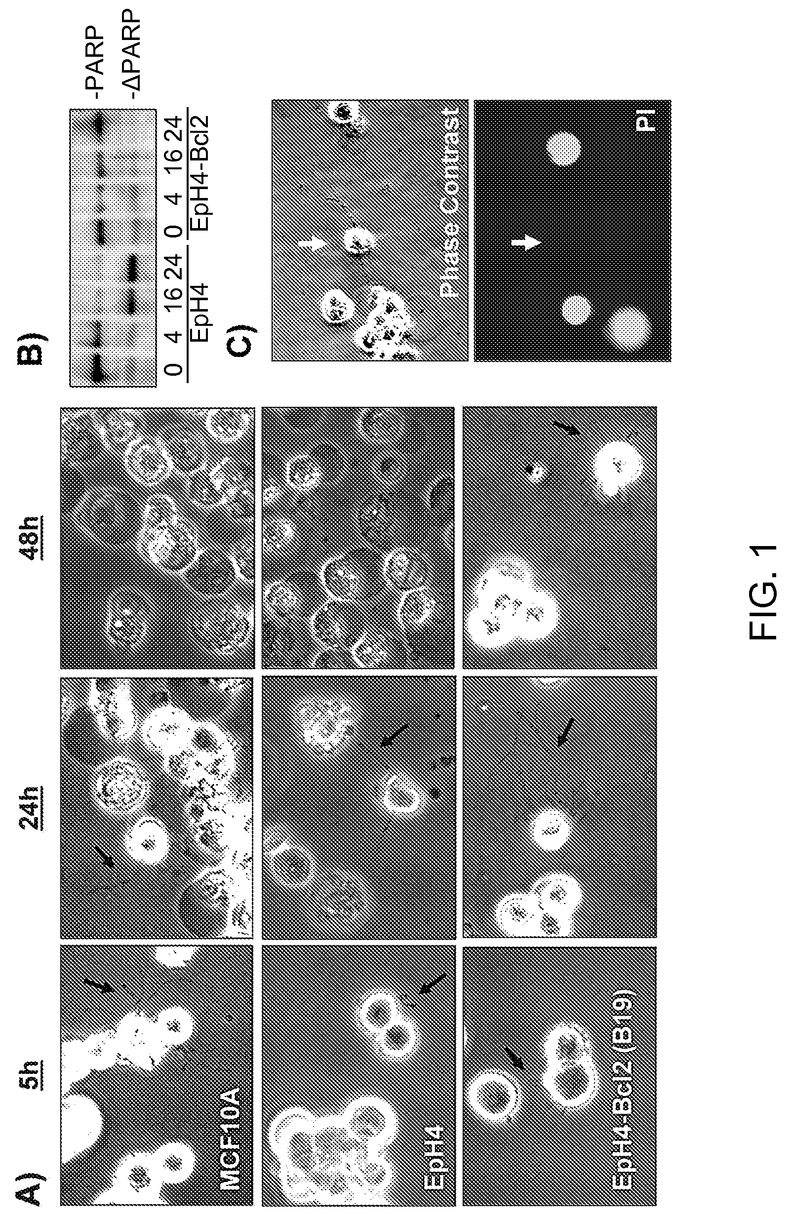
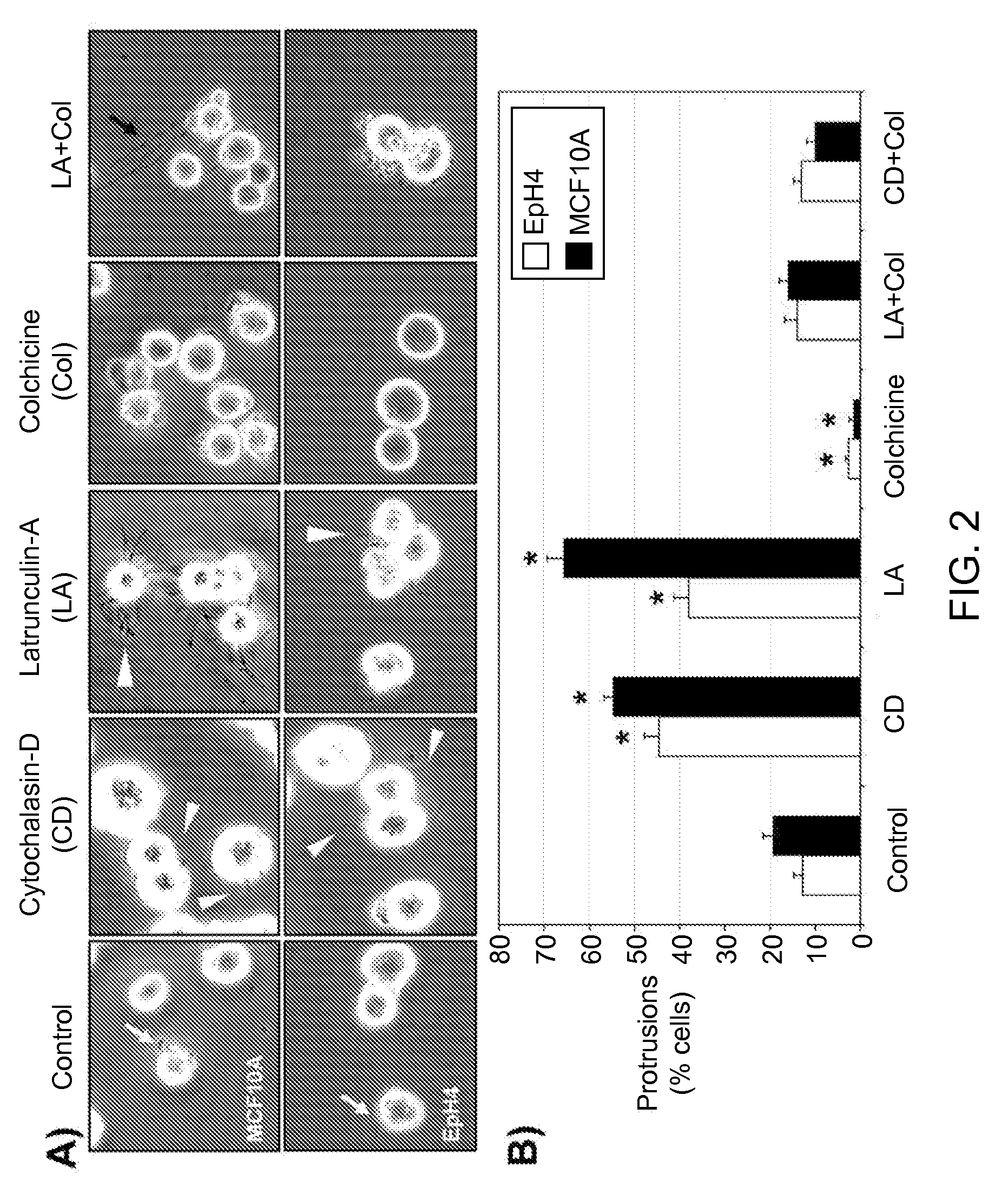
[0230]When detached from extracellular matrix, the nontumorigenic MCF10A human mammary epithelial cell line and EpH4 mouse mammary epithelial cell line produce protrusions of the plasma membrane (FIG. 1A, black arrows). Time-lapse video microscopy detects rapid motion in these protrusions and transient probing contact with surfaces. This response is short-lived, as EpH4 or MCF10A cells die efficiently by apoptosis within 24 hours following detachment (Pinkas et al., 2004; Martin and Leder, 2001), and complete cellular fragmentation by 48 hrs. terminates these protrusions (FIG. 1A). However, when EpH4 cells were used that overexpress Bcl-2 (B 19) and are highly resistant to apoptotic challenge (Pinkas et al., 2004), many of the cells remain intact after 48 hours and continue to generate protrusions (FIG. 1A). Similar resistance and protrusions are observed in EpH4 or MCF10A cells overexpressing either Bcl-2 or Bcl-xL (...

example 3

Protrusions are Increased in Metastatic Breast Tumor Cell Lines

[0235]FIG. 7 shows that metastatic breast tumor cell lines have more frequent and numerous protrusions. The nontumorigenic human mammary epithelial cell line (MCF10A) or the indicated human breast tumor cell lines were grown in suspension for 30-60 minutes, and protrusions were counted in random fields with phase-contrast microscopy. Values represent the mean±S.D. of three experiments in which at least 100 cells were counted. Treatment of MCF10A cells with the exemplary actin inhibitor, Latrunculin-A (LA, 5 mM), increases protrusions, as observed previously. Human tumor cell lines show high levels of protrusions even in the absence of LA treatment, indicating that depolymerization of actin is not required for tumor lines to produce microtubule protrusions. In addition to more frequent protrusions, metastatic tumor cell lines often displayed a greater number of protrusions per cell. This effect was most apparent in the hi...


PUM

PropertyMeasurementUnit
Massaaaaaaaaaa

Abstract

The present invention generally concerns microtubule protrusions in cancer cells, including detached cancer cells, and inhibition of the protrusions. In particular aspects, the inhibition of the protrusions interferes with attachment of the cell to a vessel wall, and in further aspects the cell is killed by forcing it to enter capillaries and be destroyed, for example by shearing. Inhibition by a variety of agents and methods is contemplated.

Description

[0001]The present invention claims priority to U.S. Provisional Patent Application Ser. No. 60 / 780,468, filed Mar. 8, 2006, which is incorporated by reference herein in its entirety.STATEMENT REGARDING FEDERALLY SPONSORED RESEARCH OR DEVELOPMENT[0002]The present invention was developed at least in part with funds from the National Cancer Institute Howard Temin Career Award (K01-CA096555-01A1) and from the Department of Defense, Breast Cancer Concept Award (BC045699). The United States Government may have rights in the invention.FIELD OF THE INVENTION[0003]The present invention generally concerns at least the fields of cell biology, molecular biology, cancer, and medicine. In particular aspects, the present invention concerns the field of cancer therapy and / or prevention.BACKGROUND OF THE INVENTION[0004]Breast tumor cells can disseminate prior to significant primary tumor growth and remain dormant in distant tissues for extended periods of time (Naumov, MacDonald et al. 2001; Naumov,...

Claims


Application Information

Patent Timeline
IPC IPC(8): A61K38/17A61K31/7105A61K31/675A61P35/00
CPCC07K14/47A61P35/00
Inventor MARTIN, STUARTWHIPPLE-BETTES, REBECCA
Owner UNIV OF MARYLAND
Learn More