
Methods for determining the redox status of proteins

a protein and redox technology, applied in the field of determining the redox status of proteins, can solve the problems of poor understanding of the complexity of the system, detriment to cell viability, lack of knowledge, etc., and achieve the effect of further reducing the reliability of other proteomic techniques

Active Publication Date: 2008-12-11
TWO TAG HOLDINGS PTY LTD
View PDF0 Cites 7 Cited by
  • Summary
  • Abstract
  • Description
  • Claims
  • Application Information

AI Technical Summary

Benefits of technology

"The present invention provides a method for determining the redox status of a protein sample by measuring the levels of reduced and oxidised cysteine groups. This method can be used to determine the redox status of new proteins or proteins that have been modified from their naturally occurring redox status. The invention also provides a database of redox values for protein samples. The method can be performed using a kit and can be used to assess ROS-related pathologies and the efficacy of therapeutic interventions."

Problems solved by technology

ROS are often though of as being directly detrimental to cell viability because they can irreversibly damage key macromolecules such as proteins, nucleic acids and lipids. however there is now evidence that low levels of at least one ROS, hydrogen peroxide (H2O2) has been implicated in the regulations of signal transduction pathways linked to the control of cell proliferation, cell growth and cell death.
However, despite great interest, only in a few cases has the biological significance of these modifications been identified.
This is in part due to a poor understanding of the complexity of the system, a lack of knowledge of the relationship between the thiol redox system and other antioxidant systems and the difficulty in identifying specific biological effects.
For example, it is difficult to establish that specific thiol group modifications exist in vivo and are not simply a manifestation of the unnatural oxidising conditions of in vitro systems.
Furthermore, the current methods for determining the redox status of biological systems e.g. proteins, cells, tissues in vivo (and even in vitro) lack sufficient sensitivity, reproducibility or specificity and do not allow the detailed investigation of the effects of changes in redox status on cell function, viability and survival.
A more sensitive assessment of thiol redox changes comes from measuring the oxidised disulfides, but this increases the complexity of the method.
Furthermore, such methods of measuring oxidised disulfides are not particularly precise and are technically demanding and time consuming.
Furthermore, the GSSG / GSH analytical techniques are not suitable for the analysis of mitochondria, cells or tissues where only limited sample is available.
The relatively poor precision of protein assays reduces the precision in assessing thiol redox changes.
However, this method does not allow the concurrent measurement of both the reduced and oxidised cysteine residues of a protein.
The lack of precision of this approach is further accentuated by variations inherent with PAGE and other systems for visualising the results of the methods.
This greatly reduces the practical utility of these methods where only one signal is measured at a time.

Method used


Image

  • Methods for determining the redox status of proteins
  • Methods for determining the redox status of proteins
  • Methods for determining the redox status of proteins

Examples


example 1

Detecting Protein Modifications in a Biological Sample

Materials / Methods

(i) Labels

[0114]Fluorescent Label 1 (F1) was BODIPY TMR cadaverine-IA (TMR, excitation 545 nm; emission 571 nm) and Label 2 (F2) was BODIPY FL C1-IA (FL, excitation 503 nm; emission 512 nm) (Molecular Probes). Each fluorescent label contains an iodoacetamide group covalently linked to a fluorescent BODIPY moiety. A carboxamidomethylation reaction of the iodoacetamide group to the free thiol groups of cysteine residues was carried out at pH 7.5 followed by covalent linkage.

(ii) Labelling

[0115]Jurkat cells+ / −2 mM H2O2 for 5 min in HBS buffer were extracted with RQB buffer (20% TCA in acetone, to trap thiol redox state) by resuspending the cells, using sonication (1×10 sec burst) and incubating the samples at −20° C. for 2 h. The RQB was removed by washing the protein pellet with ice cold acetone and protein samples were resuspended in MSS buffer (8M urea, 10 mM EDTA, 50 mM HEPES, 4% CHAPS and 1% pH3-10 IPG buffer a...

example 2

Analysis of Protein Modification in a Standard Protein Mixture

[0122]A set of standard proteins with known amounts of natural reduced and oxidised cysteine residues were analysed using an embodiment of the present invention.

Materials / Methods

[0123]A mixture of 25 μg each of BSA, AD, CA, OV, PEP, CytC, Cat, SOD and Lyz, were initially labelled with 1 mM label F1 for 2 h. F1 was removed and then the proteins were reduced using 5 mM TCEP and labelled with the 1 mM label F2 (in the presence of 5 mM TCEP).

[0124]Table 2 sets out the thiol properties of these proteins.

TABLE 2FWTotalProteinAbbrev(kDa)S—HS—SS—HpIcytochrome cCytC11.60128.5-10 (bovine)lysozymeLyz14.304811.4(chick egg white)superoxide dismutaseSOD15.51135.4-6.4(bovine)carbonic anhydraseCA29.00006-7(bovine)alcohol dehydrogenaseAD36.78085.4-5.8(yeast)pepsinPEP41.41373(ovine)ovalbuminOV42.84165(chick)catalaseCat57.64045.4(bovine)albuminBSA69.3117355.3(bovine serum)

Results

[0125]ProXpress fluorescence scans of an SDS-PAGE show that th...

example 3

Identification of Proteins Undergoing Redox Changes Following a Change in the Oxidising Environment

Materials / Methods

[0126]The fluorescent dyes, BODIPY® FL N-(2-aminoethyl)maleimide (FLm, Tag 1) and BODIPY® TMR C5-maleimide (TMRm, Tag 2) were used in a method to identify proteins undergoing thiol redox changes following a change in the oxidising environment.

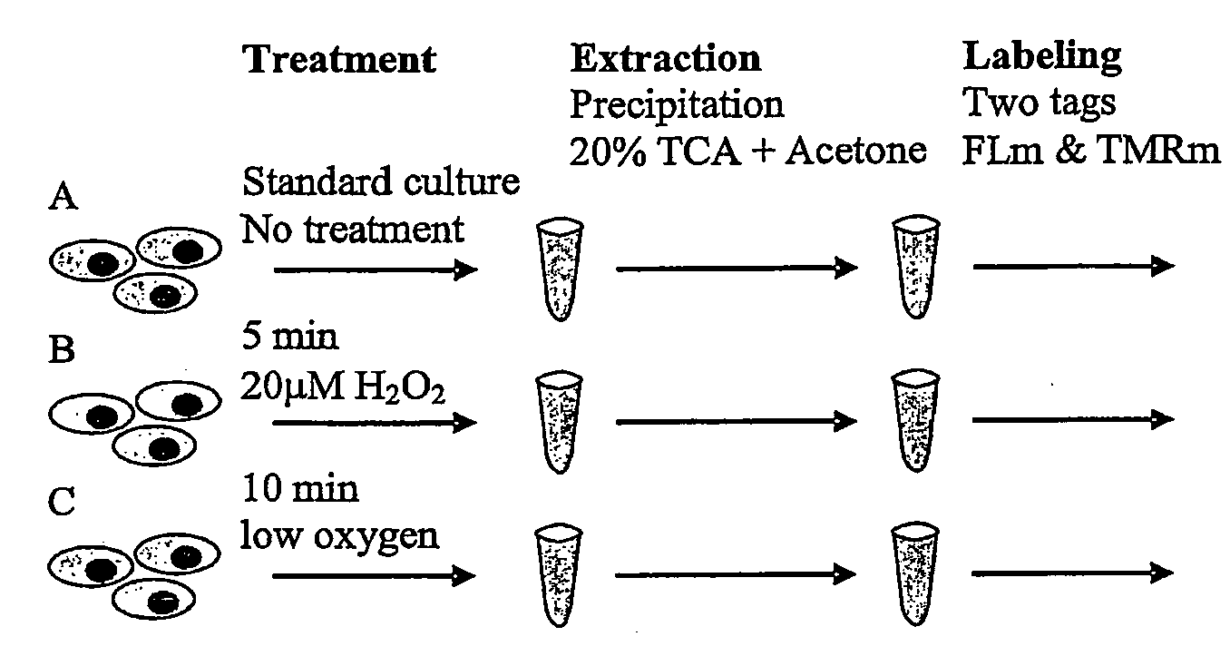
[0127]Cells were divided into three groups and treated as follows:[0128]A. standard culture conditions[0129]B. exposed to oxidising conditions (20 μM H2O2 for 5 minutes); and[0130]C. exposed to a reduced oxygen concentration (10-20 μM) relative to standard culture conditions (where oxygen concentration is 203 μM) for 10 minutes.

[0131]The protocol, schematically reproduced in FIG. 3, was repeated over four separate experiments: With respect to the different treatments, three comparisons are possible: standard conditions to low oxygen (protocol A to protocol C); oxidising treatment to standard conditions (protocol B to protocol A); ...


PUM

PropertyMeasurementUnit
pHaaaaaaaaaa
pack heightaaaaaaaaaa
volumeaaaaaaaaaa

Abstract

A method for determining the redox status of a protein sample, the method comprising the steps of: a) contacting the sample with a first label adapted to bind to at least one reduced cysteine group therein; b) contacting the sample with a reducing agent to reduce at least one oxidized cysteine group therein; c) contacting the sample with a second label adapted to bind to any reduced cysteine groups produced in step (b); and d) determining the ratio of the signal from the first label to the signal from the second label wherein the ratio indicates the redox status.

Description

RELATED APPLICATIONS[0001]This is a continuation patent application that claims priority to PCT patent application number PCT / AU2006 / 001757, filed on Nov. 21, 2006, which claims priority to Australian patent application number. 2005906469, filed on Nov. 22, 2005, the entirety of which are herein incorporated by reference.FIELD OF THE INVENTION[0002]The present invention relates to a method for determining the redox status of a protein. The invention also relates to a method for detecting protein modification, particularly modification caused by reactive oxygen specifies (ROS) and to kits and other uses of the methods described herein.BACKGROUND ART[0003]The term reactive oxygen species (ROS) is a generalized description for a collection of reactive oxygen molecules of biological significance. These include: superoxide (O2), hydroxyl radical (OH.), peroxyl radical (ROO), alkoxyl radical (RO), hydroperoxyl radical (HOO), hypochlorous acid (HOCl), hydrogen peroxide (H2O2), ozone (O3), ...

Claims


Application Information

Patent Timeline
Patent Type & Authority Applications(United States)
IPC IPC(8): G01N33/53G01N33/00C12Q1/02
CPCG01N33/58G01N33/68G01N33/6803G01N33/6815G01N33/6848
Inventor LIPSCOMBE, RICHARD J.ARTHUR, PETER G.LUI, JAMES K.
Owner TWO TAG HOLDINGS PTY LTD
Learn More