
Vascularized human skin equivalent

a human skin and equivalent technology, applied in the field of tissue grafting, can solve the problems of poor endothelial cell survival, hampered current efforts, and moderate success of clinical use of engineered skin for burn treatment, and achieve the effect of accelerating the rate of vascularization and enhancing clinical utility

Inactive Publication Date: 2007-09-06
YALE UNIV
View PDF0 Cites 20 Cited by
  • Summary
  • Abstract
  • Description
  • Claims
  • Application Information

AI Technical Summary

Benefits of technology

[0011] This invention solves the above needs known in the art and provides grafts which can be transplanted, subcutaneously or orthotopically, and which aid in the treatment of wounds due to burns, trauma, surgical incisions, non-healing ulcers and blistering diseases. The invention also provides methods of making the grafts and methods of using the grafts. The grafts of the invention are comprised of one or more of human keratinocytes, human autologous epithelial cells, and HUVECs. The HUVECs, autologous umbilical cord blood cells and / or adult peripheral blood cells, are optionally transduced with Bcl-2. The autologous skin grafts of the invention represent a major improvement over skin grafts currently used in the art due to their accelerated rate of vascularization, thus resulting in enhanced clinical utility.

Problems solved by technology

The clinical use of engineered skin for treatment of burns has met with moderate success, but is limited by the lack of vascularization and consequent sloughing of the synthetic dermal and epidermal layers.
Stimulation of angiogenesis by the introduction of endothelial cells into these compromised tissues shows promise but current efforts are hampered by poor endothelial cell survival, and a lack of maturation of the primitive vascular tubes they form.
Reports of models useful for discerning the mechanisms of incorporation of mesenchymal cells into mature vessels have been limited.
Major limitations in the construction of human synthetic microvessels include the apoptotic response of cultured human endothelial cells before or after the formation of immature tubes (Ilan et al.
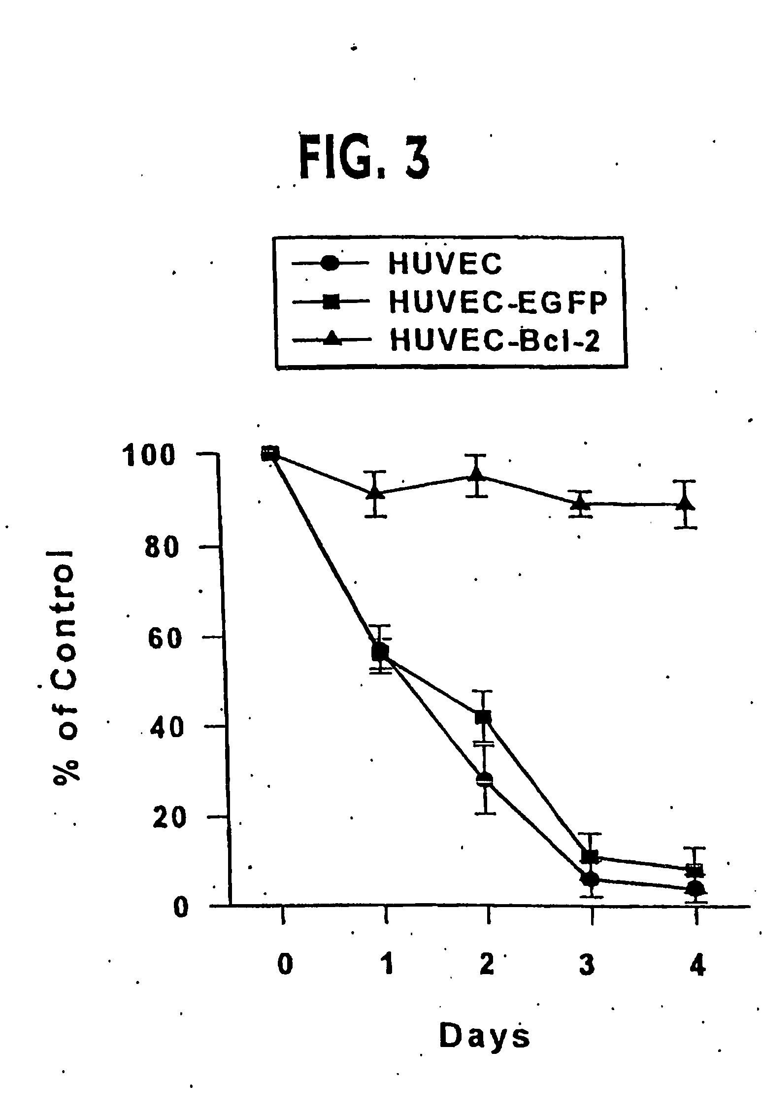
Furthermore, overexpression of caspase-resistant Bcl-2 in these cells results in the formation perfused vascular structures invested by mouse pericyte / smooth muscle cells, that remodel into mature vessels.

Method used


Image

  • Vascularized human skin equivalent
  • Vascularized human skin equivalent
  • Vascularized human skin equivalent

Examples


example 1

Isolation and Culture of HUVEC Cells

[0243] HUVEC were isolated by collagenase treatment of human umbilical veins as previously described (Gimbrone (1976) Prog. Hemostasis Thromb. 3, 1-6) and cultured on 0.2% gelatin-coated plastic in Medium 199 with 20% FCS, 50 μg / ml endothelial cell growth supplement (ECGS) (Collaborative Research / Becton Dickinson), 100 μg / ml heparin (Sigma), 2 mM L-glutamine, 100 U / ml penicillin, and 100 μg / ml streptomycin. All of the EC used in these experiments were at passage levels 1 through 6. Such cultures are homogeneous for EC markers (von Willebrand factor, CD31, inducible E-selectin) and are free of contaminating CD45+ leukocytes.

example 2

Construction of the Retroviral Vector Expressing Caspase Resistant Bcl-2

[0244] The D34A caspase-resistant form of Bcl-2 DNA (SEQ ID NO: 1) in the pSG5 expression Vector has been described (Cheng et al. (1997) Science 278, 1966-1968). The 800 bp cDNA insert was isolated by PCR and subcloned into the pCRII vector. DNA sequence of the insert of subclone #10 indicated the following terminal sequences:

(SEQ ID NO: 3)5′-AATTCGGATCACGGTCA CCATGGCGCACGCT(SEQ ID NO: 4). . . CTGAGCCACAAGTGAGTCGACCTCGAGGAATTC-3′.

EcoRI sites (GAATTC and the translation start (ATG) and stop (TGA) codons are underlined. The EcoRI excisable DNA insert was subcloned into the LZRSpBMN-Z retroviral vector. This retroviral vector DNA containing the caspase resistant form of Bcl-2 DNA was directly transfected into the Phoenix-Ampho packaging cell line by lipofection and puromycin-resistant cells were derived which served as the source of retroviral stocks.

[0245] To generate a control retroviral vector, Enhanced Gr...

example 3

Stable Transduction of Caspase-Resistant Bcl-2 or Control DNA

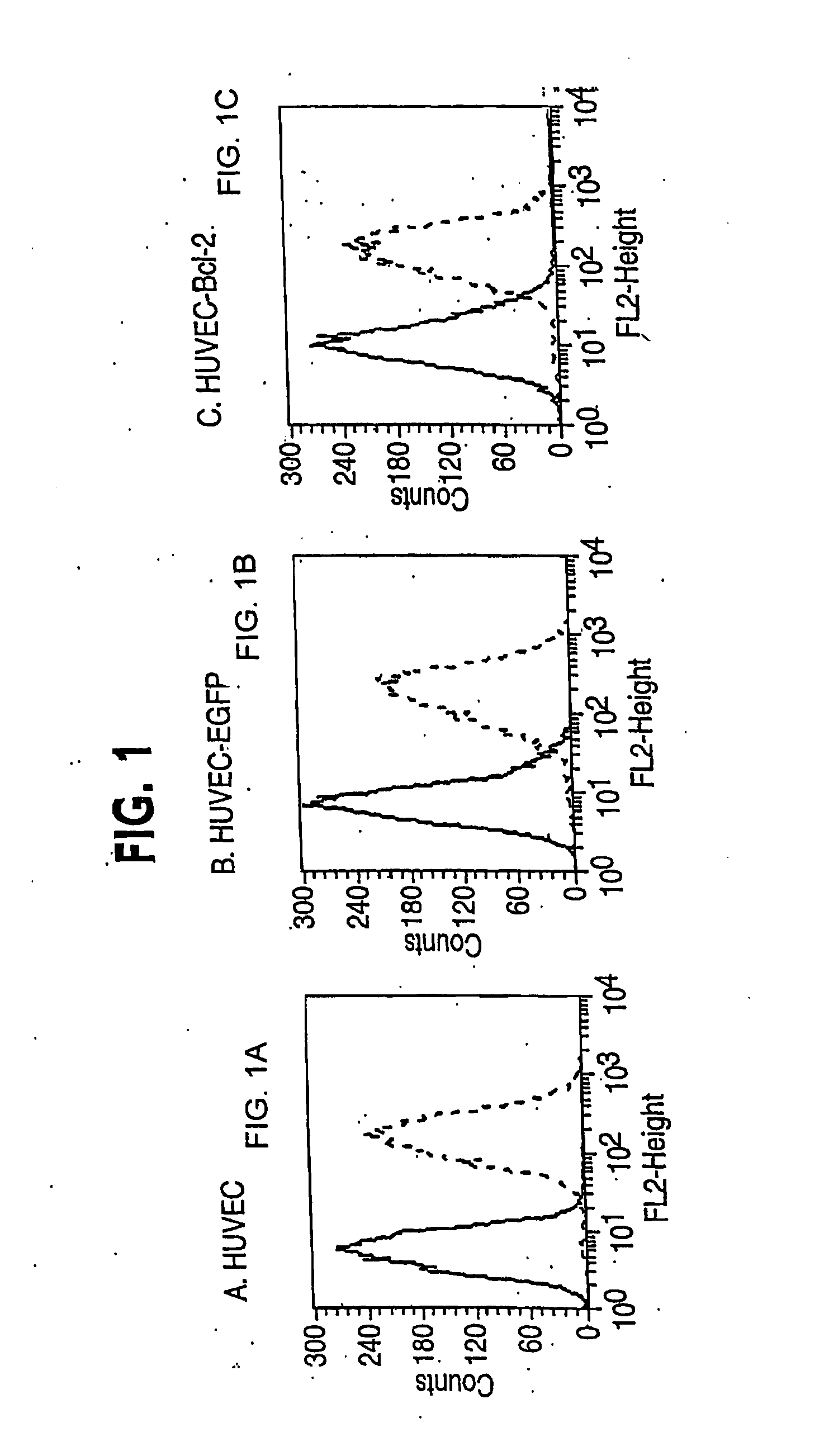
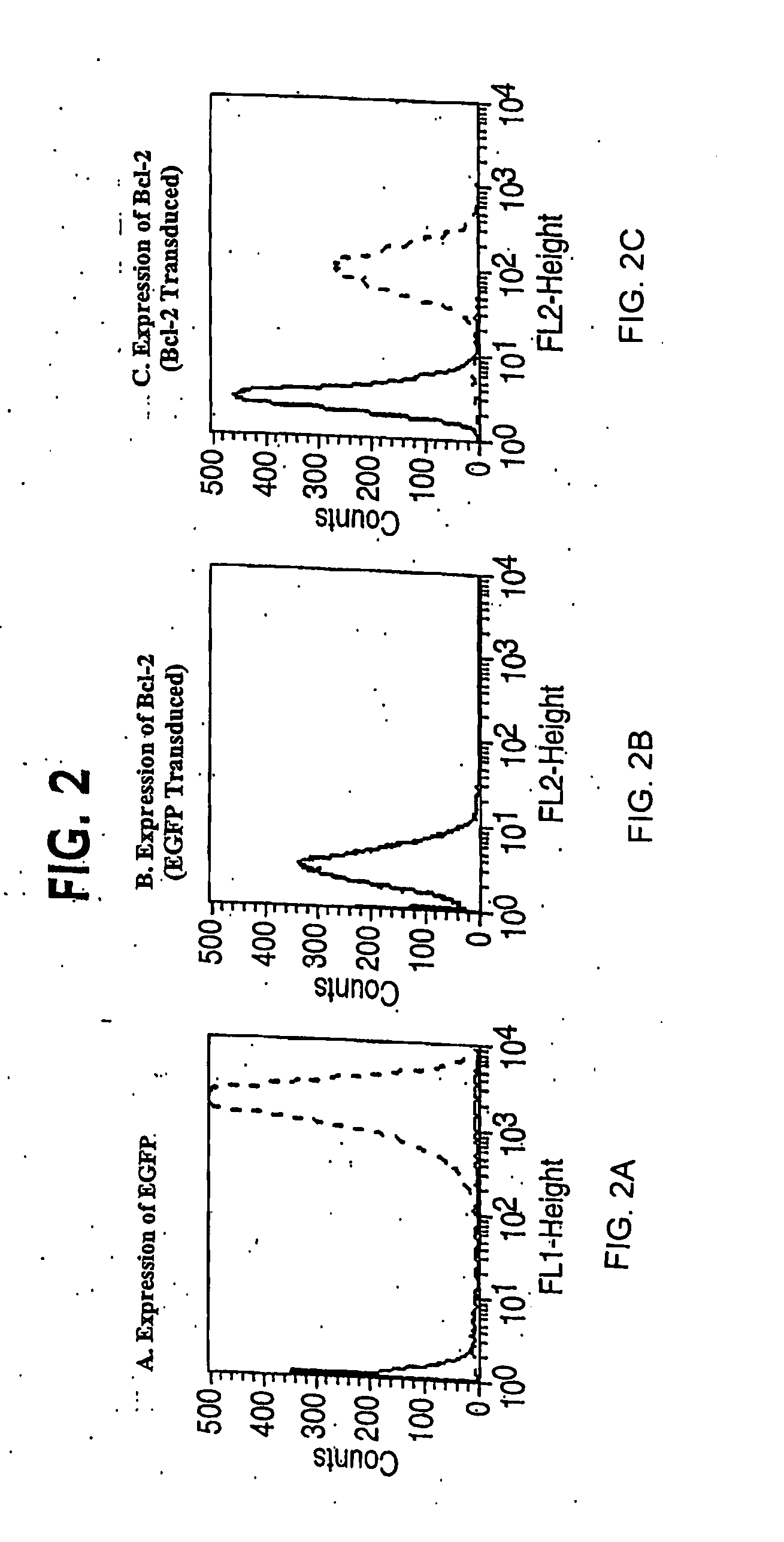
[0246] Infection of HUVEC was accomplished by four serial infections over two weeks without drug selection (Inaba et al. (1997) J. Surg. Res. 78, 31-36). In brief, standard viral infections in the presence of polybrene (5 μg / ml) were performed for six hours with 1×10⁵ HUVEC at passage one. The normal growth medium was replaced and cells were maintained overnight. The infection was repeated the next day. Cells were carried in culture for a week and then the process of double infection repeated starting with 1×10⁵ cells. Control transductions used the EGFP-encoding retroviral vector, or no retroviral vector. In general, each single retroviral infection produced 30-50% stably transduced cells. By performing two double cycles of infection, early passage HUVEC lines were reproducibly generated of which at least 95% of the cells expressed the expected cDNA.

[0247] Alternatively, the infection of HUVEC can be accomplished by se...


PUM

PropertyMeasurementUnit
Vascularizationaaaaaaaaaa

Abstract

Clinical performance of currently available human skin equivalents is limited by failure to develop perfusion. To address this problem we have developed a method of endothelial cell transplantation that promotes vascularization of human skin equivalents in vivo. Living skin equivalents were constructed by sequentially seeding the apical and basal surfaces of acellular dermis with cultured human keratinocytes and Bcl-2 transduced HUVEC or umbilical cord cells sequentially. After orthotopic implantation of grafts comprising cultured human keratinocytes and Bcl-2 transduced HUVEC cells onto mice, the grafts displayed both a differentiated human epidermis and perfusion through the HUVEC-lined microvessels. These vessels, which showed evidence of progressive maturation, accelerated the rate of graft vascularization. Successful transplantation of such vascularized human skin equivalents should enhance clinical utility, especially in recipients with impaired angiogenesis.

Description

CROSS-REFERENCE TO RELATED APPLICATIONS [0001] This application claims the benefit under 35 U.S.C. § 119(e) of U.S. Provisional Appl. No. 60 / 371,677, filed Apr. 12, 2002, the contents of which are incorporated herein in their entirety.STATEMENT REGARDING FEDERALLY-SPONSORED RESEARCH OR DEVELOPMENT [0002] This invention was made with support under grant number GM-R01 HL51044 (J.S.P), R01 HL51448 (A.L.M.B.), P30 AR4192, and K08 AR02134 (J.S.S.) awarded by the National Institutes of Health. The U.S. government has certain rights in the invention.FIELD OF THE INVENTION [0003] This invention is generally in the field of tissue grafting and relates in particular to the field of synthetic skin grafts and use of the grafts to treat wounds due to burns, trauma, surgical excisions, non-healing ulcers and blistering diseases. The present invention is further in the field of treatment of recipients with impaired angiogenesis. The invention also relates to methods of identifying genes and gene p...

Claims


Application Information

Patent Timeline
IPC IPC(8): A61K35/36C12N5/08A61K35/12C07K14/47C12N5/071
CPCA61K35/12C07K14/4747C12N2502/28C12N2502/094C12N2502/1323C12N5/0698
Inventor BOTHWELL, ALFRED L.M.POBER, JORDAN S.SCHECHNER, JEFFREY S.HERRICK, CHRISTINA
Owner YALE UNIV
Learn More