Container for lyophilization and storage of tissue

a tissue and container technology, applied in the field of tissue container for lyophilization and storage of tissue, can solve the problems of cumbersome packaging of bone implant forms, difficult to easily remove the implant form from such packaging, and little consideration has been spent on packaging design and specific problems involved in packaging bone tissue materials

Inactive Publication Date: 2007-04-05
MUSCULOSKELETAL TRANSPLANT FOUND INC
View PDF19 Cites 58 Cited by
  • Summary
  • Abstract
  • Description
  • Claims
  • Application Information

AI Technical Summary

Benefits of technology

[0018] It is an object of the invention to provide for lyophilization of bone tissue material while the same is contained within the package container while preventing microbes from escaping or entering the container and to resist absorption and discoloration from lipids found in the bone tissue material.
[0019] It is another object of the invention to provide a packaging system which holds sterile bone tissue material in a sterile condition which allows a user easy access to the sterile bone tissue material.
[0021] It is another object of the invention to provide a medical package in which an implant tissue form can be maintained in a sterile condition when it is presented to the surgeon for implantation.
[0023] It is yet another object of the invention to provide a package for storing bone tissue implant material whereby the bone tissue implants material can be removed from the package in a sterile condition.
[0024] It is still another object of the invention to provide a sterile bone tissue package which prevents cross contamination while being lypholized and stored.
[0025] It is yet another object of the invention to provide a sterile tissue package which is easy to use during the processing phase and by the end user in the operating room.

Problems solved by technology

Unfortunately, while large amounts of time have been spent on development and processing of tissue implant forms and materials, little consideration has been spent on the package design and the specific problems involved with packaging bone tissue material.
Packaging that is currently used for bone implant forms is cumbersome and it is often difficult to easily remove the implant form from such packaging while wearing gloves during a surgical procedure.
The lipids can be absorbed by or cause discoloration of the packaging material.
If the package material forms part of the package seal and that material absorbs the lipids the seal will be destroyed causing failure of the sterile barrier.
When the material is only discolored or does not form part of the sterility seal, the result is a visually unattractive package, which while causing no harm to the package or tissue gives the appearance that the product is damaged or spoiled.
However there are several drawbacks to this design.
The first drawback is since the stopper is in the up position for lyophilization and since many donors are processed in one freeze drying cycle, there is a potential for cross contamination between containers.
Secondly, the jars are subject to breakage during shipping and thirdly, the metal crimp cap is difficult to remove and can tear the gloves of the operating room personnel opening the container, causing possible contamination and the need to destroy the tissue.
It is not effective for powder, chips and granules because the venting required in the retainers allows the small particles to move from under the retainer and fall on the TYVEK® lid.
In addition to poor presentation of the product, the lipids in the tissue could then damage the seal.
This activity causes the cover to separate from the container receptacle destroying the sterility of the package and leaving pools or rivulets of a dark oily material which is unsightly and causes the surgeon to discard or send back the tissue material.
This lipid migration is a serious problem for manufacturers of allograft bone material, and presents problems for the hospital and surgeon user.

Method used


Image

  • Container for lyophilization and storage of tissue
  • Container for lyophilization and storage of tissue
  • Container for lyophilization and storage of tissue

Examples


Embodiment Construction

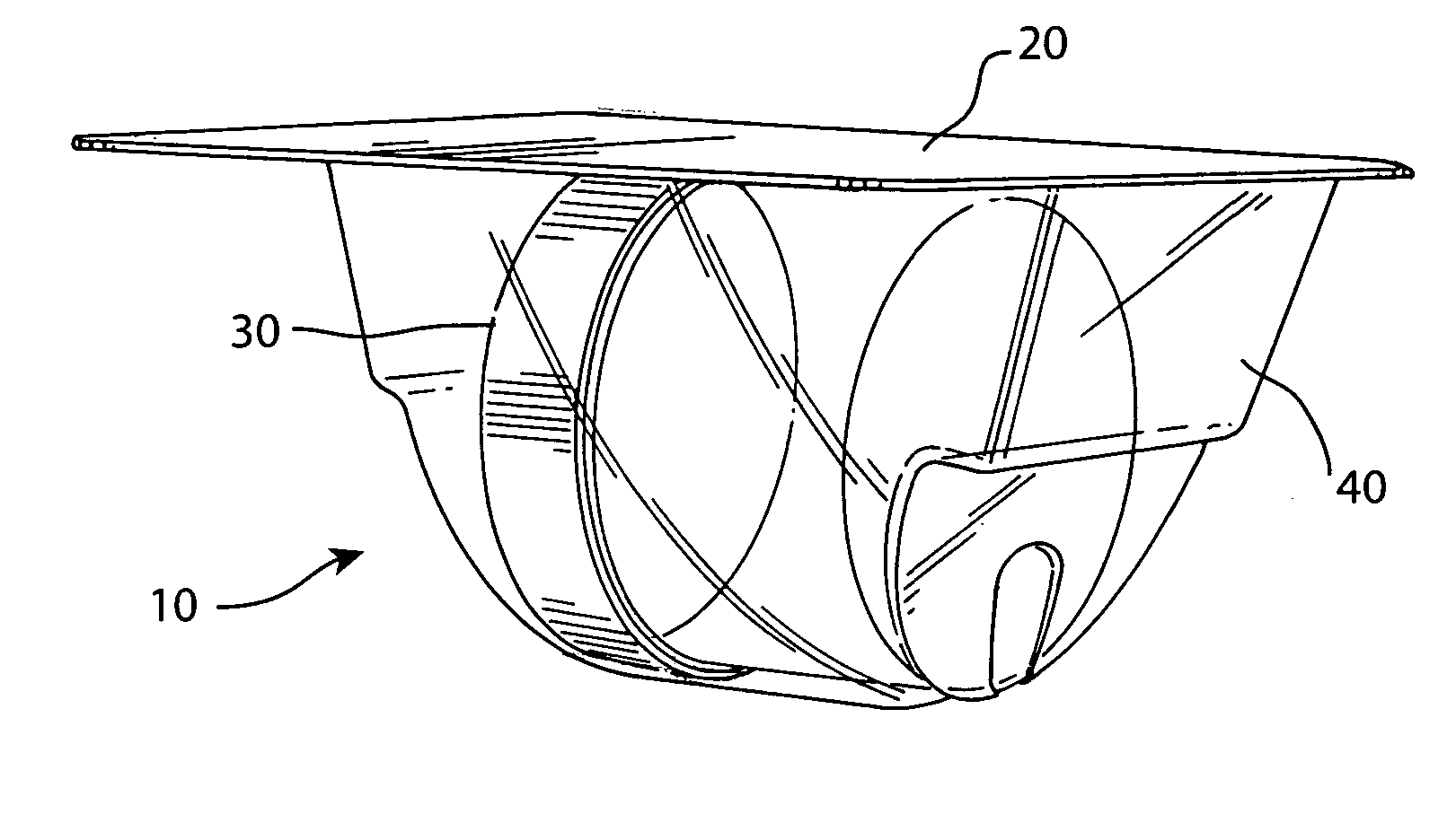
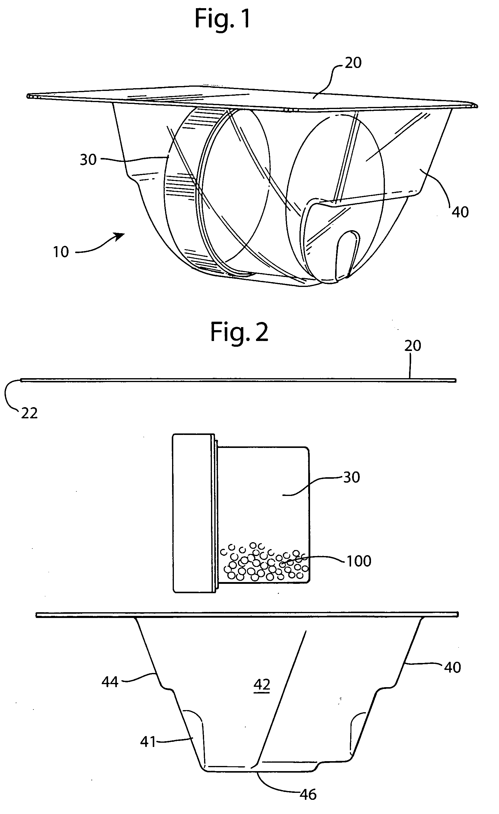
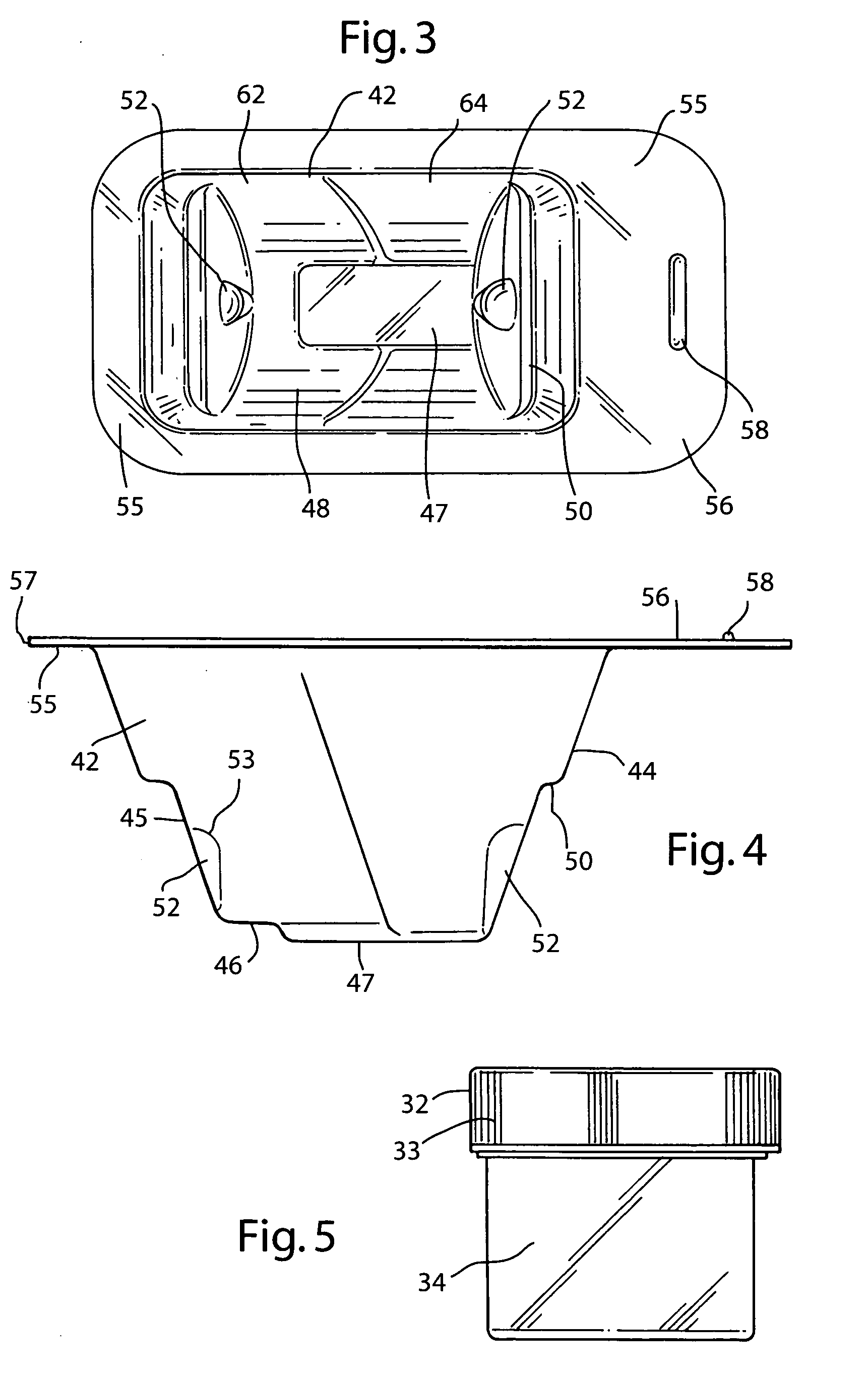
[0037] The preferred embodiment and best mode of the tissue form package invention is shown in FIGS. 1-10. The tissue package 10 comprises a blister pack 40 which holds a cylindrical container or jar 30, the blister pack outer flange 55 being covered with a cover 20. In the preferred embodiment of the present invention thin, plastic material is used that is sterilizable and can be formed by any suitable method such as for example, injection molding, pressure molding, vacuum forming and the like. The component material used for the blister pack of the allograft bone tissue package assembly 10 is preferably made in the form of a laminate made of an available material such as polytheylene terephthalateglycol (PETG) (a copolyester made by Eastman Chemical) as the inside layer, with the outside layer being another available material polycholrotrifluoroethylene (PCTFE) under the trademark ACLAR (a fluorinated-chlorinated thermoplastic made by Allied Corporation) which is impermeable to ox...


PUM


Abstract

A sterile container assembly for storing sterile allograft tissue implant material is constructed with sidewalls, end walls and a base member defining an open faced cavity and a flange surrounding and extending outward from the cavity. A step and a spacer is formed in each end wall of the container and a cylindrical implant container sized to fit into the container cavity is positioned adjacent the end wall spacers. The implant container has a housing and a threadable cap with a vent hole formed therein which is covered by an insert member constructed of sintered PTFE mounted inside the cap. A foil cover is sealed to the flange of the container covering and sealing the container cavity and the cylindrical implant container mounted therein.

Description

CROSS-REFERENCE TO RELATED APPLICATIONS [0001] There are no other applications related to the present application. TECHNICAL FIELD OF THE INVENTION [0002] The present invention generally relates to packaging for sterile tissue specimens for use in tissue transplant and more specifically to packages for allograft implant tissue forms in a specifically designed sterile package or container which allows the implant to be effectively lyophilized while preventing cross contamination by lipids or microbial agents released from the tissue during lyophilization or storage. BACKGROUND OF THE PRIOR ART [0003] Allograft tissue forms are useful in orthopaedic and neurosurgery. In practice, processed human tissue is delivered to the hospital and eventually to the surgeon in a form useful for surgical implantation in a sterile package. Unfortunately, while large amounts of time have been spent on development and processing of tissue implant forms and materials, little consideration has been spent...

Claims


Application Information

Patent Timeline
IPC IPC(8): A61B19/02
CPCA01N1/02A01N1/0263A61B19/44A61B2019/0218A61B2019/0219A61F2/0095A61F2/28A61F2002/2835A61F2310/00359A61B2050/0064A61B2050/0065A61B90/90
Inventor MERBOTH, BARBARA L.STOFFEL, FLORENCE
Owner MUSCULOSKELETAL TRANSPLANT FOUND INC
Why Eureka
Try Eureka