Inflatable cardiac device for treating and preventing ventricular remodeling

Inactive Publication Date: 2007-03-29
PARACOR MEDICAL
View PDF33 Cites 25 Cited by
  • Summary
  • Abstract
  • Description
  • Claims
  • Application Information

AI Technical Summary

Benefits of technology

[0031] The present invention provides a fluid filled prosthetic device that reduces wall stress by maintaining compressive contact against the epicardium over a significant portion of the cardiac cycle. The device is safely implantable over extended periods of time. Preferably, the device does not need to be removed unless there is a complication. The inflatable device may include an exterior surface having an increased coefficient of friction, whereby the inflatable chamber resists sliding over the epicardial surface of the heart. The invention in at least one embodiment provides anchoring members on the exterior of an inflatable chamber. The invention does not require an additional support, frame, or shell to stay in place against the heart.
[0032] In at least one embodiment, the invention provides a fluid filled prosthetic device wherein pressures exerted against the myocardium may be easily adjusted during or after implantation. The clinician can inject or withdrawn fluid from within the device during implantation of the device or at a later time. Changes in fluid volume within the device can allow for a corresponding change in the pressures exerted against the myocardium. An injection port or a reservoir are provided in some embodiments so that filling substance may be conveniently added or removed from the device without requiring a subsequent invasive procedure.
[0033] In at least one embodiment, the invention provides a prosthetic device that reduces focal wall stresses by maintaining compressive contact against a designated part of the epicardium. Furthermore, the device advantageously is capable of applying pressure against the heart without the need to apply a circumferential harness. Circumferential dissection around the heart is not needed to place the inflatable cardiac device. The inflatable device can be made in various sizes and shapes. In some embodiments, multiple compartments allow flexibility in applying compression to only a limited portion of the heart. In at least one embodiment, multiple channels may extend between the epicardial and the pericardial surfaces of the inflatable device, thereby allowing tissue growth through the channels. The channels may also be beneficial in permitting defibrillation of the heart by providing a discontinuous covering over the surface of the heart.
[0035] In certain preferred embodiments, the interior of the inflatable device chamber may be filled with various quantities and types of liquids, gases, or gels. The interior of the inflatable device may also be filled with a sponge. The volume occupied by the device and pressures delivered by the device will vary depending on the amount and type of the filling substance used to fill the chamber, the elasticity of the chamber walls, and the compliance of the inflatable device. In at least one embodiment, the device is filled with normal saline. In other embodiments, the device is filled with a gel, for example, a silicone gel. In other embodiments, the device is filled with a gas for increased elasticity. In yet other embodiments, the device is filled with a polymer or a sponge. The device may come pre-filled from the manufacturer, or could be filled by the surgeon, at the time of implantation. In some embodiments, it is advantageous to include filling catheters to allow the clinician to fill or remove filling substance via subcutaneous access, or exchange one type of filling substance for another.
[0039] In yet another embodiment, a unidirectional valve may be placed between the inflatable chamber and the reservoir. The addition of a valve permits fluid to flow in only one direction within a catheter. An advantage of a valve, in some embodiments, is that fluid in the reservoir may be forced into or removed from the inflatable chamber, utilizing only fluid already in the system, and thus avoiding the need for needle puncture of the reservoir. In other embodiments, valves that allow flow only at predetermined pressures can be used to regulate pressure within the inflatable chamber.

Problems solved by technology

The blood clots may thromboembolize and clog peripheral arteries with potential of stroke among other serious vascular problems.
In addition, splint placement requires perforation of the ventricular wall, which may lead to leakage problems such as hemorrhage or hematoma formation.
Furthermore, because one end of the splint extends to the epicardial surface of the left ventricle, options for the orientation of the splint are limited.
However, this may create an irregular ventricular wall contour.
Consequently, this may lead to aneurysm formation, fibrosis, and impairment of the contractility and compliance of the ventricle.
Also, the resulting irregular contour of the endocardial surface of the left ventricle may lead to localized hemostasis or turbulence, which may in turn lead to thrombus formation and possible thromboembolism.
Thus, a vicious cycle can result, in which dilatation leads to further dilatation and greater functional impairment.
On a cellular level, unfavorable adaptations occur as well.
This further compounds the functional deterioration.
In fact, imposing a rigid barrier to limit distension or to squeeze the heart can be potentially dangerous.
Cardiac function can be adversely affected with just a slight increase in pericardial constraint.
The presence of the device would likely cause the left ventricle to reverse-remodel and its dimensions to stabilize and even shrink.
Consequently, a device with sufficient elasticity for treating dilated hearts in established heart failure may not be able to treat a heart of normal dimensions that has suffered a myocardial infarction.
This may attenuate infarct expansion and therefore limit the extent of remodeling that further ensues.
However, an improvement in survival has yet to be consistently demonstrated.
Furthermore, perhaps due to their frail condition, NYHA class IV patients have not fared well with the procedure.
This has limited its use to NYHA class III patients.
It appears that the skeletal muscle wrap, probably because of its deterioration over time, does not provide sustained squeezing of the heart over time.
However, in a clinical setting, the surgery required to dissect and attach the muscle around the heart is very extensive and traumatic.
Even if such a therapy were proven clinically efficacious, this factor limits its potential acceptance.
Once the cardiac harness is in place, the pressure that the harness exerts upon the myocardium cannot be adjusted.
This is a significant disadvantage, requiring manipulation of anterior, posterior, and lateral surfaces of the heart.
The device is inserted to temporarily massage the heart over the short term, but is not designed to be implantable.
The frame or containment structure is not elastic and cannot be delivered with minimally invasive surgery.
Furthermore, the inflation pockets and recoil balloons are in a fixed configuration within the containment structure, therefore limiting the clinicians flexibility in the positioning of the device against chosen areas of the heart.
Cardiac harness or jackets therefore have a potential disadvantage of providing a circumferential tension or pressure around the entire heart and are not capable of applying tension or pressure to selected areas of the heart.
Another disadvantage of harnesses or jackets is that they cover the coronary arteries, making surgical access to the coronary arteries difficult of impossible at a later date.

Method used


Image

  • Inflatable cardiac device for treating and preventing ventricular remodeling
  • Inflatable cardiac device for treating and preventing ventricular remodeling
  • Inflatable cardiac device for treating and preventing ventricular remodeling

Examples


Embodiment Construction

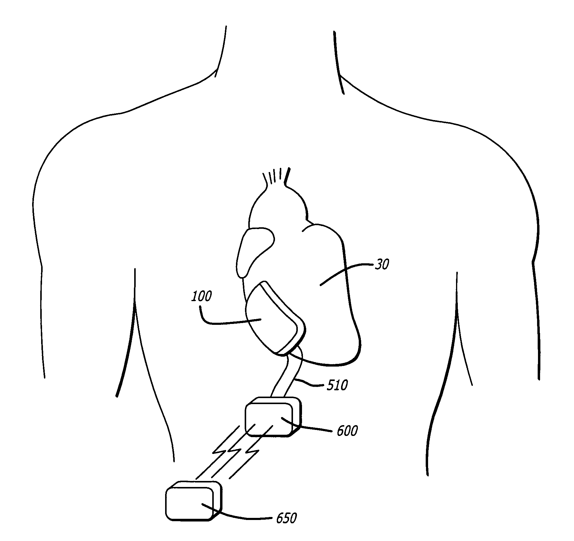
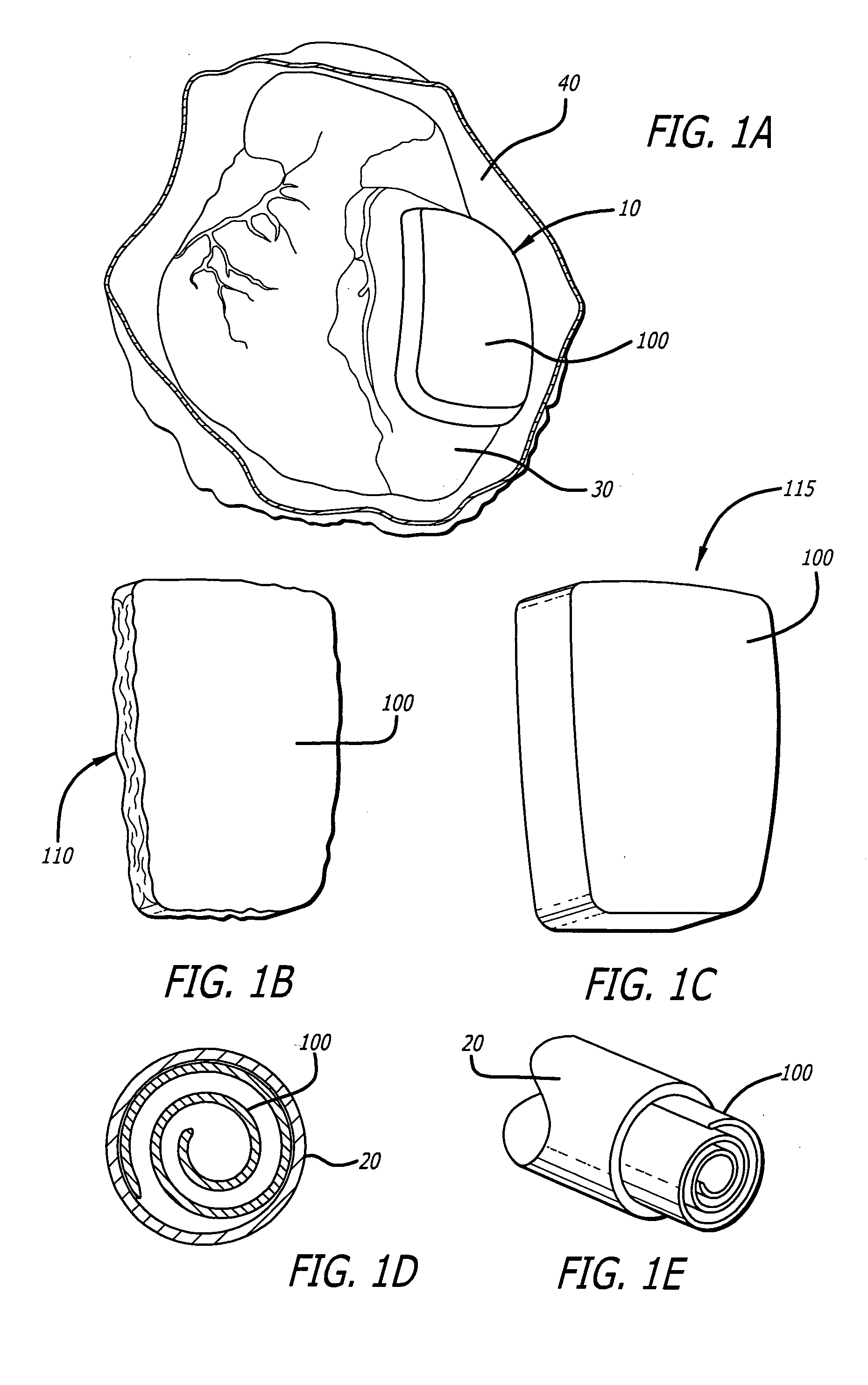
[0078] Referring first to FIG. 1A, the present invention is an inflatable cardiac device 10 including an inflatable chamber 100 for implantation adjacent to the heart. The inflatable cardiac device 10 is designed for treating remodeling of the heart and preventing or reversing deleterious effects on cardiac function associated with congestive heart failure. The inflatable cardiac device is configured to attenuate and / or reverse remodeling. The inflatable cardiac device in at least one embodiment is implanted between the epicardium 30 and the pericardium 40, within the pericardial sac. As illustrated in FIG. 1B, the inflatable chamber 100 has a first collapsed or deflated configuration 110 for minimally invasive delivery and as illustrated in FIG. 1C, a second expanded configuration 115. The inflatable chamber 100 is preferably expanded following placement in the pericardial sac, although it may also be expanded before placement in the pericardial sac. In at least one embodiment, the...


PUM


Abstract

An inflatable cardiac device for placement in the pericardial sac is described. The inflatable cardiac device is useful for treatment of congestive heart failure and for attenuating or reversing remodeling of the heart. In some embodiments, the device further includes filling catheters connected with reservoirs, on-off devices, or pressure transducers. The device may also include a drug delivery system. At least one embodiment includes electrodes capable of connection with an implantable electronic medical device such as a pacemaker or defibrillator.

Description

FIELD OF THE INVENTION [0001] The present invention relates to mechanical systems for treating congestive heart failure. Specifically, the invention relates to devices that interface mechanically with a patient's failing heart in order to improve intrinsic cardiac function. BACKGROUND OF THE INVENTION [0002] Congestive heart failure (“CHF”) is characterized by the failure of the heart to pump blood at sufficient flow rates and pressures to meet the metabolic demand of tissues, especially the demand for oxygen. Historically, congestive heart failure has been managed with a variety of drugs. There is also a considerable history of the use of devices to improve cardiac output. For example, physicians have employed many designs for powered left-ventricular assist pumps. Multi-chamber pacing has been employed to optimally synchronize the beating of the heart chambers to improve cardiac output. [0003] Various skeletal muscles have been investigated as potential autologous power sources fo...

Claims


Application Information

Patent Timeline
IPC IPC(8): A61M31/00
CPCA61B17/00491A61B2017/00243A61F2250/0003A61F2/2481A61B2017/00557
Inventor LAU, LILIPMAR, CRAIGTRUONG, ANH
Owner PARACOR MEDICAL
Why Eureka
Try Eureka