
Functional navigator

a navigator and functional technology, applied in the field of medicine, can solve the problems of not easy to locate the depth of a particular ganglion, navigators that obtain information, and the technique is rudimentary, and achieve the effect of greater security in extraction

Inactive Publication Date: 2007-01-18
CONSEJO SUPERIOR DE INVESTIGACIONES CIENTIFICAS (CSIC) +1
View PDF22 Cites 111 Cited by
  • Summary
  • Abstract
  • Description
  • Claims
  • Application Information

AI Technical Summary

Benefits of technology

[0017] A particular object of the present invention is the use of the surgical navigator for the positive and precise detection of carcinogenic tumours in surgery and its combination with the surgical instruments.
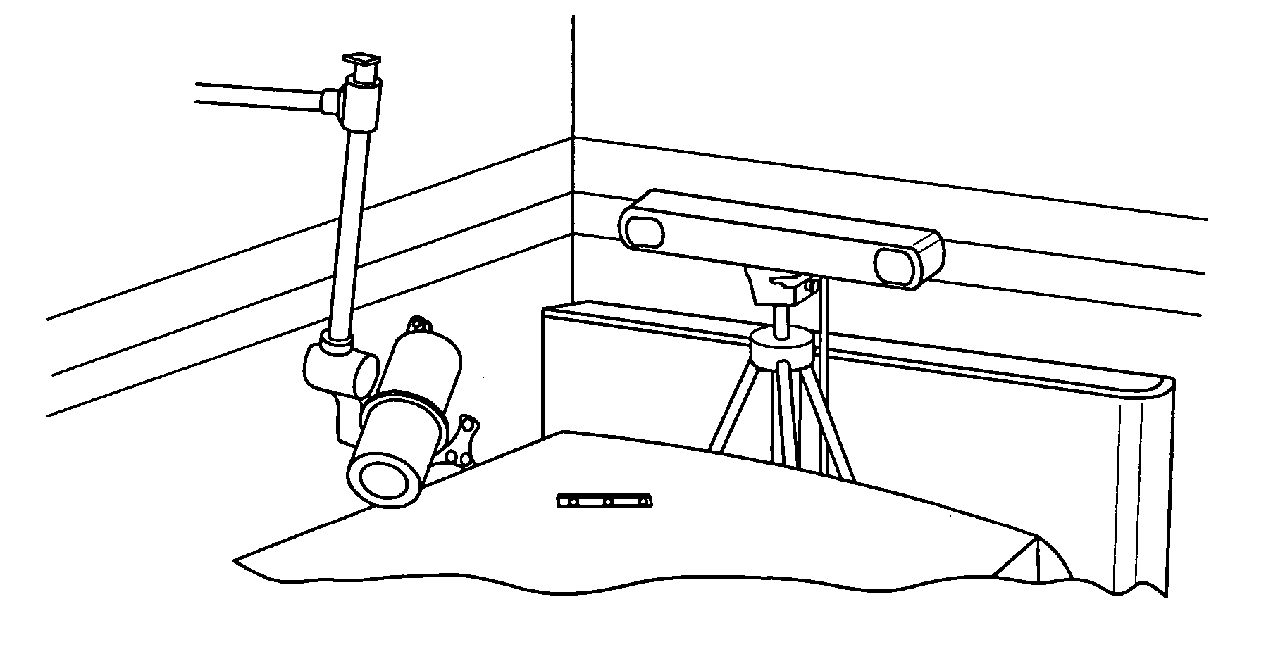
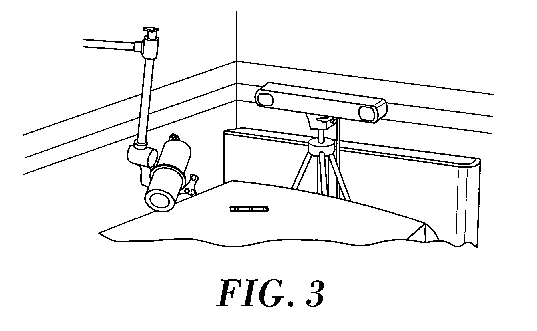
[0022] The camera is coupled to the navigator so that they can act jointly and in coordination. The camera provides functional information in real time which can be combined by means of a specific software program with the morphological information possessed by the navigator in order to form a complete morphological and functional image, which is of extraordinary utility for the surgeon.
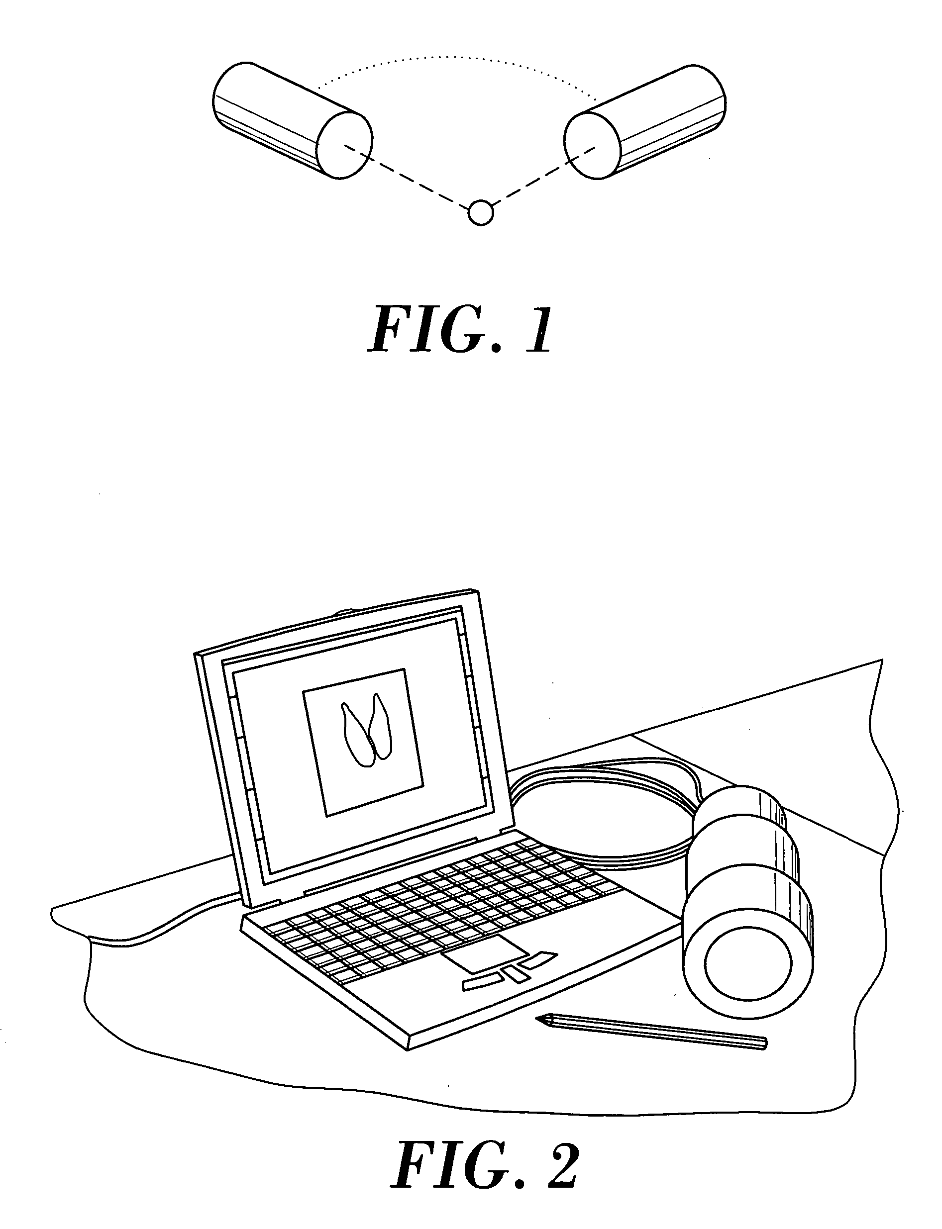
[0027] The position of the ganglion is determined by triangulation, taking the intersection of the lines determined by the camera from two different positions separated by 90° (see FIG. 1) or three, separated by 120° (surrounding the patient). Two different lead collimators have been used for the Gamma cameras: a pinhole and a parallel ray collimator. If the region under study is limited, the parallel ray one is used, since its resolution is better and independent of the distance to the ganglion (unlike in the case of the pinhole). Obviously, in either of the cases, the camera has to be located as close as possible to the region under study in order to increase the sensitivity and improve the spatial resolution.
[0030] A Gamma and PET camera currently provide an image in two dimensions. In order to obtain three-dimensional images, two different methods are used: a single camera which acquires images in two different planes (for example, locating the portable camera in different positions at 90° with respect to each other) in order to thereby obtain a stereoscopic image; or two cameras joined by means of a mechanical system in order to keep them at a fixed angle with regard to each other but variable with regard to the axis of the patient. This latter model is faster but implies a higher cost of the equipment.
[0036] The obtaining of functioning images by means of a Gamma or PET camera during the surgical operation provides essential information for deciding on the area to cut out during the actual moment of the operation and checking whether a tumour has been removed correctly and in its entirety. Also, the functional navigator solves the problem of movements of the patient's organs during the time that passes from taking the images to the surgical operation, so there is greater security in extraction of, for example, carcinogenic cells during the surgery.
[0042] It decreases the extent of the surgery, saving time, costs and any complications deriving from an aggressive extirpation of ganglia (lymphoceles, lymphatic oedema, etc.).

Problems solved by technology

Nevertheless, this technique is somewhat rudimentary since it does not allow the ganglia to be located accurately and it is not applicable when the ganglia are to be found at a certain depth from the surface of the patient.
The problem of this technique is that the images obtained are two-dimensional projections of the field of view of the camera and in many cases it is not easy to locate the depth of a particular ganglion that is inflamed.
So far, there do not exist any navigators that obtain information from functional images.
Also, the information obtained is acquired prior to the operation, which means that the surgeon cannot make checks in real time (at the moment of the operation) on the effectiveness of his work, in other words, whether he has still left any tumour cells of the patient unextracted.
So, there does not currently exist instruments permitting the ganglia and tumours to be precisely located at the moment of the operation.

Method used


Image

  • Functional navigator
  • Functional navigator
  • Functional navigator

Examples


example 1

Embodiment of the Functional Navigator with Gamma Cameras

[0050] For this, a description is given first of all of a Gamma mini-camera and then of the surgical navigator used.

[0051] The Gamma mini-camera has a total size of diameter 90 mm and length 200 mm, and a total weight of somewhat less than 2 kg. The two characteristics, small dimensions and light weight, ensure mobility of the system (see FIG. 2). The Gamma mini-camera has a spatial resolution of about 2 mm. This Gamma camera is used for visualising small organs.

[0052] The design of a portable Gamma mini-camera is optimised for the radioactive source most widely used in medical explorations, that of 99m-Tc, which emits gamma rays of 140 keV. The camera consists of a single position-sensitive photomultiplier tube (PSPMT, HR2486 (from Hamamatsu Photonica)) coupled to a scintillation crystal, an electronic system and a high voltage source, with lead collimators being able to be coupled easily.

[0053] Physical measurements have...


PUM


Abstract

The invention relates to a device which is used intra-operatively to localise carcinogenic tumours and ganglia precisely and in a three-dimensional manner and to correlate the surgical instruments with the images in real time. The structural elements of the functional navigator include: a surgical navigator, a camera which is used to obtain functional images, the position-finding elements of the camera and the surgical instruments and the specific software program which combines all of the information from the aforementioned elements. The surgical navigator is essentially characterised in that the position of the cameras which acquire the functional images is controlled by the navigator by means of emitters or reflectors which are disposed in said cameras. The information relating to the camera images and the position of same, which is determined by the navigator, is analysed by a specific software program and combined with the position of the surgeon's surgical instruments in order to obtain images.

Description

RELATED APPLICATIONS [0001] The present application is a Continuation of co-pending PCT Application No. PCT / ES2003 / 000497, filed Oct. 1, 2003, which in turn, claims priority from Spanish Application Serial No. P200202220, filed Oct. 1, 2002. Applicants claim the benefits of 35 U.S.C. §120 as to the PCT application and priority under 35 U.S.C. §119 as to said Spanish application, and the entire disclosures of both applications are incorporated herein by reference in their entireties.TECHNICAL FIELD OF THE INVENTION [0002] The invention comes within the sector of Medicine, and in particular Nuclear Medical Physics. It is a device which permits the precise location and extirpation of carcinogenic tumours at any point of the human body BACKGROUND OF THE INVENTION [0003] Cancer is a disease that is produced as a result of an uncontrolled development of abnormal or defective cells, which tend to infiltrate, spread and metastasise, or invade, other tissues, whether surrounding or distant f...

Claims


Application Information

Patent Timeline
IPC IPC(8): A61B5/05A61B19/00
CPCA61B19/5212A61B19/5244A61B6/4258A61B2019/5289A61B6/037A61B2019/5255A61B2034/2055A61B34/20A61B2090/364A61B90/361
Inventor BENLLOCH BAVIERA, JOSE MARIAALCANIZ RAYA, MARIANOMARTINEZ, FILOMENO SANCHEZCOLOMER, VICENTE GRAU
Owner CONSEJO SUPERIOR DE INVESTIGACIONES CIENTIFICAS (CSIC)
Learn More