
Pegylated g-csf polypeptides and methods of producing same

a technology of gcsf and polypeptides, which is applied in the direction of peptide/protein ingredients, immune disorders, extracellular fluid disorders, etc., can solve the problems of insufficient uniformity of peg moieties bound to groups other than those intended, complex characterization of such proteins, and inability to meet the requirements of characterization, etc., to achieve the effect of increasing the stability and uniformity of a pegylated polypeptid

Inactive Publication Date: 2007-01-18
MAXYGEN HLDG
View PDF0 Cites 2 Cited by
  • Summary
  • Abstract
  • Description
  • Claims
  • Application Information

AI Technical Summary

Benefits of technology

"The present invention relates to a method for increasing the stability and uniformity of a PEGylated polypeptide, specifically a PEGylated granulocyte colony stimulating factor (G-CSF) polypeptide. The method involves subjecting the polypeptide to an altered pH for a period of time to remove PEG moieties attached to a hydroxyl group and then adjusting the pH to a suitable level for long-term storage. The invention also provides a composition comprising a mixture of positional PEG isomers of the polypeptide and a method for producing such a composition. The PEGylated G-CSF polypeptides produced using the methods described herein have improved stability and can be used to increase the level of neutrophils in a patient."

Problems solved by technology

As a result, PEGylated proteins prepared using this technology may not have a sufficient degree of uniformity and may contain a number of different PEG isomers other than those that were intended.
This is undesired for various reasons, including the fact that it makes characterization of such proteins more complicated.
Further, PEG moieties bound to groups other than those intended may be relatively unstable.
Depending on the formulation and storage conditions, this may lead to an undesired loss of these labile PEG groups and thus potentially to a change in the properties of the PEGylated protein over time.
Further, there may be potential regulatory or safety issues for a PEGylated protein pharmaceutical in which there is a risk that one or more of the PEG moieties may detach from the protein in the body after it is administered to a patient.

Method used


Image

  • Pegylated g-csf polypeptides and methods of producing same
  • Pegylated g-csf polypeptides and methods of producing same
  • Pegylated g-csf polypeptides and methods of producing same

Examples


example 1

Preparation and Analysis of a Partially de-PEGylated G-CSF Variant

Sample Preparation and de-PEGylation

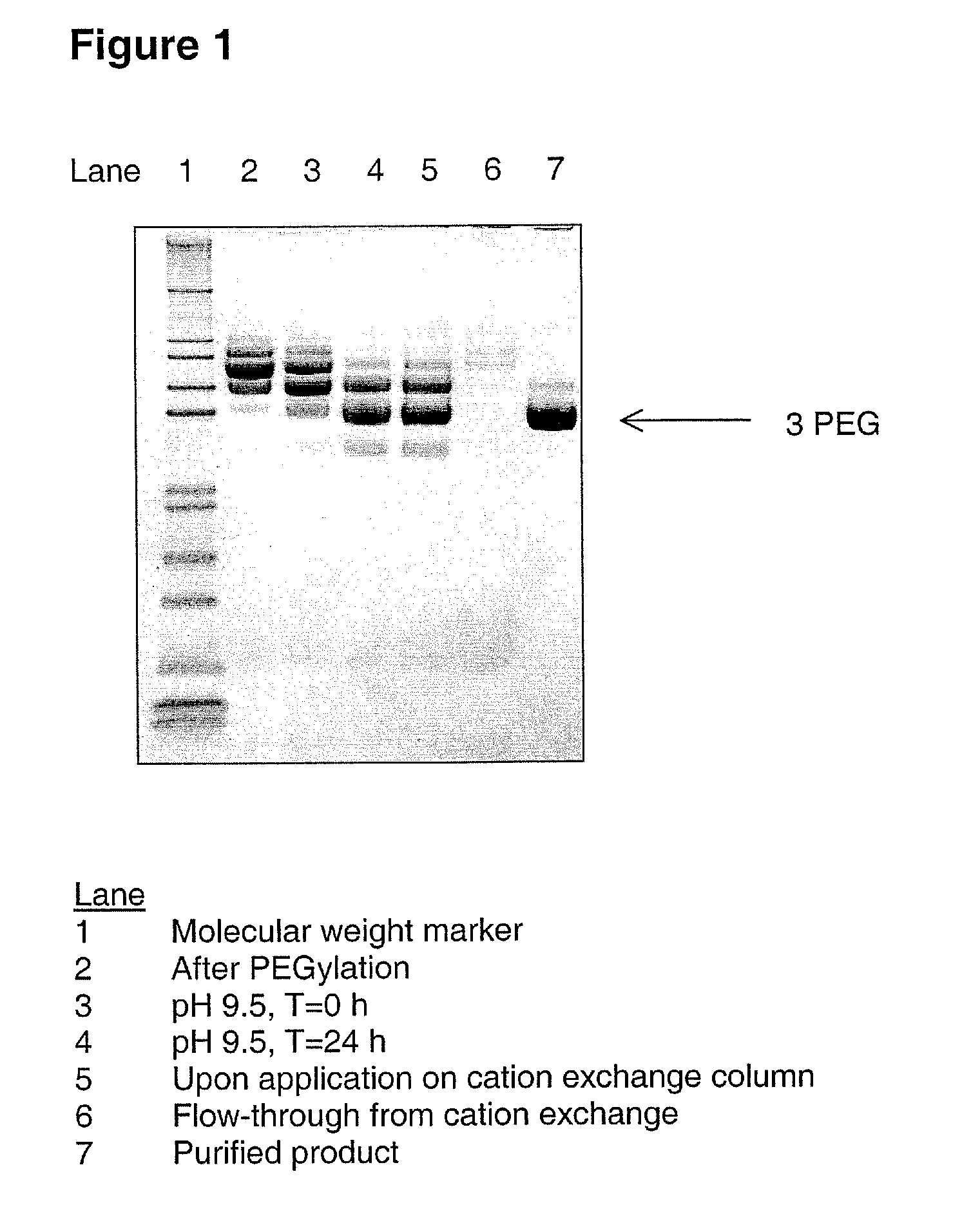
[0087] A G-CSF variant having the substitutions K16R, K34R, K40R, T105K and S159K compared to wild-type human G-CSF (SEQ ID NO:1) was produced from CHO-K1 cells substantially as described in WO 03 / 006501. 200 mL of 4.5 mg / mL of the G-CSF variant (900 mg G-CSF) was PEGylated using mPEG-SPA 5000 (Nektar Therapeutics). Briefly, 100 mL of a 13.2% (w / w) solution of mPEG-SPA 5000 was added over a period of 10 minutes to the 200 mL of G-CSF solution and gently stirred to ensure sufficient mixing. The sample was allowed to incubate for 44 minutes at 21±3° C., pH 8.5, with gentle stirring. The sample mixture was subsequently adjusted to pH 9.5 using a stock solution of 800 mM boric acid pH 10.0. The sample was then incubated at 21±3° C. for 24 hours without stirring. The sample was then diluted approx. 2.5 fold with 100 mmol / kg citric acid, 20 mmol / kg NaOH, pH 2.5, to a final pH of 3.5. ...

example 2

Preparation and Analysis of a Partially de-PEGylated G-CSF Variant

Sample Preparation, de-PEGylation and Purification

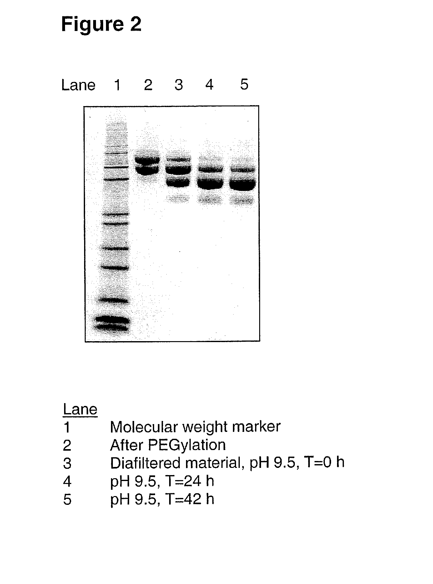
[0101] A G-CSF variant having the substitutions K16R, K34R, K40R, T105K and S159K compared to wild-type human G-CSF (SEQ ID NO:1) was produced from CHO-K1 cells and subsequently PEGylated using mPEG-SPA 5000 substantially as described in Example 1 above, resulting in 29 mL of 3.5 mg / mL PEGylated G-CSF variant. This was diafiltrated into 150 mM sodium borate, pH 9.5, using a Vivaspin filter device equipped with a 10 kDa MWCO filter. The final sample concentration was 4.7 mg / mL. The sample was then incubated at 21±3° C. for 42 hours.

[0102] After incubation, the sample was prepared for cation exchange chromatography by dilution with 30 mM citric acid, 10 mM NaOH, pH 2.9. The sample was then applied onto an XK 16 / 20 column (Amersham Biosciences) packed with 28 mL of SP-Sepharose HP resin. The column was equilibrated with an equilibration buffer of 20 mM citric acid, 1...

example 3

Comparative Example

[0110] For comparative purposes, FIG. 4 shows a cation exchange chromatogram of a purified, PEGylated G-CSF variant that was not subject to de-PEGylation according to the present invention. The G-CSF variant in this case had the same five substitutions compared to native human G-CSF as indicated above in Examples 1 and 2, i.e. K16R, K34R, K40R, T105K and S159K. It was prepared and PEGylated with mPEG-SPA 5000 substantially as described in Example 1, with the exception of the fact that it was not subjected to incubation at pH 9.5 for removal of labile PEG moieties. As a result, the PEGylated variant comprised a mixture of a number of different PEG isomers having 2-6 attached PEG moieties. This mixture of positional isomers was purified using cation exchange chromatography substantially as described in Example 1. A fraction containing primarily 4-5 attached PEG moieties was isolated and, after ultrafiltration and diafiltration substantially as described in Example ...


PUM

PropertyMeasurementUnit
molecular weightaaaaaaaaaa
temperatureaaaaaaaaaa
pHaaaaaaaaaa

Abstract

A method for increasing the stability and uniformity of a PEGylated G-CSF polypeptide having at least one PEG moiety attached to the epsilon amino group of a lysine residue or the N-terminal amino group and at least one PEG moiety attached to a hydroxyl group, comprising subjecting the polypeptide to an elevated pH of above 8.0 for a period of time suitable to remove PEG moieties attached to a hydroxyl group, and reducing the pH to about 8.0 or lower; as well as PEGylated G-CSF polypeptides and compositions produced according to the method and methods for increasing neutrophil levels in a patient using the PEGylated G-CSF polypeptides and compositions.

Description

CROSS-REFERENCE TO RELATED APPLICATIONS [0001] Pursuant to 35 U.S.C. §119(e), this application claims the benefit of U.S. Provisional Application Ser. No. 60 / 686,726 filed on Jun. 1, 2005, the disclosure of which is incorporated by reference herein in its entirety for all purposes.FIELD OF THE INVENTION [0002] The present invention relates to a method for removing labile PEG moieties from PEGylated G-CSF proteins to increase stability and uniformity, and to the resulting PEGylated G-CSF proteins. The invention also relates to pharmaceutical compositions comprising the PEGylated proteins and methods of treatment by administering the pharmaceutical compositions. BACKGROUND OF THE INVENTION [0003] The covalent attachment of polyethylene glycol (PEG) moieties to proteins or polypeptides (“PEGylation”) is a well-known technique for improving the properties of such proteins or polypeptides, in particular pharmaceutical proteins, e.g. in order to improve circulation half-life and / or to shi...

Claims


Application Information

Patent Timeline
Patent Type & Authority Applications(United States)
IPC IPC(8): A61K38/19C07K14/535
CPCA61K38/193C12P21/02C07K14/535A61K47/48215A61K47/60A61P31/00A61P31/10A61P31/18A61P35/02A61P37/04A61P43/00A61P7/00A61K47/50
Inventor GERMANSEN, CARSTENSONI, BOBBYRASMUSSEN, GRETHE
Owner MAXYGEN HLDG
Learn More