
Sample mounts for microcrystal crystallography

a microcrystal and crystallography technology, applied in the direction of radiation beam directing means, optical radiation measurement, supporters, etc., can solve the problems of reducing the total area of polyimide, reducing fluid resistance and flow disturbance, etc., and achieves excellent mechanical properties, low density, and low atomic number elements.

Active Publication Date: 2006-04-27
CORNELL RES FOUNDATION INC
View PDF25 Cites 10 Cited by
  • Summary
  • Abstract
  • Description
  • Claims
  • Application Information

AI Technical Summary

Benefits of technology

[0019] The present invention addresses the foregoing need through provision of microcrystal sample mounts in which a thin plastic film is employed to support a crystal to be analyzed. The film has a thickness of 50 μm or less and is preferably made from polyimide. A key feature of the invention is that a curvature is imparted to the film to increase its structural rigidity substantially. The increase in rigidity enables the use of thinner films with thicknesses on the order of 3-15 μm (or even thinner, provided other mount dimensions are correspondingly reduced), which reduces background scattering of the X-rays.
[0020] To provide the requisite curvature, a number of techniques can be employed, depending upon the type of crystallography application. In cryocrystallography applications, where capillary tubes are not employed, the polyimide film is preferably mounted either to an external curved surface of a rod or pin, the internal curved surface of a hollow rod or sleeve, or between both a sleeve and a rod. In these embodiments, the rod and / or hollow sleeve preferably have beveled top ends to maximize the viewing angle of the sample position so that one can see crystals and any crystal aperture when the rod, sleeve and mount are angled relative to the horizontal. Alternatively, the film can be attached to a rod having a conical end such that the plastic mount is tilted relative to the rod, so that it is easier to scrape a crystal off a flat surface and the crystal is located on axis, which makes alignment even easier.
[0022] Polyimide is a good choice for the film material because of its excellent mechanical properties; because it has a low density and is composed of low atomic number elements so that it scatters X-rays very weakly; and because its gold hue provides good optical contrast with macromolecular crystals. The polyimide films are microfabricated to have a tapered tip end for holding a crystal to be examined. This tapering minimizes the volume of the film in the X-ray beam (whose size is usually matched to or smaller than the crystal size) when the plane of the film is oriented parallel to the beam. Preferably, a first sample aperture is disposed at the tip end for reception of the crystal, though the aperture is not essential. This aperture allows the crystal to be precisely located, and further minimizes the volume of the film in the X-ray beam and thus the background scatter from the film. For mounts intended for cryocrystallography of macromolecular or virus crystals grown in solution, a small channel is preferably disposed in the film that connects the sample aperture to a larger aperture which facilitates wicking of any excess fluid from the sample aperture with little risk of touching the crystal. The large aperture also reduces the total area of polyimide and thus reduces the fluid resistance and flow disturbances caused as the mount is moved through a crystal-containing drop. The channel and large aperture are not necessary for “dry” crystals of inorganic or small-molecule organic materials where excess fluid is not an issue.
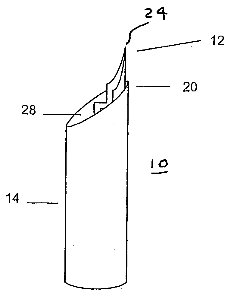
[0023] The film preferably has a small fixed width (5-100 μm, depending on the size of the crystals to be examined) surrounding the sample aperture that reduces scattering from the polyimide film (and any adsorbed fluid) when the plane of the film is oriented parallel to the X-ray beam. An overall triangular shape of a top portion of the film that is intended to extend beyond the rod or sleeve to which it is attached, together with a tapered or beveled shape of the rod or sleeve, provides a good aerodynamic profile that minimizes sample “flutter” in a gas cooling stream (relevant for lowest mosaicity crystals).
[0024] The present invention thus provides an alternative to conventional loops that retains all of their advantages (including complete compatibility with existing and developing technologies for high-throughput crystallography), but that resolves most if not all of the problems in mounting smaller crystals. Their potential advantages include completely reproducible sample “loop” sizes down to 3 micrometers or even smaller if thinner, shorter films with larger curvature are used, accurate and reproducible sample positioning, good sample-mount contrast, easier removal of excess liquid, minimal thermal mass and more rapid flash cooling; reduced background scattering, and easy design customization and mass production. In addition, the color, rigidity and reproducibility of the mounts should make it feasible to retrieve crystals using robots, with large savings in labor. In contrast, the use of loop type mounts requires that all crystals be retrieved by hand from crystallization drops, because loops are floppy and have little contrast with drop solutions.

Problems solved by technology

The large aperture also reduces the total area of polyimide and thus reduces the fluid resistance and flow disturbances caused as the mount is moved through a crystal-containing drop.

Method used


Image

  • Sample mounts for microcrystal crystallography
  • Sample mounts for microcrystal crystallography
  • Sample mounts for microcrystal crystallography

Examples


Embodiment Construction

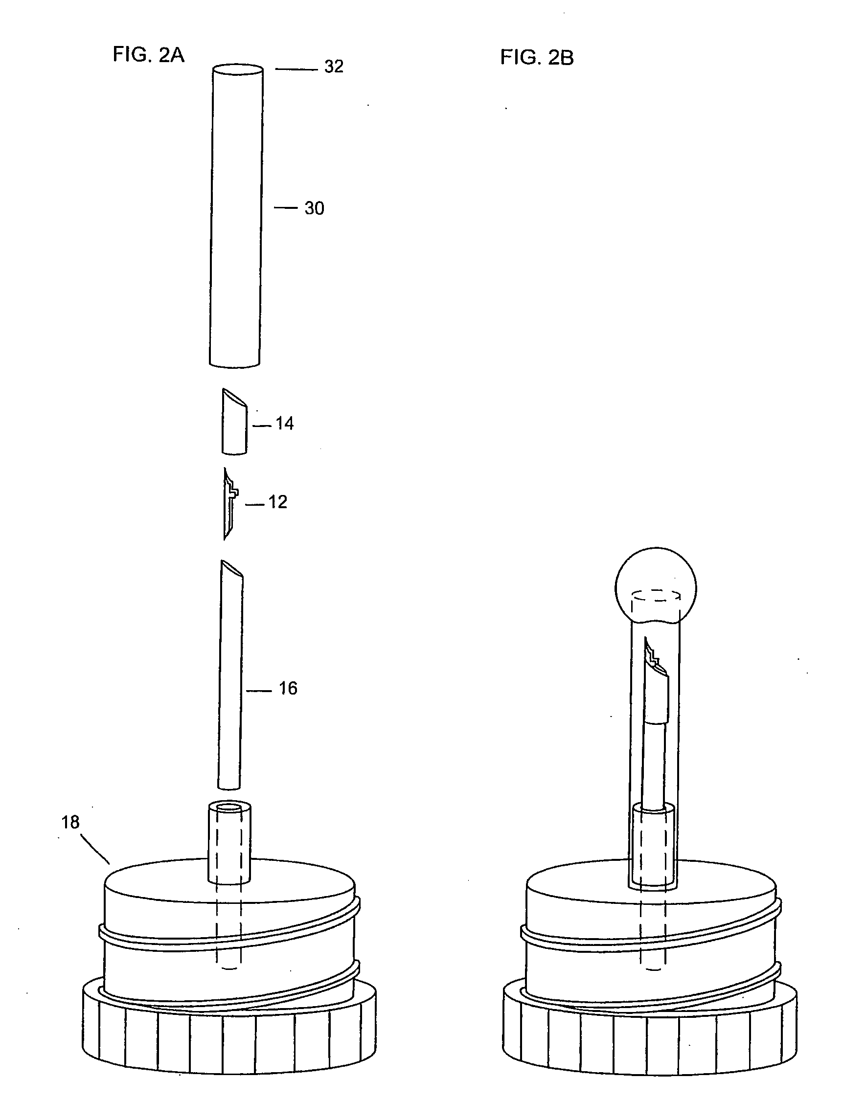
[0029] With reference to FIGS. 1A-1D, several variations of a microcrystal mount 10 are illustrated that are constructed in accordance with four preferred embodiments of the invention and are each designed particularly for use with a goniometer or other standard sample holder / positioner used in X-ray crystallography. In each embodiment, the mount 10 includes a microfabricated plastic film 12 that is attached to a structure having a curved surface such that curvature is imparted to the film 12. In the embodiment of FIG. 1A, the film 12 is attached by any suitable means, such as adhesive, tape, thermal bonding or the like, to a small diameter cylindrical plastic or metal post 16. As illustrated in FIG. 2A, the post 16 is sized to be inserted into standard plastic or metal magnetic goniometer head mount 18, such as those sold by Hampton Research or being developed by the macromolecular crystallography community for high-throughput automated crystallography, or into keyed variants of th...


PUM

PropertyMeasurementUnit
widthaaaaaaaaaa
thicknessaaaaaaaaaa
diameteraaaaaaaaaa

Abstract

Sample mounts (10) for mounting microcrystals of biological macromolecules for X-ray crystallography are prepared by using patterned thin polyimide films (12) that have curvature imparted thereto, for example, by being attached to a curved outer surface of a small metal rod (16). The patterned film (12) preferably includes a tapered tip end (24) for holding a crystal. Preferably, a small sample aperture is disposed in the film for reception of the crystal. A second, larger aperture can also be provided that is connected to the sample aperture by a drainage channel, allowing removal of excess liquid and easier manipulation in viscous solutions. The curvature imparted to the film (12) increases the film's rigidity and allows a convenient scoop-like action for retrieving crystals. The polyimide contributes minimally to background and absorption, and can be treated to obtain desired hydrophobicity or hydrophilicity.

Description

CROSS REFERENCE TO RELATED APPLICATIONS [0001] This application is a continuation under 35 U.S.C. 120 and 365(c) of International Application No. PCT / US2004 / 006088, filed Mar. 22, 2004, which claims the benefit under 35 U.S.C. 119(e) of U.S. Application No. 60 / 455,853, filed Mar. 20, 2003.BACKGROUND OF THE INVENTION [0002] 1. Field of the Invention [0003] The present invention relates in general to sample mounts for mounting and manipulating macromolecular and virus crystals and other samples for X-ray crystallography, and methods of using the same. [0004] 2. Description of the Background Art [0005] One of the most common ways of mounting crystals for X-ray data collection and structure determination is to insert them into thin-walled (typically 10 micrometer) glass or quartz capillaries. These thin capillaries are X-ray transparent and produce relatively little background scatter. They can be sealed at both ends, providing a stable environment for the crystal. This is particularly ...

Claims


Application Information

Patent Timeline
Patent Type & Authority Applications(United States)
IPC IPC(8): C30B27/02C30B15/00C30B21/06C30B28/10C30B30/04G01N23/20
CPCC30B29/58C30B35/00G01N23/20G01N23/20025Y10T117/1036Y10S269/90Y10S269/909Y10T29/53961Y10S269/908
Inventor THORNE, ROBERT E.STUM, ZACHARYO'NEILL, KEVINKMETKO, JAN
Owner CORNELL RES FOUNDATION INC
Learn More