
Blood cell separating system

a blood cell and lectin technology, applied in the field of blood cell separation systems, can solve the problems of inability to extract lymphocytes having a y chromosome from an entire group of similar lymphocytes, high risk of miscarriage, and inability to achieve the effect of nylon wool columns or density centrifugation

Inactive Publication Date: 2005-09-29
NETECH
View PDF7 Cites 13 Cited by
  • Summary
  • Abstract
  • Description
  • Claims
  • Application Information

AI Technical Summary

Benefits of technology

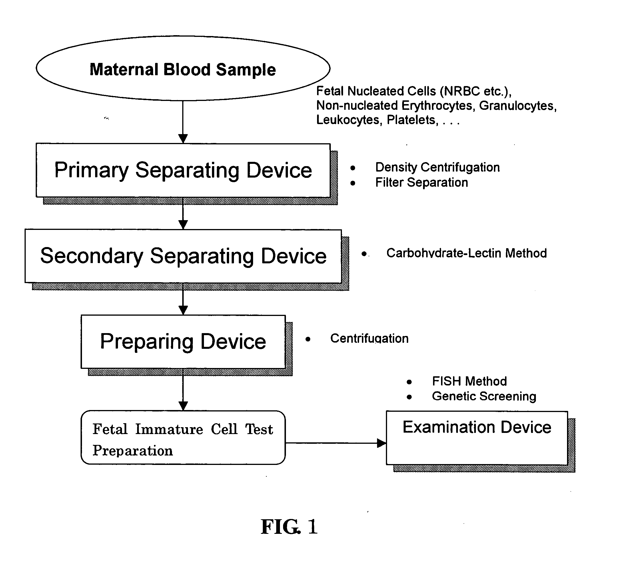
[0013] (1) comprises a primary (crude) separating device for removing mainly non-nucleated erythrocytes, leukocytes and platelets from a blood sample taken from a pregnant woman to obtain a primary (crude) separated sample, said primary separating device separating and removing mainly leukocytes which are nucleated cells, non-nucleated erythrocytes, platelets and the like under conditions which highly prevent loss of fetal nucleated cells among the various types of blood cells included in a maternal blood sample;

Problems solved by technology

The genetic diagnosis methods which are practiced clinically at present are invasive procedures such as amniocentesis, villus sampling and fetal blood collection, of which sampling of fetal cells by amniocentesis in particular allows for a positive diagnosis, but also carries a high risk of miscarriage at about 1 / 300.
However, the work of extracting lymphocytes having a Y chromosome from an entire group of similar lymphocytes is extremely difficult, and attempts to separate and concentrate them by means of a nylon wool column or density centrifugation did not succeed in yielding a practical solution.
Additionally, the presence of fetal cells remaining from past pregnancies was also considered to be a problem, and the development of a safe method of positive fetal diagnosis allowing for testing of a desired neonatal fetus did not progress any further.
However, the proportion of their presence in the maternal blood is extremely low at 1 / 105 to 1 / 107 of all nucleated cells, and just as with fetal cell diagnosis by lymphocytes, there are technical obstacles to achieving a process of separating and detecting a sufficient number of fetal nucleated erythroblasts from maternal nucleated cells such as leukocytes.
While attempts have been made to separate the erythroblasts by means of flow cytometry using transferin receptor antibodies or magnetic beads, the problem of specificity of antibodies has precluded the efficient detection of erythroblasts having Y chromosomes.
On the other hand, there are collection methods in which, instead of performing cell separation, the erythroblasts are identified morphologically and picked out one at a time by micromanipulation, but the process of discriminating erythroblasts from among masses of maternal nucleated cells requires considerable technical expertise and is extremely time-consuming.
Thus, this method requires special equipment, and for example, can take several days to process a single specimen.
However, at least 30 fetal cells are considered to be necessary in order to establish a general and practical fetal testing method for testing genes and chromosomes in a statistically conclusive manner, which makes this sampling method unrealistic when bearing in mind that the quantity of blood which may be safely collected from a pregnant woman is at most 10 cc, so that even to this day, there is a strong demand for a method for quickly and conveniently separating and concentrating erythroblasts at a high yield.
When separating and recovering blood cells such as erythroblasts, the peripheral blood is generally first pretreated using density centrifugation, but the erythroblasts were found to have been damaged during this pretreatment stage by density centrifugation.
Furthermore, in order to know of chromosome abnormalities and the like in nucleated erythroblasts separated and purified in this way, they must be inspected by the FISH (Fluorescence In Situ Hybridization) method or the like, but this gives rise to practical problems which need to be overcome during the process of preparing specimens appropriate for such testing.

Method used


Image

  • Blood cell separating system
  • Blood cell separating system
  • Blood cell separating system

Examples


example 1

Primary Separation by Density Centrifugation

[0064] Histopaque (Sigma) was obtained to use as a density centrifugation reagent, to which sodium diatrizoate was added and 6 types of density gradient fluid with specific gravities adjusted to 1.077-1.105 were prepared. 7 cc of venous blood were taken from women who were 10-20 weeks pregnant, then centrifugated for 30 minutes in each density gradient fluid (20° C., 1500 rpm). The cells collected around the boundary between the density gradient fluid and plasma component (upper layer) were recovered, then centrifugally rinsed with a biological buffer solution to obtain a crude separated sample with most of the non-nucleated erythrocytes and platelets removed.

[0065] The samples primarily purified under the various density conditions were secondarily separated by the carbohydrate-lectin method. As the substrate, a plastic chamber slide (2 wells, product of Nalgenunc) was used. The glycoconjugate polymer coated onto the substrate was PVMeA...

example 2

Additional Separation by Panning

[0067] A plastic chamber slide (4 wells, product of Nalgenunc) was treated with FCS or a 0.01 wt % aqueous solution of a glycoconjugate polymer (PV-Sugar) (product of Netech). As the glycoconjugate polymer, those having the structures of glucose, maltose, gluconic acid, N-acetylglucosamin, mannose, lactose or melibiose were used.

[0068] Density gradient centrifugation was performed on umbilical blood recovered after birth according to a standard method using Histopaque (d, 1.095), and the cells aggregating near the boundary between the Histopaque and plasma were collected. The samples were resuspended in RPMI1640 to which 10 wt % FCS was added, and inoculated onto the above-described wells whose surfaces were coated with FCS or glycoconjugate polymers. After incubation for 30 minutes at 37° C., the unattached cells were recovered in the form of a cell suspension fluid, and the cells attached to the wells were stained with a Pappenheim stain to identi...

example 3

Primary Separation by Filter Separation

[0073] Instead of the density centrifugation method of Example 1, a primary separation was performed using a filter comprising an unwoven polyester fabric with an average pore size of 8 μm. A sample of maternal blood was diluted with a biological buffer solution containing 1 wt % BSA, then passed through the filter by natural dripping. Next, the buffer solution alone was passed through a filter to rinse away the residual erythrocytes in the filter. Subsequently, the buffer solution was passed in the opposite direction with a syringe pump, and the unattached cells which did not pass through the filter were recovered. The cell fraction which did not pass through the filter but did not strongly adhere to the filter was taken as the primary separated sample, which was secondarily separated by the carbohydrate-lectin method. Table 4 shows the results with a primary separated sample obtained from the above-described filter, in the form of a comparat...


PUM

PropertyMeasurementUnit
densityaaaaaaaaaa
temperatureaaaaaaaaaa

Abstract

A blood cell separating system is offered for precisely separating and concentrating rare fetal nucleated cells intermixed in the blood of a pregnant woman, to conveniently obtain test preparations capable of being used for prenatal chromosomal / genetic diagnosis. A blood cell separating system is characterized by comprising (1) a primary separating device for removing mainly non-nucleated erythrocytes, leukocytes and platelets from blood samples taken from a pregnant woman to obtain a primary separated sample, (2) a secondary separating device for using a carbohydrate-lectin method to remove residual non-nucleated erythrocytes and leukocytes from the primary separated sample obtained by the primary separating device to obtain a secondary separated sample with concentrated fetal nucleated cells, and (3) preparing device for preparing the secondary separated sample obtained by the secondary separating device.

Description

TECHNICAL FIELD [0001] The present invention relates to a system for the separation of blood cells using lectins, particularly to a system for efficiently removing non-nucleated erythrocytes and mature leukocytes, to separate and concentrate fetal nucleated erythroblasts, from samples containing nucleated erythroblasts which are fetal nucleated cells found in the maternal peripheral blood or umbilical blood of pregnant women, and further, a system for testing the chromosomes and genes thereof. BACKGROUND ART [0002] In the field of genetic diagnosis, the development of methods for prenatal diagnosis which do not endanger the embryo, i.e. the fetus, has been long anticipated. The genetic diagnosis methods which are practiced clinically at present are invasive procedures such as amniocentesis, villus sampling and fetal blood collection, of which sampling of fetal cells by amniocentesis in particular allows for a positive diagnosis, but also carries a high risk of miscarriage at about 1...

Claims


Application Information

Patent Timeline
Patent Type & Authority Applications(United States)
IPC IPC(8): C12N5/07C12N5/073C12N5/078G01N33/537G01N33/569G01N33/68
CPCG01N33/537G01N33/56966G01N2800/368G01N2400/00G01N33/689G01N33/48G01N33/49
Inventor YURA, HIROFUMISAITO, YOSHIOKITAGAWA, MICHIHIRO
Owner NETECH
Learn More