
Search for cancer markers by a novel screening method

a cancer marker and screening method technology, applied in the field of peptide concentration methods, can solve the problems of low specificity of pancreatic cancer markers, unsatisfactory treatment results of pancreatic cancer, and low usefulness in early diagnosis, and achieve the effect of efficient screening novel pancreatic cancer markers

Inactive Publication Date: 2005-05-19
JAPAN REPRESENTED BY DIRECTOR GEN OF AGENCY OF NAT CANCER CENT +1
View PDF1 Cites 0 Cited by
  • Summary
  • Abstract
  • Description
  • Claims
  • Application Information

AI Technical Summary

Benefits of technology

"The present invention provides a method for efficiently screening novel pancreatic cancer markers by combining a one-step sample preparation and matrix assisted mass spectrometry. This method allows for the discovery of low molecular weight peptides that can serve as pancreatic cancer markers. The invention also provides markers for diagnosing pancreatic cancer, as well as antibodies that can be used in diagnostic tools. The method involves concentrating low molecular weight peptides in serum-free cultured cells and comparing them using a mass spectrometer. The invention also provides a diagnostic kit or reagent for detecting or quantitating the peptides. Overall, the invention provides a more efficient and effective way to screen for pancreatic cancer markers."

Problems solved by technology

Thus, conventionally, it was often found at a stage when treatment is difficult, and was considered an intractable tumor.
Despite these advances in diagnostic methods, treatment results of pancreatic cancer have been below expectations.
Furthermore, though a sugar chain antigen such as CA19-9, a tumor marker, is useful as a marker for the diagnosis and monitoring of progressive pancreatic cancer, its usefulness in early diagnosis is not satisfactory.
The problem, therefore, is the low specificity of markers for pancreatic cancer.
Thus, it was only possible to detect substances that are present in large quantities in test samples.
However, the method that discovered the usefulness of this proGRP as a tumor marker is one in which sera of patients with SCLC and sera of patients with non-SCLC are measured by RIA, and thus it is necessary to generate many different antibodies and to carry out the determination of a multitude of samples, requiring a large amount of time and effort.
In addition, new peptide candidates cannot be found by this method.

Method used


Image

  • Search for cancer markers by a novel screening method
  • Search for cancer markers by a novel screening method
  • Search for cancer markers by a novel screening method

Examples


example 1

Preparation of a Serum-free Culture Supernatant

[0099] A cell line was cultured to confluence in a RPMI1640 medium containing 10% fetal bovine serum (Life Technologies). After aspirating the medium, it was washed three times in 10 ml each of a serum-free medium, and further cultured at 37° C. for 1 hour. After two more washings, 3 ml of the medium per 10 cm culture dish was added and cultured for 48 hours.

[0100] After removing the cells in a centrifuge, the culture supernatant was transferred to a polypropylene tube and stored at 4° C. until analysis. The protein concentration was determined using the Protein Assay kit (Bio-Rad) with immunoglobulin as a standard. As a control, a medium containing 10% serum was added to an unused dish and cultured for 72 hours, which was then washed as described above and used. The medium obtained is termed a “cell-free” culture supernatant. For all cell lines, trypan blue dye exclusion test was performed, confirming that 95% or more cells survived ...

example 2

Optimization of SELDI Analysis for Profiling Low Molecular Weight Proteins

[0101] Mass was determined using Ciphergen SELDI Protein Biology System 2 (Ciphergen Biosystems, Inc., CA) as a mean of 110 laser shots. For spectral analysis, monovalent and divalent molecular ions of bovine insulin were used to carry out external calibration (m / z 5734.56 and 2867.78). Values of m / z when referred to in the text were rounded to three-place or four-place significant figures. Precision of mass was better than 1000 ppm (0.1%) in the analytical range.

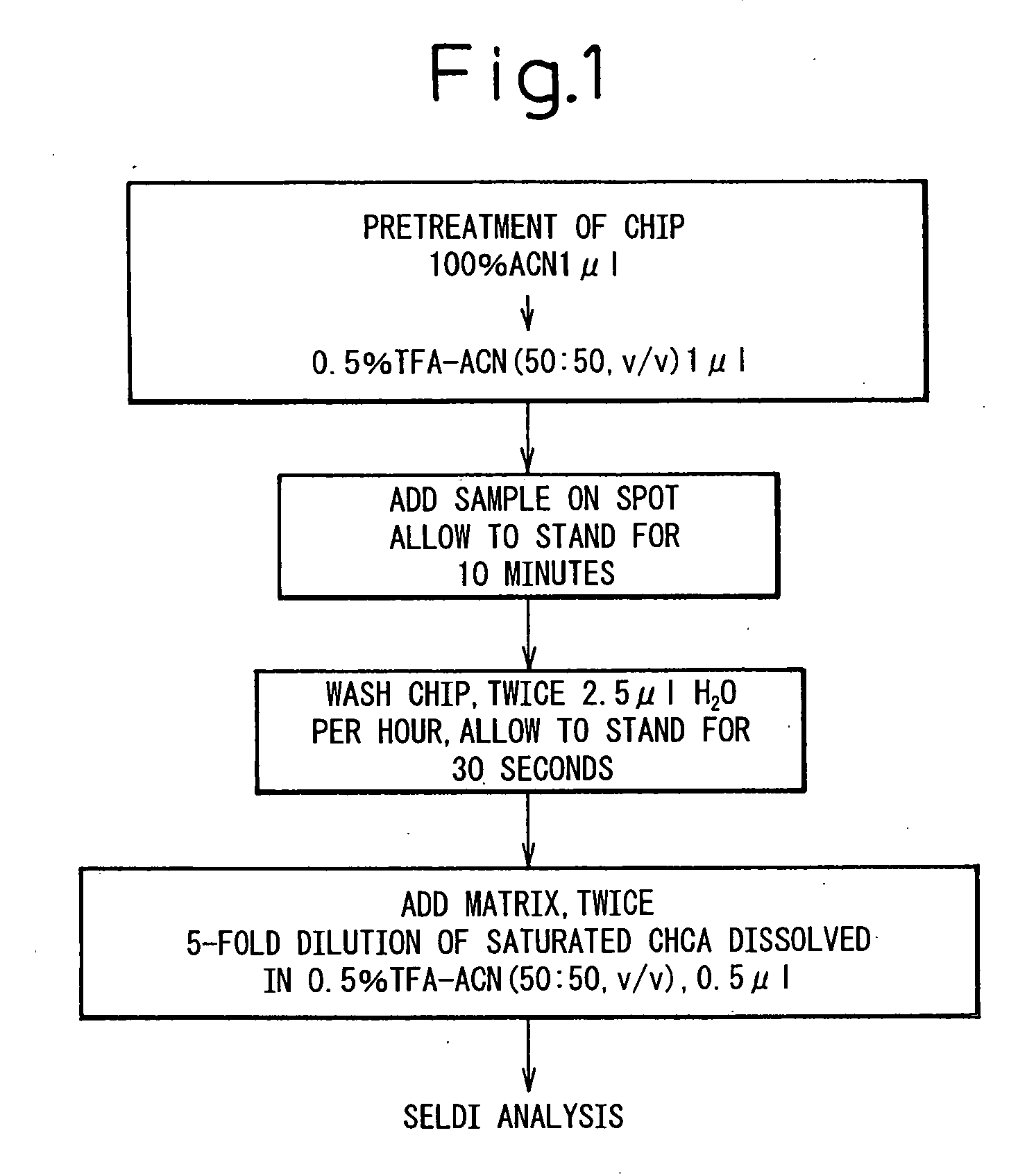
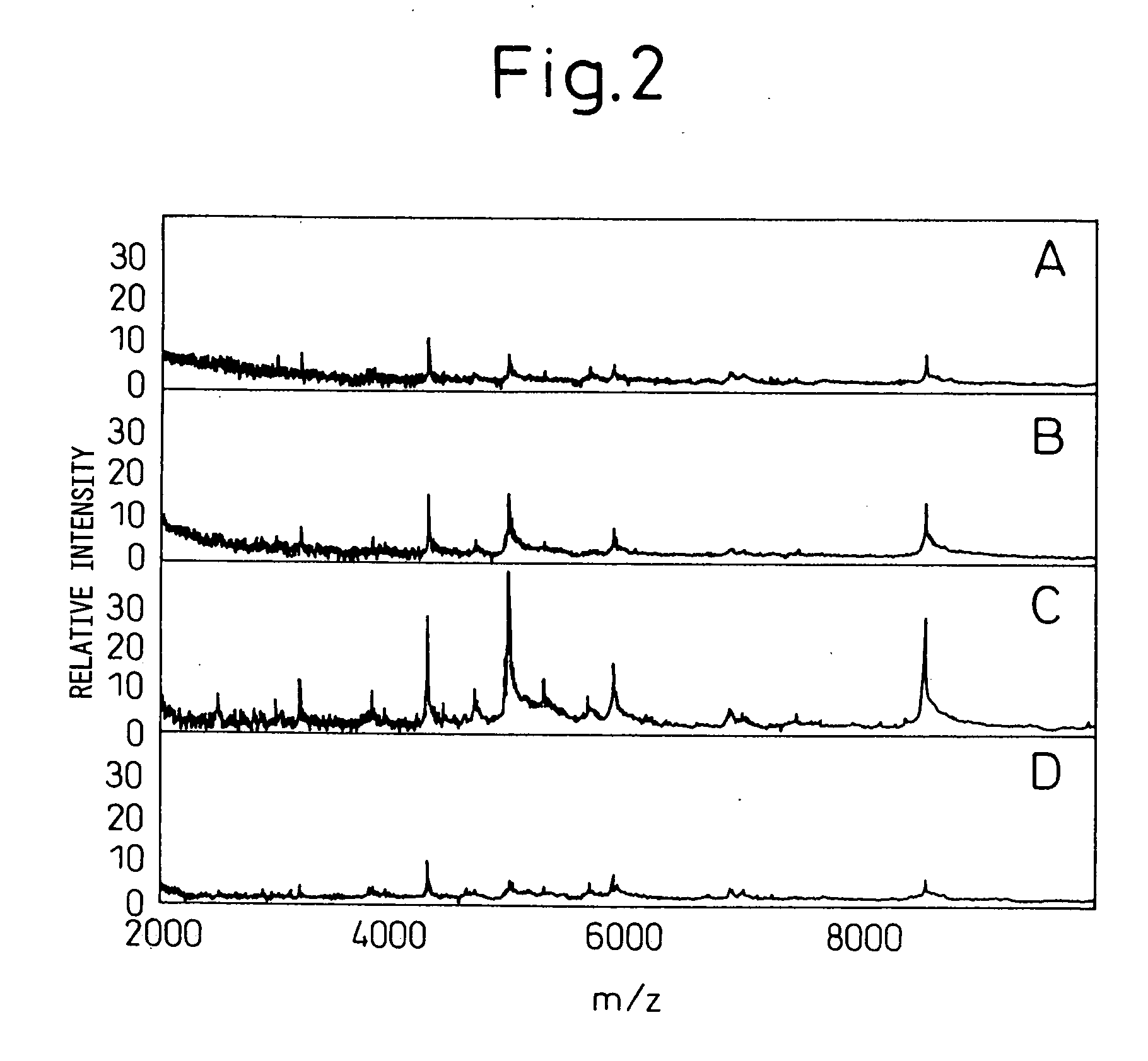
[0102] In order to analyze low molecular weight proteins, the H4 protein chip array having a chemical surface that captures protein with hydrophobic interaction was used. The experimental conditions for sample binding and matrix cocrystals were optimized to obtain spectra. As the application of mass spectrometry in protein profiling of biological materials is at a development stage and thus there are no established protocols, the optimization thereo...

example 3

Comparison of Tricine Electrophoresis and SELDI Analysis

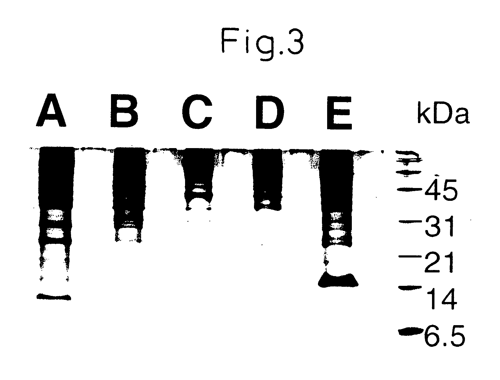
[0105] Using the protocol in Example 2, low molecular weight molecules were analyzed for five cell lines using 5 μl each. Protein concentration varied depending on the cell line (0.05-0.8 mg / ml), being equivalent to 250 ng to 2 μg analyzed per spot as the amount of total protein. Data were compared with those obtained by tricine SDS-PAGE that has been established as a technique for protein separation [Schagger et al., Anal. Biochem. 166:368-379 (1987)]. In order to perform tricine SDS polyacrylamide gel electrophoresis, 100 μl of the culture supernatant was concentrated to 20 μl, which was then dissolved in the sample buffer.

[0106] The gel composition used was 16.5% T-3% C. The bands were visualized by silver staining. Even when 100 μl of sample was used, electrophoresis detected only a small number of bands in the range of molecular weight 20000 or lower (FIG. 3). In the SELDI analysis, on the other hand, many signals were f...


PUM

PropertyMeasurementUnit
volumeaaaaaaaaaa
concentrationaaaaaaaaaa
v/vaaaaaaaaaa

Abstract

The present invention relates to a method of concentrating low molecular weight peptides in the supernatant of serum-free cultured cells, said method comprising allowing the peptides to bind to a strong cation exchanger under an acid condition, and eluting them under an alkali condition to concentrate the peptide. Furthermore, peptides having the amino acid sequence as set forth in SEQ ID NO: 1 or 2, and a method of screening cancer markers using antibody to these peptides, are disclosed.

Description

FIELD OF THE INVENTION [0001] The present invention relates to a peptide concentration method, based on a one-step sample preparation method, for the detection of low molecular weight peptides secreted from cultured cells into a serum-free medium, and a differential display method based on matrix assisted mass spectrometry, a novel method of screening cancer markers using such a method, peptides that could be pancreatic cancer markers detected by said screening method, antibodies to said peptides, hybridomas that produce said antibodies, a diagnostic reagent comprising said antibody, and the like. BACKGROUND ART [0002] Pancreatic cancer is a tumor that develops at the retroperitoneal space, a position clinically difficult to be detected. Thus, conventionally, it was often found at a stage when treatment is difficult, and was considered an intractable tumor. Diagnostic methods have made a great progress due to diagnostic imaging such as US, CT and MRI, and endoscopy based on recent t...

Claims


Application Information

Patent Timeline
Patent Type & Authority Applications(United States)
IPC IPC(8): C07K1/18C07K14/47C07K14/705C07K16/30C12P21/08G01N33/574
CPCC07K14/47C07K14/4748C07K14/705Y10T436/141111C07K16/303G01N33/574C07K16/30
Inventor SASAKI, KAZUKISATO, KAEYAMAGUCHI, KEN
Owner JAPAN REPRESENTED BY DIRECTOR GEN OF AGENCY OF NAT CANCER CENT
Learn More