Cytotoxicity mediation of cells evidencing surface expression of CD44

a cytotoxicity and surface expression technology, applied in the field of cancer diagnosis and treatment, can solve the problems of insufficient evidence, inconsistent findings, and increased likelihood of tumor invasiveness and induction of angiogenesis through the ecm

Inactive Publication Date: 2005-05-12
F HOFFMANN LA ROCHE & CO AG
View PDF26 Cites 12 Cited by
  • Summary
  • Abstract
  • Description
  • Claims
  • Application Information

AI Technical Summary

Benefits of technology

[0037] The instant inventors have previously been awarded U.S. Pat. No. 6,180,357, entitled “Individualized Patient Specific Anti-Cancer Antibodies” directed to a process for selecting individually customized anti-cancer antibodies, which are useful in treating a cancerous disease. For the purpose of this document, the terms “antibody” and “monoclonal antibody” (mAb) may be used interchangeably and refer to intact immunoglobulins produced by hybridomas (e.g. murine or human), immunoconjugates and, as appropriate, immunoglobulin fragments and recombinant proteins derived from said immunoglobulins, such as chimeric and humanized immunoglobulins, F(ab′) and F(ab′)2 fragments, single-chain antibodies, recombinant immunoglobulin variable regions (Fv)s, fusion proteins etc. It is well recognized in the art that some amino acid sequence can be varied in a polypeptide without significant effect on the structure or function of the protein. In the molecular rearrangement of antibodies, modifications in the nucleic or amino acid sequence of the backbone region can generally be tolerated. These include, but are not limited to, substitutions (preferred are conservative substitutions), deletions or additions. Furthermore, it is within the purview of this invention to conjugate standard chemotherapeutic modalities, e.g. radionuclides, with the CDMAB of the instant invention, thereby focusing the use of said chemotherapeutics. The CDMAB can also be conjugated to toxins, cytotoxic moieties, enzymes e.g. biotin conjugated enzymes, or hematogenous cells, whereby an antibody conjugate is formed.

Problems solved by technology

Also, the intracellular degradation of HA by lysosomal hylauronidases after internalization of the CD44-HA complex can potentially increase the likelihood of tumor invasiveness and induction of angiogenesis through the ECM.
However, inconsistent findings were observed in a majority of tumor types and this is probably due to a combination of reagents, technique, pathological scoring and cell type differences between researchers.
Taken together, there is no evidence that antibodies such as these are suitable for use as cancer therapeutics since they either are not directed against cancer (e.g. activate lymphocytes), induce cell proliferation, or when used with cytotoxic agents inhibited drug-induced death of cancer cells.
Survival of the treated animals was concomitantly increased.
The antibody was only effective if administered before lymph node colonization, and was postulated to interfere with cell proliferation in the lymph node.
None of these studies demonstrated human utility for this antibody.
One notable feature of this antibody was that it recognized all isoforms of CD44, which suggests limited possibilities for therapeutic use.
Further limiting the application of BIWA 1 is the immunogenicity of the murine antibody (11 of 12 patients developed human anti-mouse antibodies (HAMA)), heterogenous accumulation throughout the tumor and formation of antibody-soluble CD44 complexes.
Although deemed to be acceptable in both safety and tolerablility for the efficacy achieved, these studies have higher rates of adverse events compared to other non-radioisotope conjugated biological therapies in clinical studies.
Although there were some effects on tumor size these effects did not fulfill the criteria for objective responses to treatment.
Thus, it can be seen that although Mab U36 is a highly specific antibody the disadvantage of requiring a radioimmunoconjugate to achieve anti-cancer effects limits its usefulness because of the toxicity associated with the therapy in relation to the clinical effects achieved.
These results, though they are encouraging and support the development of anti-CD44 monoclonal antibodies as potential cancer therapeutics, demonstrate limited effectiveness, safety, or applicability to human cancers.
More specifically the inventors point out that this antibody does not recognize human cancers.

Method used


Image

  • Cytotoxicity mediation of cells evidencing surface expression of CD44
  • Cytotoxicity mediation of cells evidencing surface expression of CD44
  • Cytotoxicity mediation of cells evidencing surface expression of CD44

Examples


example 1

[0076] The hybridoma cell line H460-16-2 was deposited, in accordance with the Budapest Treaty, with the American Type Culture Collection, 10801 University Blvd., Manassas, Va. 20110-2209 on Sep. 4, 2002, under Accession Number PTA-4621. In accordance with 37 CFR 1.808, the depositors assure that all restrictions imposed on the availability to the public of the deposited materials will be irrevocably removed upon the granting of a patent.

Antibody Production:

[0077] H460-16-2 monoclonal antibody was produced by culturing the hybridoma in CL-1000 flasks (BD Biosciences, Oakville, ON) with collections and reseeding occurring twice / week. The antibody was purified according to standard antibody purification procedures with Protein G Sepharose 4 Fast Flow (Amersham Biosciences, Baie d'Urfé, QC). It is within the scope of this invention to utilize monoclonal antibodies that are human, humanized, chimerized or murine antibodies.

example 2

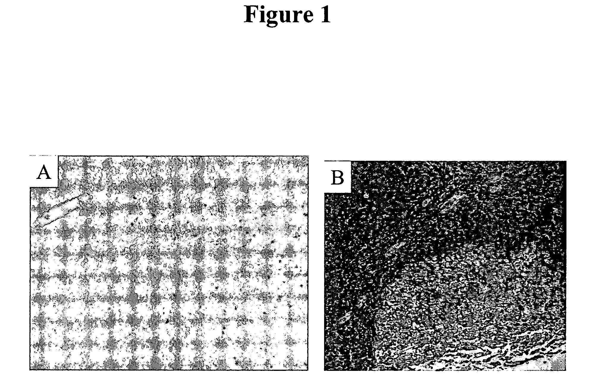
Normal Human Tissue Staining

[0078] IHC studies were previously conducted to characterize H460-16-2 antigen distribution in humans (Ser. No. 10 / 603,000) and in comparison to L178 (Ser. No. 10 / 647,818). The current studies compare H460-16-2 to another antibody directed against CD44 (BU75) since the H460-16-2 antigen may be a cancer variant of CD44 as determined previously by biochemical methods. Binding of antibodies to 59 normal human tissues was performed using a human normal organ tissue array (Imgenex, San Diego, Calif.). All primary antibodies (H460-16-2; BU75 anti-CD44 (BIOCAN Scientific Inc., Mississauga, ON); and mouse IgG, negative control (Dako, Toronto, ON)) were diluted in antibody dilution buffer (Dako, Toronto, ON) to a concentration of 5 μg / ml (found to be the optimal concentration in previous optimization steps). The negative control antibody has been shown to be negative to all mammalian tissues by the manufacturer. The procedure for IHC is as follows.

[0079] Tissue...

example 3

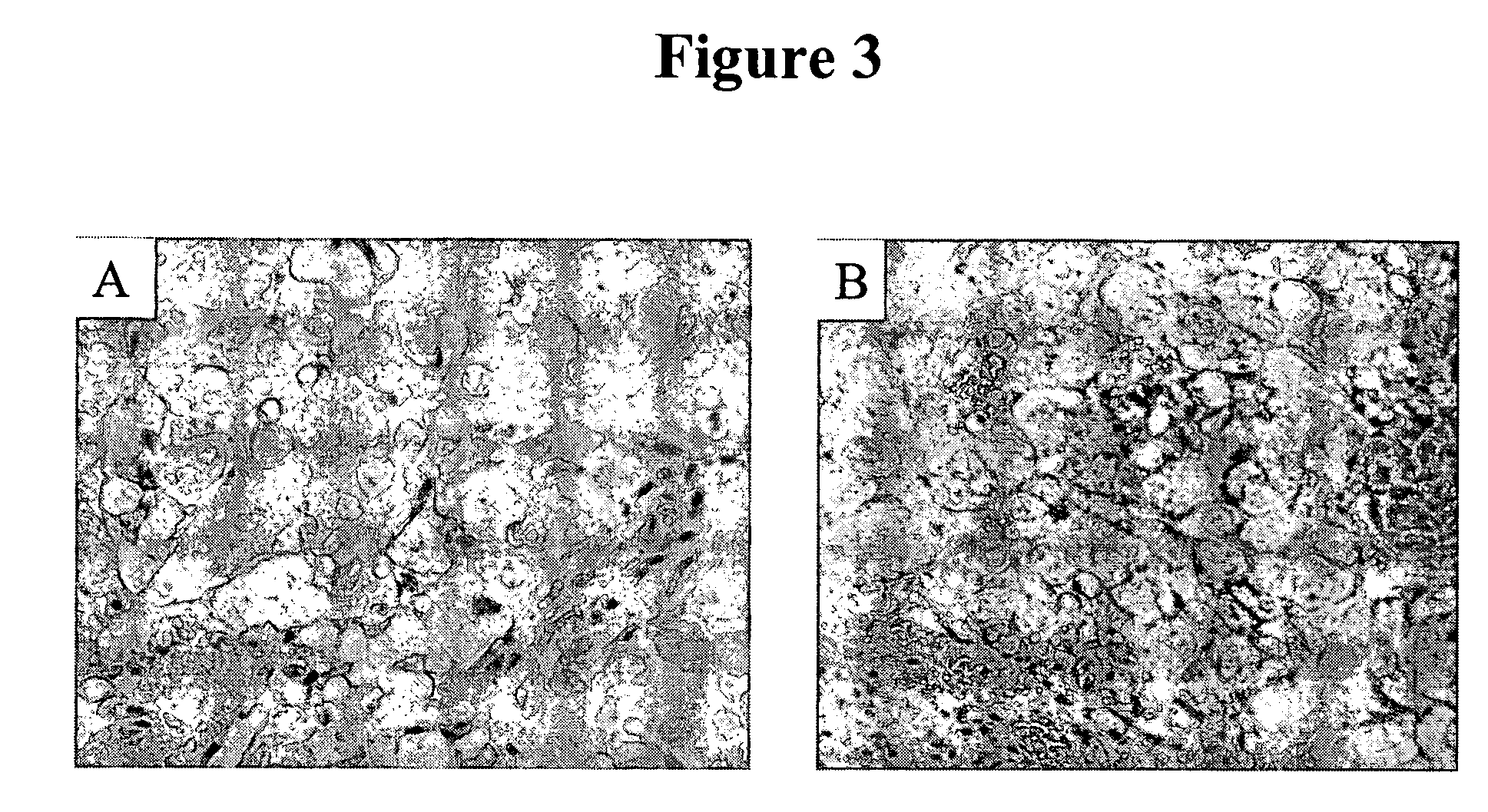
Human Breast Tumor Tissue Staining

[0082] Previous IHC studies were undertaken to determine the cancer association of the H460-16-2 antigen with human breast cancers and whether the H460-16-2 antibody was likely to recognize human cancers (Ser. No. 10 / 603,000) and how it compared to anti-CD44 staining with L178 (Ser. No. 10 / 647,818). Currently, a comparison was made for BU75 anti-CD44 staining, c-erbB-2 anti-Her2 and an antibody directed towards Aspergillus niger glucose oxidase, an enzyme which is neither present nor inducible in mammalian tissues (negative control). A breast cancer tissue array derived from 50 breast cancer patients and 10 samples derived from non-neoplastic breast tissue in breast cancer patients was used (Imgenex Corporation, San Diego, Calif.). The following information was provided for each patient: age, sex, American Joint Committee on Cancer (AJCC) tumor stage, lymph node, estrogen receptor (ER) and projesterone receptor (PR) status. The procedure for IHC f...


PUM

PropertyMeasurementUnit
concentrationaaaaaaaaaa

Abstract

This invention relates to the diagnosis and treatment of cancerous diseases, particularly to the mediation of cytotoxicity of tumor cells; and most particularly to the use of cancerous disease modifying antibodies (CDMAB), optionally in combination with one or more chemotherapeutic agents, as a means for initiating the cytotoxic response. The invention further relates to binding assays which utilize the CDMABs of the instant invention.

Description

REFERENCE TO RELATED APPLICATIONS [0001] This application is a continuation-in-part of application Ser. No. 10 / 647,818 filed Aug. 22, 2003, which is a continuation-in-part of application Ser. No. 10 / 603,000, filed Jun. 23, 2003, which is a continuation-in-part of application Ser. No. 09 / 727,361, filed Nov. 29, 2000, which is a continuation-in-part of application Ser. No. 09 / 415,278, filed Oct. 8, 1999, now U.S. Pat. No. 6,180,357 B 1, the contents of each of which are herein incorporated by reference.FIELD OF THE INVENTION [0002] This invention relates to the diagnosis and treatment of cancerous diseases, particularly to the mediation of cytotoxicity of tumor cells; and most particularly to the use of cancerous disease modifying antibodies (CDMAB), optionally in combination with one or more chemotherapeutic agents, as a means for initiating the cytotoxic response. The invention further relates to binding assays, which utilize the CDMAB of the instant invention. BACKGROUND OF THE INV...

Claims


Application Information

Patent Timeline
Patent Type & Authority Applications(United States)
IPC IPC(8): A61K39/395A61K47/48A61K49/00A61P35/00C07K16/00C07K16/28C07K16/30G01N33/50G01N33/566G01N33/567G01N33/569G01N33/574
CPCC07K16/3015G01N33/57449A61K39/39558A61K45/06A61K47/48561A61K49/0058A61K51/1027A61K2039/505B82Y5/00C07K16/00C07K16/2884C07K16/30G01N33/57484C07K2316/95G01N33/5011G01N33/5014G01N33/5082G01N33/566G01N33/56972G01N33/574G01N33/57415G01N33/57419G01N33/57423A61K2300/00A61K47/6849A61P35/00C07K2317/73
Inventor YOUNG, DAVID S.F.HAHN, SUSAN E.FINDLAY, HELEN P.
Owner F HOFFMANN LA ROCHE & CO AG
Why Eureka
Try Eureka