Improved in vitro method of culturing mammalian cells for autologous cell implantation/transplantation methods
- Summary
- Abstract
- Description
- Claims
- Application Information
AI Technical Summary
Benefits of technology
Problems solved by technology
Method used
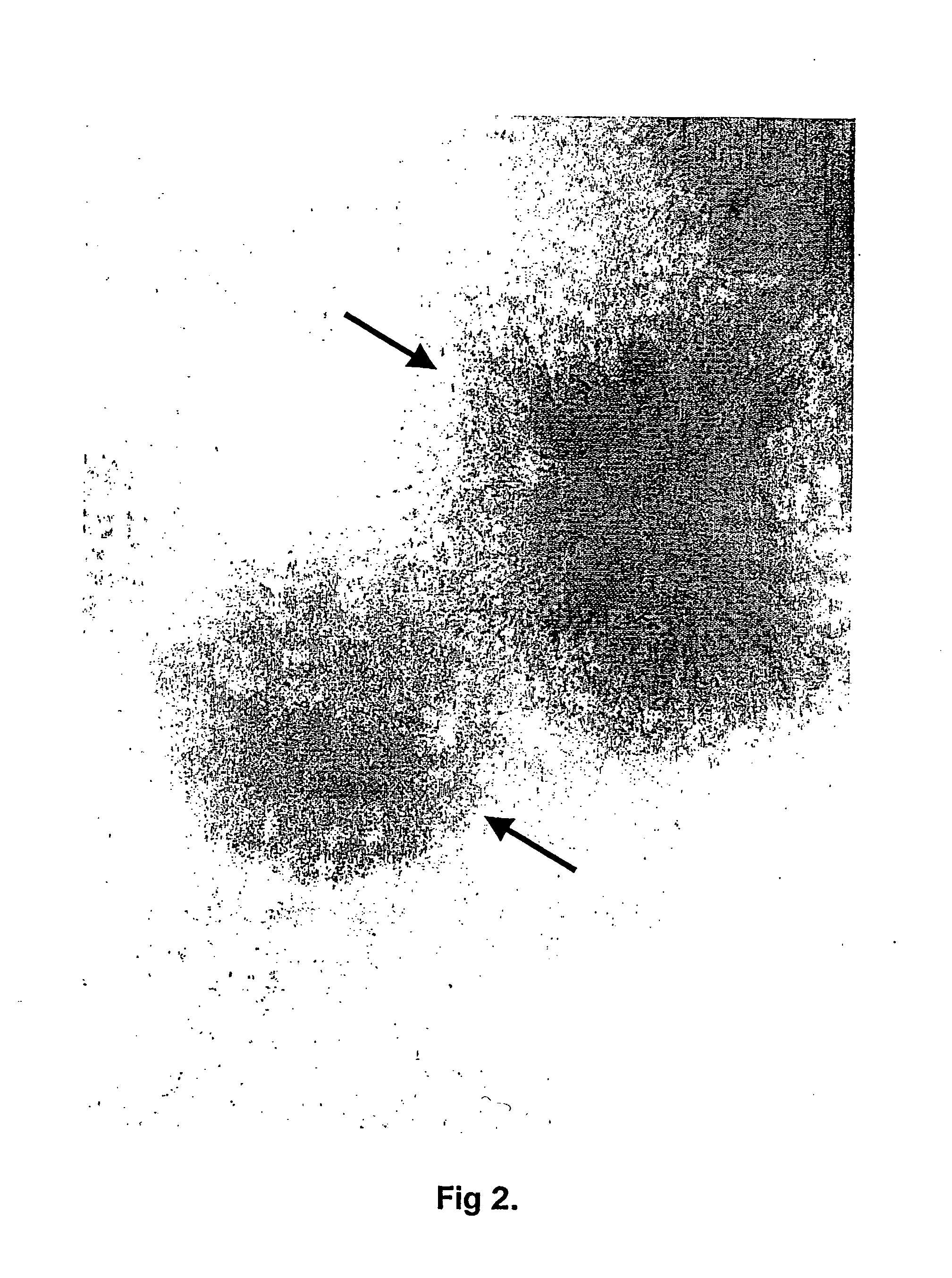
Image


Examples
example 1
[0143] Protocol for Culturing Cartilage Explants and Producing Cell Colony Forming Unit(s) in vitro for Autologous Chondrocyte Implantation (ACI) Use
[0144] The cartilage explant system is currently been used in the company's cell production unit related to the clinical trial: IBO-ACI-02, investigating the repair efficiency of cultured autologous chondrocytes implanted into articular cartilage defects in the knee. Until December 2001, the cartilage biopsies have been obtained from seven patients suffering from knee problems. The seven biopsy explants have each been cultured to about 11 million cells and implanted into the patients successfully at two major Danish hospitals in the Copenhagen area. The first outcome of the clinical investigations will be present during 2003 / 4.
[0145] In short, a cartilage biopsy was harvested from the patient's knee and immediately transferred into sterile growth medium, supplemented with L-ascorbic acid 2-phosphate [50 .mu.g / ml (300 .mu.mol / l)] and gen...
example 2
[0149] Differential Isolation of Chondrogenic Cell Lines in the Cartilage Explant System
[0150] A cartilage biopsy was harvested from the patient's knee with a needle or a scalpel and transferred into sterile growth medium as described above. The cartilage piece was cut horizontal (tangential to the cartilage surface) with a sharp sterile scalpel into various zones. The cutting process was done under a microscopy in the laminar airflow hood in order to secure the proper separation and selection. The cartilage piece can be cut horizontal several times into various zones or the cartilage biopsy can be cut just one time into two layers. After separating the various cartilage layers into sterile tubes by cutting several times, the cartilage pieces were finally washed several times with PBS buffer with magnesium and calcium ions before adding the cartilage pieces to the tissue culture flasks. The further cell culturing process took place in a similar fashion as described in the protocol i...
example 3
[0151] Partial Enzymatic Treatment of Cartilage Explants With Crude Collagenase to Obtain CFU According to the Invention
[0152] A cartilage biopsy in the weighting of 100 mg was harvested from the patient's knee and transferred into sterile growth medium with antibiotic and fungizone as described in Example 1. The cartilage biopsy was washed in PBS with magnesium and calcium buffer and dissected vertical and horizontal with a sharp sterile scalpel into 24 mm cartilage pieces (explants). The cartilage explants in 25 ml growth medium were added to a 75 cm.sup.2 tissue culture flask together with 10 U / ml (10 U / ml.times.25 ml=250 Units) crude collagenase from Clostridium Histolyticum (type 1A (C9891) or type 1 (C0130), or type VIII (C2139), type II (C6885) or type IV (C5138) or type V (C9283) (all obtained from Sigma chemicals). The tissue culture flask was then incubated at 37.degree. C. with 5% CO.sub.2 with low shaking for 18 hours.
[0153] After 18 hours of incubation with crude collag...
PUM
 Abstract
Description
Claims
Application Information
 - R&D
- Intellectual Property
- Life Sciences
- Materials
- Tech Scout
- Unparalleled Data Quality
- Higher Quality Content
- 60% Fewer Hallucinations
Browse by: Latest US Patents, China's latest patents, Technical Efficacy Thesaurus, Application Domain, Technology Topic, Popular Technical Reports.
© 2025 PatSnap. All rights reserved.Legal|Privacy policy|Modern Slavery Act Transparency Statement|Sitemap|About US| Contact US: help@patsnap.com