Method and apparatus for isolating and detecting biological and other particles

a biological and other particle technology, applied in the field of biological particle isolating and detecting devices, can solve the problems of ineffective cyto-centrifuges, inability to exclude the presence of organisms not targeted, inappropriate anti-microbial therapy, etc., to accelerate the turn-around time of microbiology laboratories, reduce the risk of cross contamination, and be ergonomically convenient

Active Publication Date: 2022-09-20
AINCOBIO LLC
View PDF25 Cites 0 Cited by
  • Summary
  • Abstract
  • Description
  • Claims
  • Application Information

AI Technical Summary

Benefits of technology

[0024]Many molecular assays, “lab-on-chip” devices, and point-of-care technologies have failed to supplant routine blood cultures for the detection and quantification of pathogens. The present design and process was meticulously crafted by in-depth field research together with expertise in the art and novel and inventive techniques that centered around the deficiencies encountered by technicians “in the field” and an imaginative approach to correcting these inadequacies. The present invention has the additional advantage not only because it uniquely blends cultivation methods and new sample preparation technologies to accelerate microbiology laboratory turn-around-time but also because it integrates long-felt needs in specimen processing for microorganism detection, differentiation and analysis into the existing workflow to create a system that is ergonomic, efficient and less prone to cross contamination.
[0025]The present invention pushes the limits of rapid and non-destructive detection of low-level microbes. Electrical impedance spectroscopy (EIS) is an established method for detecting microorganisms. Several instruments commercially available employ this technology: Bactometer™ (Biomerieux), RABIT™ (DW Scientific) Malthus™ (Malthus Instrument), BacTrac™ (SyLab). However, these commercial instruments lack concentration and isolation steps needed to achieve rapid detection of low-level microorganisms. The present invention harnesses macro- and microscale physics to exploit the biophysical properties of microbes to clean up the sample and concentrate them to boost sensitivity of the detector.
[0027]Concisely, preferred embodiments of the present invention have several distinct advantages over conventional indirect and direct in vitro detection methods. First, it makes it possible to concentrate target subpopulations of particles within the sample without the need for bioengineered targeting probes. In this way, it allows users to remain hypothesis-independent when beginning the analysis of the sample. Secondly, by integrating low volume physical chambers (from picoliters up to several hundred microliters) within a larger volume vessel, the device bridges a critical technological gap which hitherto has hindered the use of microscale physical phenomena in analyzing large volume specimens typically required in industrial, environmental, or clinical applications. The design and operation of the device allow for an operator to analyze large volumes demanded by these applications within a much shorter interval than can be realized using existing technologies. Importantly, the methods discussed here accomplish this accelerated processing time within a single, disposable device and without the needed for complicated manual steps and risk of sample contamination. Yet another advantage to the current device is its ability to seamlessly integrate into current laboratory workflow: Clinical labs are understaffed, overtasked, and technician turnover rates are high. Several compelling technologies require numerous manual fluid transfer and washing steps, making these solutions less likely to be adopted in clinical labs. Preferred embodiments of the present invention comprise an automated, self-contained (no manual fluid transfer steps) device, and require minimal training.

Problems solved by technology

However, in many applications the cyto-centrifuge is not useful due to the presence of background particulate matter (debris, or perhaps other cell types) that prevent or interfere with the direct observation of target biological particles.
The problem with this approach is that the operator needs to design the antibodies against specific targets.
However, a negative result does not exclude the possibility that organisms which were not targeted are present.
In the context of bloodstream infections, the negative result does not provide a complete diagnosis, and leads to inappropriate antimicrobial therapy.
Many molecular assays, “lab-on-chip” devices, and point-of-care technologies have failed to supplant routine blood cultures for the detection and quantification of pathogens.
However, these commercial instruments lack concentration and isolation steps needed to achieve rapid detection of low-level microorganisms.

Method used


Image

  • Method and apparatus for isolating and detecting biological and other particles
  • Method and apparatus for isolating and detecting biological and other particles
  • Method and apparatus for isolating and detecting biological and other particles

Examples


example 1

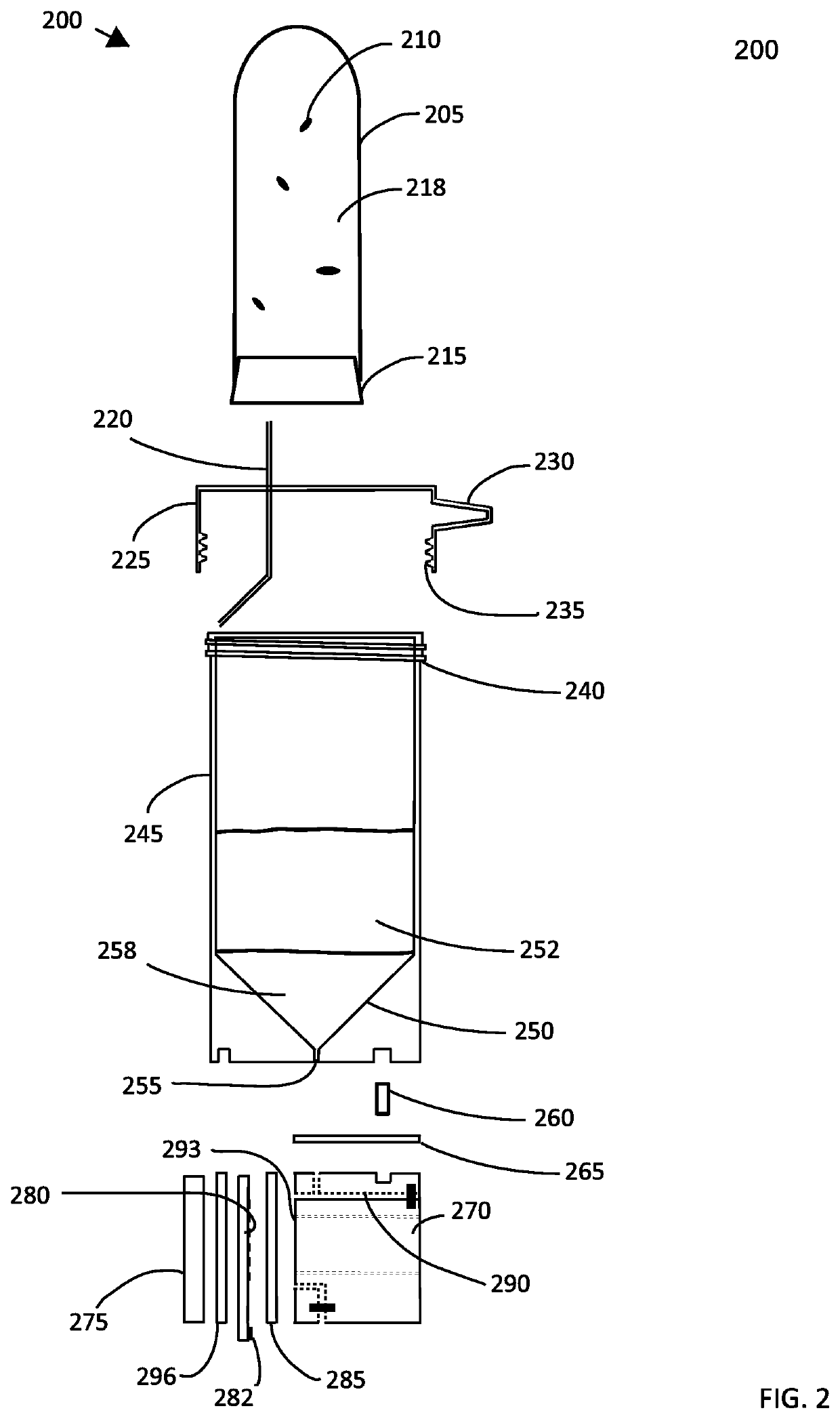
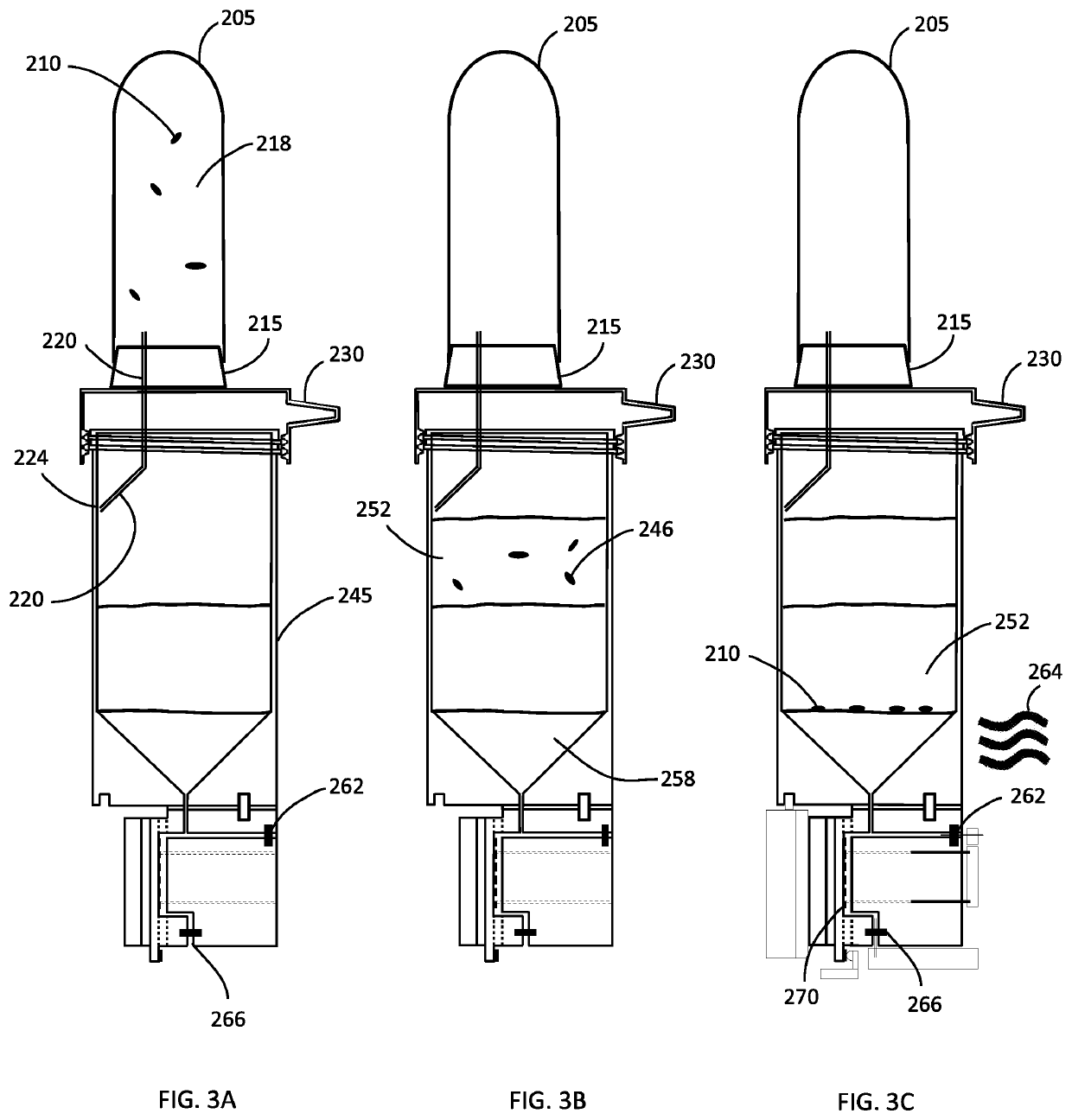
[0135]Firstly, the use of a density gradient medium as a barrier to the ions in the original sample was tested and shown to work successfully. Briefly, a substance called OptiPrep is created, which is about 60% iodixanol, was diluted with 5% D-mannitol to create a solution having a physical density of 1080 kg / m3. About 500 microliters of this density gradient medium (OptiPrep) is placed in a 2 mL centrifuge tube. Then, about 500 microliters of whole blood, previously treated with saponin (final concentration of saponin was ˜5 mg / mL), is spiked with bacteria (Enteroccocus faecalis) and is carefully layered on top of the density gradient medium using a pipettor equipped with a wide-bore pipette tip, so as to not disturb the heavier bottom fluid layer and cause mixing of the sample and that layer. The tube containing the sample and the separation density medium is then centrifuged at 8,000 RPM for 15 minutes using a standard bench top centrifuge having a rotor diameter of about 20 cm. ...

example 2

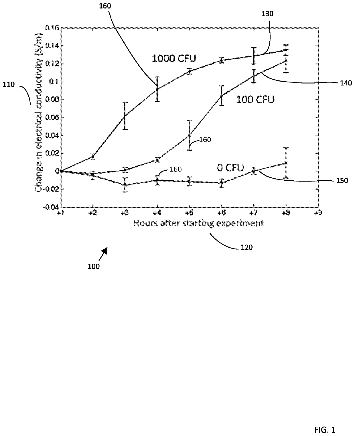
[0137]In another set of experiments, one mL of blood is placed above one mL of the density gradient medium. In this separate experiment, a larger volume allows for the electrical conductivity of the density gradient medium to be measured off-line using a flow-through probe and a bench-top conductivity meter. In three separate experiments, the highest conductivity value measured was 62.5 mS / m, which is about 24× lower than the electrical conductivity of whole blood (˜1.5 S / m). The density gradient not only presents the migration of some biological particles to the bottom of the device while still allowing sufficiently dense particles to be transported through the density medium, it also provides a barrier to the diffusion of ions from the sample into lower fluid layers.

example 3

[0138]In yet another set of experiments, a small hole was bored in the bottom of the centrifuge tube and a rubber septum having dimensions of 11 mm in diameter and about 3 mm in thickness was positioned at the bottom of the centrifuge tube. This allowed the operator to access the bottom layer without disturbing the sample and the fluid interface between the density gradient medium and the sample when extracting a sub-sample from the density gradient medium. To test this device, E. coli was grown overnight in lysogeny broth, and 100 microliters of the culture was harvested and layered on top of a density gradient medium. The gradient medium was made by mixing Optiprep and a mannitol solution to adjust the density to approximately 1080 kg / m3. After centrifuging the tube at 10,000 RPM for 10 minutes, the sample liquid appeared clarified, and a pellet formed along the surface of the septum. A needle was used to puncture the septum the plunger was withdrawn and depressed repeatedly to re...


PUM

PropertyMeasurementUnit
volumetric flow rateaaaaaaaaaa
frequenciesaaaaaaaaaa
voltagesaaaaaaaaaa

Abstract

An apparatus and method for isolating bacterial particles in a sample using a container with material in temporary fluid blocking position to lower orifice in the container, a separation medium having an electrical conductivity lower than and physical density greater than that of the sample above the material that supports a sample concentrate after passing through the separation medium when exposed to centrifugal force, a heating element for liquefying the material to permit flow into a chamber past an electrode array that attracts and holds subject particles. The system allows rapid detection and isolation of particles from samples from animal, human, environmental sites, a bio-industrial reactor or a food or beverage production facility requiring relatively small volumes, short incubation times resulting in structurally intact particles for further analysis. Testing may be completed in a single unit that requires decreased technician manipulation, fewer steps and a decrease in cross-contamination.

Description

CROSS REFERENCE TO RELATED APPLICATION[0001]This application claims the benefit of U.S. provisional application Ser. No. 62 / 622,391, entitled “Method and Apparatus for Isolating, Detecting, and Characterizing Biological Particles” filed on Jan. 26, 2018 which is hereby incorporated by reference as if fully set forth herein.FIELD OF THE INVENTION[0002]The present invention relates to a method and device for isolating and detecting biological and other particles generally and provides for the concentration of discrete biological particles (e.g. single cell organisms, single cells, microscopic multicellular organisms, fragments of cellular material, extracellular vesicles, subcellular organelles, viruses) and other particles, including biologically functionalized particles, such as micro-beads that are coated with a biological recognition element (eg., antibodies, oligonucleotide probes), detection of biological particle presence, a calculation of their abundance, analysis of particles...

Claims


Application Information

Patent Timeline
Patent Type & Authority Patents(United States)
IPC IPC(8): B01L3/00B01L7/00B03C5/00B03C5/02G01N1/31G01N1/40G01N15/06
CPCB01L3/502761B01L7/00B03C5/005B03C5/026G01N1/31G01N1/4077G01N15/0656B01L2200/0652B01L2200/0673B01L2200/0689B01L2300/042B01L2300/0645B01L2300/12B01L2300/16B01L2400/0409B01L2400/0424B01L2400/0677B03C2201/26B01L3/5021G01N2015/0065G01N15/1031G01N2001/4083
Inventor D'AMICO, LORENZOGASCOYNE, PETER RUSSELL CHARLES
Owner AINCOBIO LLC
Why Eureka
Try Eureka