Serologic assay of liver fibrosis
a liver fibrosis and assay technology, applied in the field of serologic assay of liver fibrosis, can solve the problems that biopsy has not yet been reached, and achieve the effect of reducing liver fibrosis and reducing liver fibrosis
- Summary
- Abstract
- Description
- Claims
- Application Information
AI Technical Summary
Benefits of technology
Problems solved by technology
Method used
Image

Examples
example 1
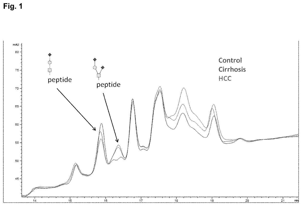
Isolation of HPX and Separation of Glycopeptides by HILIC Chromatography
[0178]HPX was purified from plasma by hemin affinity followed by C18 chromatography as previously described. Sanda et al., Electrophoresis 34: 2342-2349 (2013). Isolated HPX (20 pmol), dried in a vacuum concentrator and stored at −80° C., was suspended in 50 mM ammonium bicarbonate solution, pH 7.8 (Sigma-Aldrich, St. Louis, Mo.) with 0.05% RapiGest (Waters, Milford, Mass.), reduced, alkylated, and digested with 0.2 μg of Trypsin Gold for MS (Promega, Madison, Wis.) in a Barocycler reactor (Pressure BioSciences, South Easton, Mass.) at 37° C. for 60 minutes, also as previously described. O-glycopeptides were isolated by hydrophilic interaction chromatography (HILIC) on a ZIC-HILIC column 150×2.1 mm, 3 μm particles (EMD Merck, Germany) in a 30 min HPLC (Agilent, Santa Clara, Calif.) gradient of acetonitrile (ACN) / H2O with 0.01% trifluoroacetic acid (TFA). Starting conditions were set to 90% ACN with 0.01% TFA fol...
example 2
De-Sialylation and Exoglycosidase Treatment of O-Glycopeptides of HPX
[0179]Tryptic digest of HPX or the HILIC-enriched glycopeptide fractions of the tryptic digest were de-sialylated with 2 M acetic acid (Sigma-Aldrich) at 80° C. Completion of the de-sialylation reaction was confirmed by LC-MS / MS analysis. De-sialylated glycopeptides were evaporated in a vacuum concentrator (Labconco) and further treated with 0.1 U of 1-3 beta galactosidase (New England BioLabs, Ipswich, Mass.) in 50 mM sodium acetate, pH 6, at 37° C. for 8 hours. Products of the reaction were cleaned for further analysis by SPE C18, concentrated using vacuum concentrator, and dissolved in 0.1% formic acid for further LC-MS analysis.
example 3
LC-MS / MS Analysis
[0180]Tryptic digest of HPX, HILIC-enriched fractions, or acid- and exoglycosidase-treated glycopeptide fractions were analyzed under identical chromatographic conditions. Peptides were separated by reversed phase chromatography (Tempo Eksigent-AB Sciex, Framingham, Mass.) on a ChromXP C18-CL (3 μm, 120 Å, 180 μm, 20 mm) trap column and ChromXP C18-CL (3 μm, 120 Å, 75 μm, 150 mm) HPLC capillary chip column (Eksigent-AB Sciex) interfaced with a 5600 TripleTOF mass spectrometer (AB Sciex, Framingham, Mass.). Chromatographic method consisted of 10-min trapping / washing step (2% ACN, 0.1% formic acid (FA)) at 3 μL / min flow rate and 30 min gradient elution at a flow rate of 300 nL / min (Solvent A: 2% ACN with 0.1% FA; Solvent B: 100% ACN with 0.1% FA) using the following timetable: 5-40% Solvent B 0-23 min; 45-100% Solvent B 23-25 min; 100% Solvent B 25-30 min. Mass spectrometer was set to ion spray voltage 2,400 V, ion source gas (GS1) 13, declustering potential 90 and in...
PUM
| Property | Measurement | Unit |
|---|---|---|
| pH | aaaaa | aaaaa |
| pH | aaaaa | aaaaa |
| flow rate | aaaaa | aaaaa |
Abstract
Description
Claims
Application Information
 - R&D
- Intellectual Property
- Life Sciences
- Materials
- Tech Scout
- Unparalleled Data Quality
- Higher Quality Content
- 60% Fewer Hallucinations
Browse by: Latest US Patents, China's latest patents, Technical Efficacy Thesaurus, Application Domain, Technology Topic, Popular Technical Reports.
© 2025 PatSnap. All rights reserved.Legal|Privacy policy|Modern Slavery Act Transparency Statement|Sitemap|About US| Contact US: help@patsnap.com