Multifocal imaging systems and method

a multi-focal imaging and imaging system technology, applied in the field of multi-focal imaging systems and methods, can solve the problems of reducing the resolution of the resulting image, affecting the usefulness of tissue images, and cross-talk that can occur, so as to achieve fast imaging of the material

Active Publication Date: 2020-03-24
MASSACHUSETTS INST OF TECH
View PDF85 Cites 10 Cited by
  • Summary
  • Abstract
  • Description
  • Claims
  • Application Information

AI Technical Summary

Benefits of technology

The present invention provides systems and methods for the multifocal imaging of biological materials using optical systems with focal locations. The detector can comprise a plurality of detector elements that are correlated with the focal locations to efficiently collect light from the material being optically measured. The invention also addresses the issue of cross talk caused by the scattering of light and provides solutions to increase the resolution of the resulting images. Additionally, the invention is capable of acquiring high quality 3D datasets over 3D tissue and organ samples suitable for characterization of the vasculature and metastasis of cancer cells. The invention can also be used to stain the tissue and improve the classification and automation of the process.

Problems solved by technology

An important issue in the collection of light from discrete focal spots or locations within a turbid medium such as tissue is the cross talk that can occur due to the scattering of light.
This cross talk can substantially limit the usefulness of the images of the tissue that are produced.
By increasing the distance between adjacent focal spots such cross talk can be reduced or eliminated, however, this reduces the resolution of the resulting image or increases the time needed to scan the tissue.
Equally importantly, traditional 3D microscopes sample only tens to hundreds of cells and can never achieve comparable statistical accuracy and precision in many biomedical assays as techniques such as flow cytometry and image cytometry.

Method used


Image

  • Multifocal imaging systems and method
  • Multifocal imaging systems and method
  • Multifocal imaging systems and method

Examples


case 1

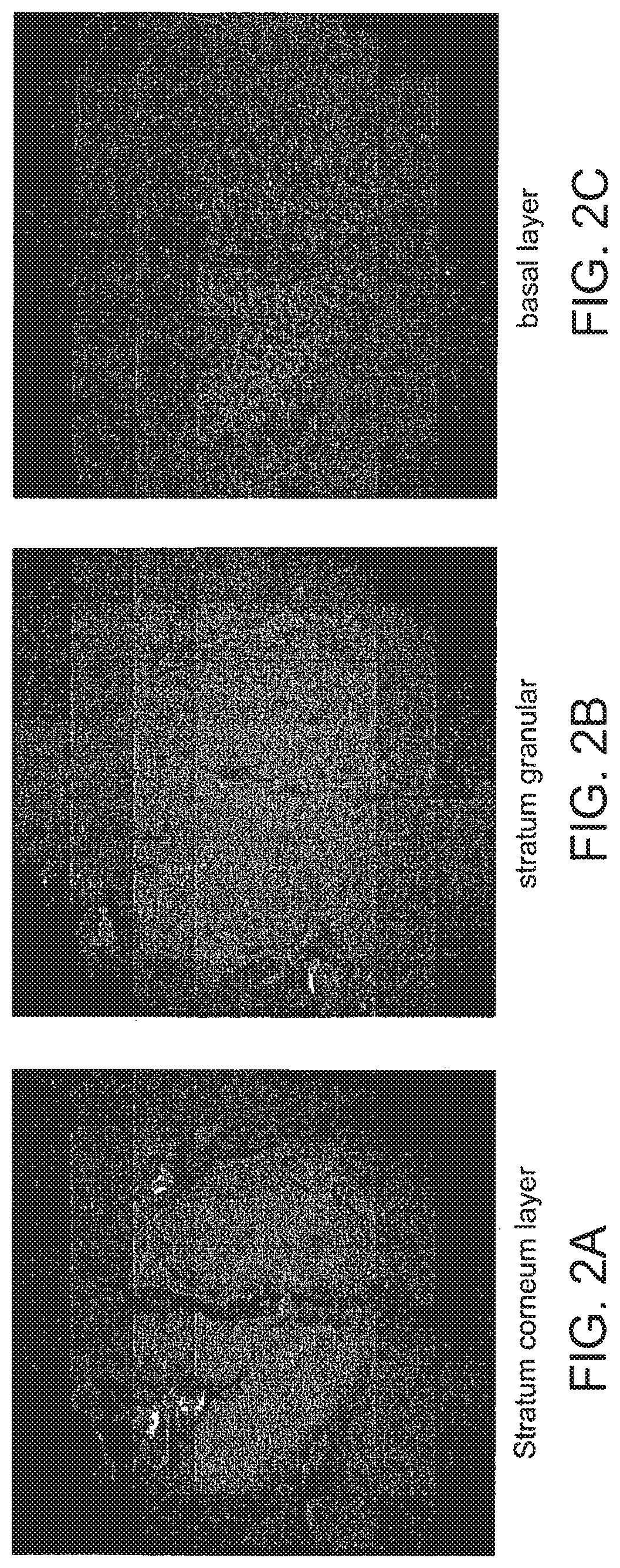
[0165]The normalized signal distribution resembles the normalized power map squared and shows an intensity drop of 45% toward the corner PMTs in respect to the center PMTs. The laser power was attenuated to 75.3 mW in the sample and can reach a maximal value of approx. 645 mW, resembling a power of approx. 15 mW for the center foci in the sample. Measured intensity profile can be generated by imaging a homogeneously distributed fluorescent dye under a cover slip. The intensity measurement is not only mapped by the power / intensity distribution of the foci, but also by the sensitivity of the detector array. As a result it resembles the “true” measured intensity distribution. The image consists of 192×192 pixels and was generated by an array of 6×6 foci which were scanned across a uniform fluorescent dye sample. 2D xy image normalization is carried out in different ways:[0166] The normalized inverse of this intensity image (from a uniform fluorescent dye) is multiplied with the yx imag...


PUM


Abstract

In the systems and methods of the present invention a multifocal multiphoton imaging system has a signal to noise ratio (SNR) that is reduced by over an order of magnitude at imaging depth equal to twice the mean free path scattering length of the specimen. An MMM system based on an area detector such as a multianode photomultiplier tube (MAPMT) that is optimized for high-speed tissue imaging. The specimen is raster-scanned with an array of excitation light beams. The emission photons from the array of excitation foci are collected simultaneously by a MAPMT and the signals from each anode are detected using high sensitivity, low noise single photon counting circuits. An image is formed by the temporal encoding of the integrated signal with a raster scanning pattern. A deconvolution procedure taking account of the spatial distribution and the raster temporal encoding of collected photons can be used to improve decay coefficient. We demonstrate MAPMT-based MMM can provide significantly better contrast than CCD-based existing systems.

Description

CROSS REFERENCE TO RELATED APPLICATIONS[0001]This application is a continuation of U.S. patent application Ser. No. 11 / 442,702, filed May 25, 2006, which claims the priority of U.S. Provisional Application No. 60 / 684,608 filed May 25, 2005 entitled, MULTI FOCAL MULTIPHOTON IMAGING SYSTEMS AND METHODS, the whole of each of these applications being hereby incorporated by reference herein.STATEMENT REGARDING FEDERALLY SPONSORED RESEARCH OR DEVELOPMENT[0002]This invention was made with government support under grant numbers R33 CA84740 and R33 CA91354 awarded by the National Institutes of Health. The government has certain rights in this invention.BACKGROUND OF THE INVENTION[0003]Systems and methods for microscopic analysis of biological material have been used for characterization and diagnosis in many applications. Fluorescence microscopy, for example, has been used for optical analysis including the histological analysis of excised tissue specimens. Optical coherence tomography has b...

Claims


Application Information

Patent Timeline
Patent Type & Authority Patents(United States)
IPC IPC(8): G01N21/64G02B21/16G02B21/00
CPCG01N21/6486G01N21/6458G02B21/002G02B21/0076G01N21/6452G02B21/0032G02B21/16
Inventor BAHLMAN, KARSTENKIM, KI-HEANRAGAN, TIMOTHYSO, PETER T. C.
Owner MASSACHUSETTS INST OF TECH
Why Eureka
Try Eureka