
Device and method for removal of blood-borne pathogens, toxins and inflammatory cytokines

a technology of inflammatory cytokines and blood-borne pathogens, which is applied in the field of devices and methods for removing blood-borne pathogens, toxins and inflammatory cytokines, can solve the problems of few treatments that attempt to reduce the inflammation caused during sepsis, infection remains a leading cause of death for battle trauma patients, and many organ failures and deaths. it achieves rapid adsorption and minimizes inflammation

Active Publication Date: 2020-01-21
EXTHERA MEDICAL
View PDF178 Cites 6 Cited by
  • Summary
  • Abstract
  • Description
  • Claims
  • Application Information

AI Technical Summary

Benefits of technology

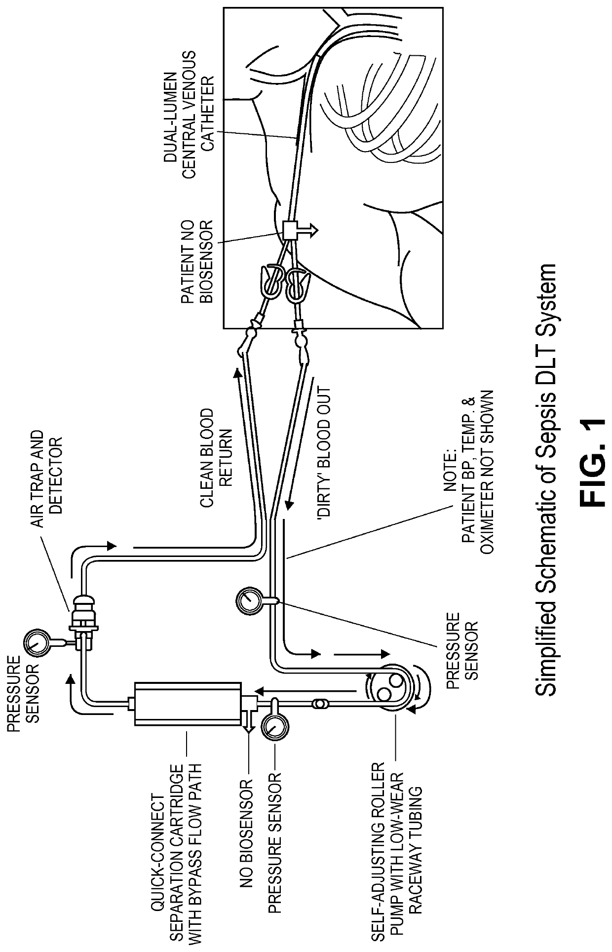
The present invention is a treatment system that avoids the pitfalls of previous therapies by using convection transport, specific capture of desired molecules, and a natural anticoagulant to minimize inflammation. The system operates without the need for systemic anticoagulation and is effective against a significant number of blood-borne factors of interest. It reduces the patient's pathogen load and includes a biosensor for nitric oxide. The system is a self-powered, safe, and efficient treatment for sepsis and dysregulated inflammation.

Problems solved by technology

Infection remains a leading cause of death for battle trauma patients.
The body's own defense system begins to attack healthy tissue, often leading to multiple organ failure and death.
Although antibiotics are generally effective in treating an infection, there are very few treatments that attempt to reduce the inflammation caused during sepsis.
Outside the military, more than 750,000 people in the United States develop severe sepsis every year, a syndrome characterized by an overwhelming systemic response to infection that can rapidly lead to organ failure and ultimately death.
Additionally, sepsis may elicit the onset of both abnormal clotting and bleeding leading to disseminated intra-vascular coagulation (DIC).
After many attempts at a pharmacological treatment, survival rates remained unimproved.
A serious limitation is the time required to perform cell cultures for pathogen identification for proper antibiotic selection.
Emboli can clog blood vessels, depriving downstream tissue of blood, with often life-threatening consequences.
The interaction of blood and a medical device is complicated further by the simultaneous initiation of the extrinsic pathway of the coagulation cascade when tissue factor is exposed.
Unfortunately, uncontrolled bleeding and spontaneous hemorrhage can occur with such procedures and is clearly a risk for wounded warriors.
Longer term use of soluble systemic heparin can lead to heparin-induced thrombocytopenia, a chronic condition involving severe platelet depletion.
Virtually all, materials used in conventional dialysis, and high-surface-area, nano-porous media separation technologies suffer from clotting risks.
Several dialysis-like therapy (DLT) separation technologies for sepsis treatment have been tested by others with little success.
Continuous veno-venous hemofiltration and high-volume hemofiltration have shown positive results for septic patients with renal failure, but conclusive evidence for benefit of patients without renal failure is lacking.
Additionally, deleterious effects may occur with these earlier technologies.
The disadvantage of current technologies include 1) reliance on slow diffusion kinetics within the media's nano-sized pores 2) indiscriminate capture, 3) lack of pathogen capture, 4) use of artificial materials that ironically increase inflammation and 5) need for systemic anticoagulation.
In addition, so-called size exclusion separation is difficult in clinical practice since many of the biomarkers (e.g. kidney-creatinine and liver-bilirubin) used by clinicians to evaluate patient status are removed by the separation process itself.
However, polymyxin B is cationic and is inherently very thrombogenic.

Method used


Image

  • Device and method for removal of blood-borne pathogens, toxins and inflammatory cytokines
  • Device and method for removal of blood-borne pathogens, toxins and inflammatory cytokines
  • Device and method for removal of blood-borne pathogens, toxins and inflammatory cytokines

Examples


example 1

Preparation of Heparin Column

[0209]Polyethylene (PE) beads, with an average diameter of 0.3 mm (lot no. 180153), are supplied by the Polymer Technology Group (Berkeley, USA) and the columns (Mobicol, 1 mL) are obtained from MoBiTec (Germany). Heparin and polyethyleneimine (PEI) are purchased from Scientific Protein Laboratories (Waunakee, Wis., USA) and BASF (Ludwigshafen, Germany) respectively. All chemicals used are of analytical grade or better.

[0210]Immobilization of heparin onto the beads was performed as described by Larm et al. (Larm O, Larsson R, Olsson P. A new non-thrombogenic surface prepared by selective covalent binding of heparin via a modified reducing terminal residue. Biomater Med Devices Artif Organs 1983; 11: 161-173).

[0211]The polymeric surface was heparinized using the general procedure described below.

[0212]The polymeric surface is etched with a oxidizing agent (potassium permanganate, ammoniumperoxidisulfate) in order to introduce hydrophilic characteristics t...

example 2

Patients

[0221]The study protocol was approved by the local ethics committee at the Karolinska University Hospital and signed informed consent was obtained from each patient. Arterial blood was drawn from the hemodialyzers of three septic (fever>38° C., chills, leukocytes>12×109 cells / L) patients (2M / 1F, aged 43, 56 and 68 years; Table 1).

[0222]

TABLE 1Clinical characteristics of patients donating blood.WhiteMeanbloodBodyarterialRespiratorycellAgetemperatureHeart ratepressureratecountPatientSex(years)(° C.)(beats / min)(mmHg)(breaths / min)(109 / L)1M4339.2°110762019.02M5638.6°90951817.53F6838.5°100892119.5

[0223]The patients are previously administered with broad-spectrum antibiotics (ceftazidime or cefuroxime together with an aminoglycoside; one dose of each) and heparin (200 IU / kg body weight at the start of the dialysis). The blood was collected in EDTA vacuum tubes and immediately transferred to an adjacent room where 1 mL was applied to the previously prepared columns and passed throug...

example 3

Quantitative Analysis of Cytokines

[0224]The amounts of cytokines are determined using photoluminescence with a plate reader (Multiskan Ascent). Each sample was measured in three wells, and the geometric mean used for analysis. The intraassay coefficient of variation was below 8% for all kits. We used Coamatic antithrombin kit (Haemochrom, cat #8211991), Quantikine Human IL-6 (R&D Systems, cat #D6050), Quantikine Human IL-10 (R&D Systems, cat #D1000B), Protein C Antigen Test 96 (HL Scandinavia AB, product #H5285), Human CCL5 / RANTES Quantikine (R&D Systems, cat #DRN00B), Quantikine Human sVCAM-1 (CD106) (R&D Systems, cat #DVC00), Quantikine Human IFN-gamma (R&D Systems, cat #DIF50), Quantikine HS TNF-alpha / TNFSF1A (R&D Systems, cat #HSTA00D) and BD OptEIA Human C5a ELISA Kit II (BD Biosciences, cat #557965).

Statistical Evaluation

[0225]Paired Kruskal-Wallis test was used to compare the before and after column blood concentrations of each cytokine, with a two-tailed p-value below 0.05 i...


PUM

PropertyMeasurementUnit
concentrationaaaaaaaaaa
pressure dropaaaaaaaaaa

Abstract

The present invention is directed to an integrated system and a method for utilizing the system to detect and remove blood-borne factors of interest, such as pathogens and / or toxins and / or cytokines, from blood or serum (blood) by contacting the blood with a solid, essentially nonporous substrate which has been surface treated with molecules or chemical groups (the adsorbent media or media) having a binding affinity for the pathogens and / or toxins to be removed (the adsorbents). The invention can be used to remove virulence factors, e.g. toxins, that are released from various pathogens. In one aspect, the invention is for the treatment of sepsis and infection, such as infections associated with battle field trauma.

Description

CROSS-REFERENCE TO RELATED APPLICATIONS[0001]This application is a continuation of PCT / US2012 / 025316, filed Feb. 15, 2012, which claims priority to U.S. Provisional Application No. 61 / 443,236, filed Feb. 15, 2011, and U.S. Provisional Application No. 61 / 471,102, filed Apr. 1, 2011, the disclosures of which are hereby incorporated by reference in their entirety for all purposes.FIELD OF THE INVENTION[0002]The present invention is directed to an integrated system and a method for utilizing the system to detect and remove blood-borne factors of interest, such as pathogens and / or toxins and / or cytokines, from blood or serum (blood) by contacting the blood with a solid, essentially nonporous substrate which has been surface treated with molecules or chemical groups (the adsorbent media or media) having a binding affinity for the pathogens and / or toxins to be removed (the adsorbents). The invention can be used to remove virulence factors, e.g. toxins, that are released from various pathog...

Claims


Application Information

Patent Timeline
Patent Type & Authority Patents(United States)
IPC IPC(8): A61B5/00A61B5/145A61B5/01A61M1/16A61B5/1468A61B5/0205A61B5/02
CPCA61B5/14546A61B5/4836A61B5/01A61B5/486A61M1/16A61B5/02055A61B5/14532A61B5/14542A61B5/0002A61B5/02028A61B5/1468A61M1/3679A61M2205/3303A61B5/6866A61B5/0215A61B5/145A61M2202/206A61M1/3673B01J20/321B01J20/3212B01J20/3251A61M2205/3368A61M2205/3331A61M1/34
Inventor MCCREA, KEITHWARD, ROBERT S.PITARRA, JR., GEORGE
Owner EXTHERA MEDICAL
Learn More