Method for preparing mB7.1-GPI fusion proteins and their uses

A fusion protein, mb7.1-gpi technology, applied in the field of bioengineering

Inactive Publication Date: 2004-11-10
ZHEJIANG UNIV
View PDF0 Cites 5 Cited by
  • Summary
  • Abstract
  • Description
  • Claims
  • Application Information

AI Technical Summary

Problems solved by technology

The method of preparing mB7.1-GPI in this study has not been reported in domestic and foreign literature

Method used


Image

  • Method for preparing mB7.1-GPI fusion proteins and their uses
  • Method for preparing mB7.1-GPI fusion proteins and their uses
  • Method for preparing mB7.1-GPI fusion proteins and their uses

Examples


Embodiment 1

[0041] Example 1 Construction of eukaryotic expression vector of mB7.1-GPI gene, expression and product purification- Construction of hPLAP-1 exon 11 cloning vector:

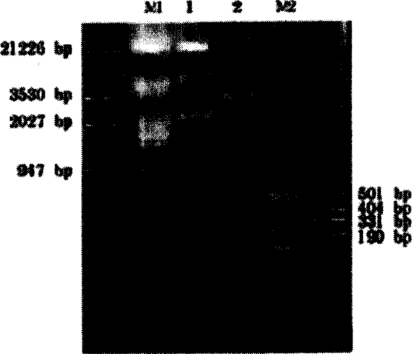
[0042] 1. Electrophoresis analysis of the amplified hPLAP-1 gene exon 11 product. The size of the PCR-amplified hPLAP-1 gene exon 11 is 307bp. The results of 1.5% agarose gel electrophoresis can be found in figure 1 , where M: 100bp DNA molecular size marker. Lane 1: PCR product of hPLAP-111 exon.

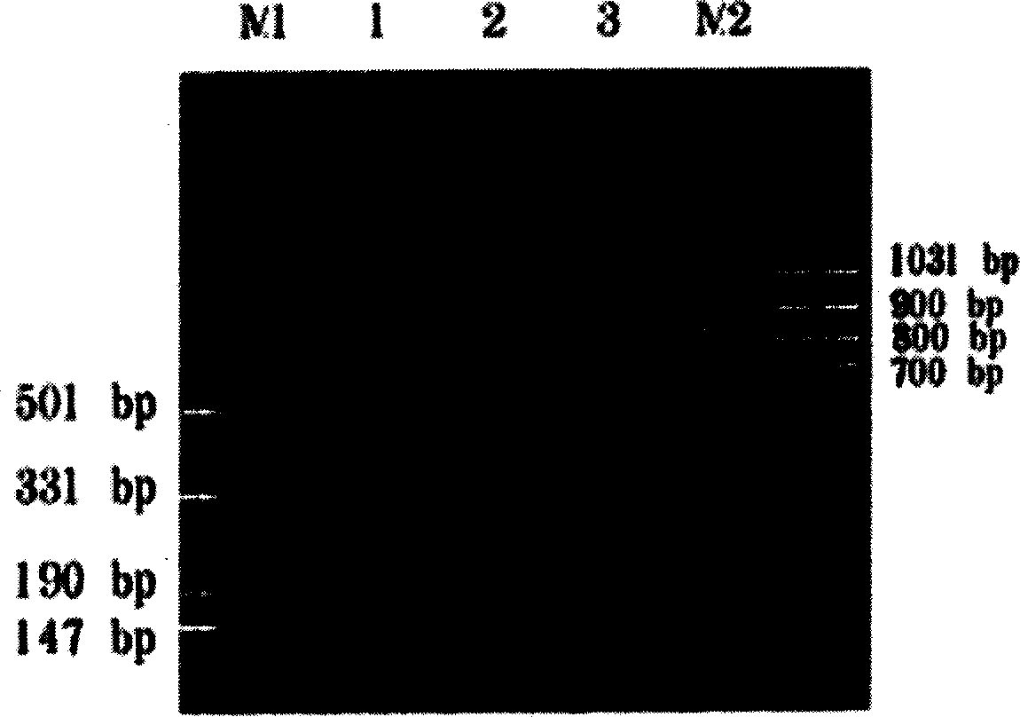
[0043] 2. Enzyme digestion and identification of recombinant pGEM-hPLAP-1 11exon plasmid DNA Recombinant pGEM-hPLAP-1 11exon plasmid DNA was double-digested with EcoRI and XhoI, and the digested product was electrophoresed on 1.5% agarose gel. The size of the target gene was 307bp , the results see figure 2 , where M1, M2: DNA molecular size markers, lane 1: the PCR product of exon 11 of hPLAP-1, lane 2: the product of enzyme digestion of exon 11 of pGEM-hPLAP-1.

[0044] 3. Sequence Analysis of Amplified Genes

...

Embodiment 2

[0066] Example 2 Application research of mB7.1-GPI fusion protein

[0067] 1. Anchoring effect of mB7.1-GPI fusion protein on tumor cell membrane

[0068] mB7.1-GPI was incubated with EL-4 tumor cells, and the immunofluorescent laser confocal microscope observation showed that the surface of the cell membrane showed green fluorescence (see Figure 14 ), indicating that mB7.1-GPI protein can be anchored to the EL-4 tumor cell membrane. mB7.1-GPI and EL-4 tumor cells were co-incubated for 4 hours, then placed at 4°C for 0h, 4h or 8h, and detected by FCM. The fluorescence intensities of samples at 0h, 4h, and 8h after anchoring were 13.7, 12.7, and 10.6, and the percentages of positive cells were 95.4%, 91.3%, and 85%, respectively. Compared with samples at 0h and 4h, the fluorescence intensity and the percentage of positive cells did not decrease significantly, indicating that the fusion protein can be anchored on the tumor cell membrane after incubation with EL-4 tumor cells,...


PUM

PropertyMeasurementUnit
molecular weightaaaaaaaaaa
fluorescenceaaaaaaaaaa

Abstract

The invention provides the preparation and application of mB7.1-GPI fusion protein, an eukaryotic expression system (pcDNA3.1(+)-mB7.1-GPI) of B7.1(mB7.1-GPI), preparing mB7.1-GPI fusion proteins. They can anchor on tumor cell membrane, and the experiment proves that the anchoring has considerably stability; keeps bioactivity of immune protein, and has the functions of stimulating multiplication of mice splenic cells and secreting IL-2 and IFN-Y genes. The mB7.1-GPIs anchor the tumor cells to make tumor vaccine able to inhibit tumor growth of tumor-bearing mice and prolong the living time of the tumor-bearing mice, and its mechanism has relation with the activity of animal immune cells NK and CTL in tumor vaccine enhancement test and the cell genes IL-2 and IFN-gama secreted.

Description

[0001] Field [0002] The invention belongs to bioengineering technology, and mainly relates to the preparation of glycosylated phosphatidylinositol-anchored mouse B7.1 fusion protein, the preparation of the fusion protein membrane surface modified tumor vaccine and the application in tumor treatment. Background technique [0003] Anti-tumor vaccines are the main strategy of tumor immune gene therapy. During the preparation of tumor vaccines, the surface of autologous or allogenic tumor cells is modified to enhance their immunogenicity and immune efficacy. Currently, tumor vaccine modification is mainly realized by gene transfection. Such as Hodge, etc. [1] Introduce the B7.1 gene into tumor cells for expression, provide co-stimulatory factors, and enhance the anti-tumor immune response of tumor vaccines; Shrayer et al. [2] The mouse B16 melanoma cells were transfected with SEA gene to express SEA, and the tumor cells were used to make tumor vaccines, which showed a strong an...

Claims


Application Information

Patent Timeline
Patent Type & Authority Applications(China)
IPC IPC(8): A61K38/16A61P35/00C12N15/62
Inventor 余海易平勇
Owner ZHEJIANG UNIV
Why Eureka
Try Eureka