Method for automatically segmenting chest anterior mediastinal focus based on CT image

A CT image and automatic segmentation technology, applied in the field of medical image processing, can solve the problem of low incidence of anterior mediastinal lesions, and achieve the effect of optimizing the lesion segmentation edge, reducing the complexity and improving the segmentation accuracy.

Pending Publication Date: 2021-11-26
北京康兴顺达科贸有限公司
View PDF0 Cites 0 Cited by
  • Summary
  • Abstract
  • Description
  • Claims
  • Application Information

AI Technical Summary

Problems solved by technology

However, due to the relatively low incidence of anterior mediastinal lesions, there are few studies on the automatic segmentation of CT images, so how to accurately segment anterior mediastinal lesions still faces great challenges

Method used


Image

  • Method for automatically segmenting chest anterior mediastinal focus based on CT image
  • Method for automatically segmenting chest anterior mediastinal focus based on CT image
  • Method for automatically segmenting chest anterior mediastinal focus based on CT image

Examples


Embodiment Construction

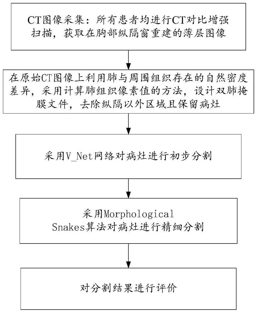
[0016] Such as image 3 As shown, this method for automatic segmentation of chest anterior mediastinal lesions based on CT images comprises the following steps:

[0017] (1) CT image acquisition: All patients underwent enhanced chest CT scans to obtain thin-layer images reconstructed in the chest mediastinum window;

[0018] (2) Using the natural density difference between the lung and the surrounding tissue on the original CT image, the method of calculating the pixel value of the lung tissue is used to design a double-lung mask file to remove areas other than the mediastinum and retain lesions;

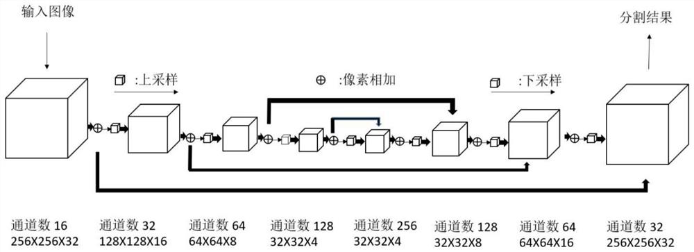
[0019] (3) Use the V_Net network to initially segment the lesion (Initial Segmentation);

[0020] (4) Using the Morphological Snakes algorithm to finely segment the lesion (AccurateSegmentation);

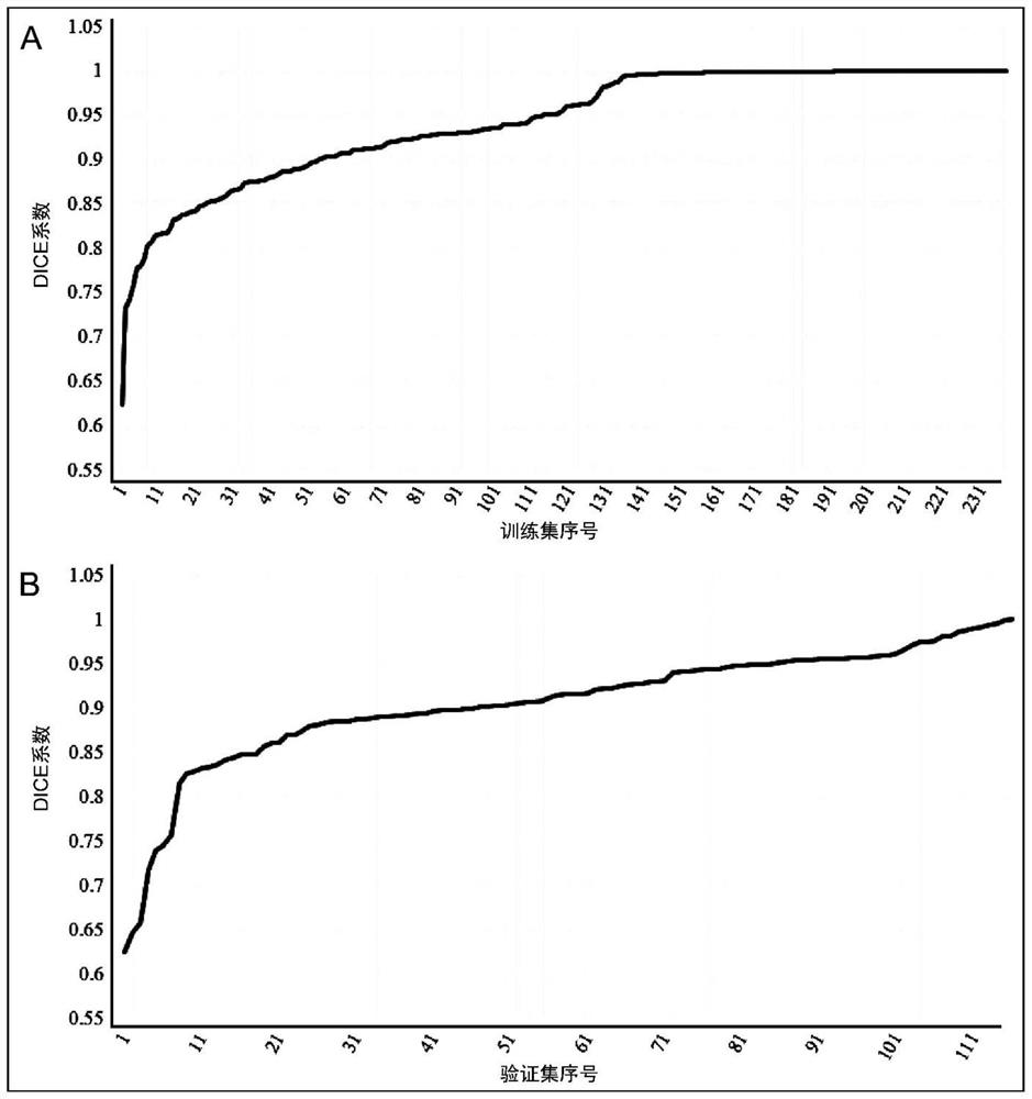
[0021] (5) Evaluate the segmentation results.

[0022] First, based on the original CT image, the present invention adopts the method of calculating lung tissue pixel values ​​to desig...


PUM


Abstract

According to the method for automatically segmenting the chest anterior mediastinal focus based on the CT image, the segmentation result is good, quantitative feature analysis of subsequent deep learning can be met, and a methodological basis is provided for automatic segmentation research of the anterior mediastinal focus. The method for automatically segmenting the chest anterior mediastinal focus based on the CT image comprises the following steps: (1) CT image acquisition: performing chest CT contrast enhancement scanning on all patients to obtain a thin-layer image reconstructed by a mediastinal window; (2) designing a double-lung mask file on the original CT image by using the natural density difference between the lung and surrounding tissues and adopting a method for calculating the pixel value of lung tissues, and removing the area outside the mediastinal septum; (3) carrying out preliminary segmentation on the focus by adopting a V_Net network; (4) carrying out fine segmentation on the focus by adopting a Morphological Snakes algorithm; and (5) evaluating a segmentation result.

Description

technical field [0001] The invention relates to the technical field of medical image processing, in particular to a method for automatically segmenting chest anterior mediastinal lesions based on CT images. Background technique [0002] The purpose of preoperative imaging examination of thoracic anterior mediastinal lesions is to initially evaluate the malignancy of the tumor, and further speculate on its histopathological changes and conduct risk assessment, so as to assist in the selection of preoperative treatment options and clinical prognosis. The evaluation of CT imaging signs is based on the lesion itself and its relationship with the surrounding tissue structure, and empirical and observational indicators are mostly used instead of quantitative indicators. Radiomics is a computerized quantitative imaging analysis method that can perform high-throughput feature extraction on medical imaging images. However, the traditional radiomics segmentation of lesions relies on m...

Claims


Application Information

Patent Timeline
IPC IPC(8): G06T7/11G06T7/12G06T7/136G06T7/187G06T7/00G06N3/04G06N3/08G16H50/20G16H50/30G16H30/20
CPCG06T7/11G06T7/12G06T7/136G06T7/187G06T7/0012G06N3/08G16H50/20G16H50/30G16H30/20G06T2207/10081G06T2207/20081G06T2207/30061G06T2207/30096G06N3/045Y02T10/40
Inventor 马国林李海梅张冰韩小伟刘秀秀
Owner 北京康兴顺达科贸有限公司
Why Eureka
Try Eureka